扩展功能
文章信息
- 米小其, 王成
- Xiaoqi MI, Cheng WANG
- 雌性虎纹毛园蛛和雄性浦那毛园蛛的新发现(蜘蛛目:园蛛科)
- First Description on the Female of Eriovixia huwena and the Male of E. poonaensis (Araneae, Araneidae)
- 四川动物, 2016, 35(5): 728-733
- Sichuan Journal of Zoology, 2016, 35(5): 728-733
- 10.11984/j.issn.1000-7083.20160107
-
文章历史
- 收稿日期: 2016-06-03
- 接受日期: 2016-06-15
The small to median-sized orb-weaver spider genus Eriovixia Archer, 1951 is characteristized by the combination of the following characters: carapace pear-shaped; abdomen ovoid, subtriangular or polygonal, female of some species having a long “tail” beyond the spinnerets; epigynum linguiform or triangular, arched medially; median ocular area of male situated on an eye tubercle; palpal patella without macroseta. A total of 20 species from Africa and Asia included in the genus (World Spider Catalog, 2016). Yin et al.(1990) described three new species from Yunnan and Hainan province in 1990. Yin and Zhao (1994) described another new species from Hubei province. Four species including a new species from the Gaoligong Mountains, western Yunnan province (Mi et al., 2010) and 6 species including 3 new species from Hainan province (Han & Zhu, 2010) were described in 2010. So far, 14 species have been recorded from China, mainly occurred in south China, especially in Yunnan and Hainan province (Schenkel, 1963; Song, 1987; Feng, 1990; Yin et al., 1990, 1997, 2012; Chen & Zhang, 1991;Yin & Zhao, 1994; Zhu et al., 1994; Song et al., 1999, 2001;Han & Zhu, 2010; Mi et al., 2010).
While examining the specimens collected from Yunnan-Guizhou Plateau, southwest China, 9 species of Eriovixia were identified: E. cavaleriei (Schenkel, 1963), E. excelsa (Simon, 1889), E. huwena Han & Zhu, 2010, E. laglaizei (Simon, 1877), E. nigrimaculata Han & Zhu, 2010, E. poonaensis (Tikader & Bal, 1981), E. pseudocentrodes (BÖsenberg & Strand, 1906), E. sticta Mi, Peng & Yin, 2010 and E. yunnanensis (Yin et al., 1990). E. poonaensis and E. huwena were re-described in this paper, with the female of E. huwena and the male of E. poonaensis were described for the first time. Diagnoses, distribution data, illustrations of the two species are provided. The specimens are deposited in Museum of Tongren University (MTU).
Material and methods
Specimens are stored in 75% ethanol. The epigynum was cleared in lactic acid for examination. Specimens were examined using an Olympus SZ30 stereo microscope. Digital photographs were taken using a Cannon Powershot G12 digital camera mounted on an Olympus SZX16, compound focus images were generated using Helicon Focus. Leg measurements are given as: total length (femur, patella + tibia, metatarsus, tarsus). All measurements are given in millimeters (mm). Abbreviations used in the text: ALE, anterior lateral eyes; AME, anterior median eyes; MOA, median ocular area; PLE, posterior lateral eyes; PME, posterior median eyes.
Taxonomy
Family Araneidae Clerck, 1757
Genus Eriovixia Archer, 1951
Eriovixia huwena Han & Zhu, 2010 (Fig. 1~6)

|
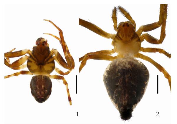
| Fig. 1~2 Eriovixia huwena Han & Zhu, 2010 1. Male habitus, dorsal view; 2. Female habitus, dorsal view; scale bars=1 mm. |
| |

|
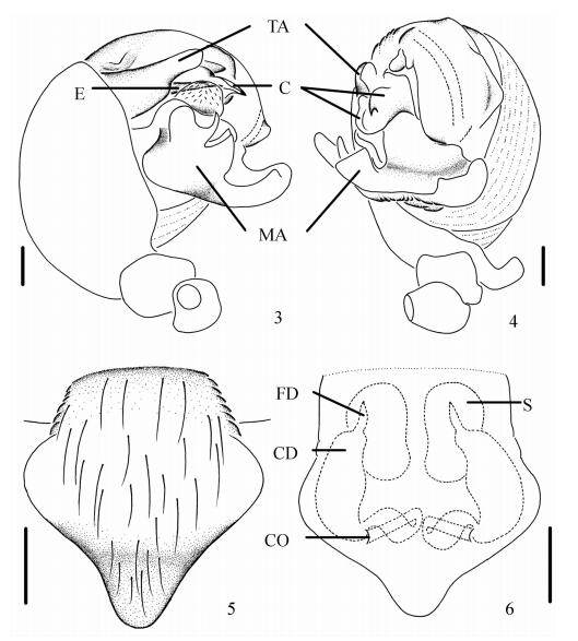
| Fig. 3~6 Eriovixia huwena Han & Zhu, 2010 3. Left palp, lateral view; 4. Left palp, ventral view; 5. Epigynum, ventral view; 6. Vulva, posterior view; C.conductor, CD.copulatory ducts, CO.copulatory openings, E.embolus, FD.fertilization ducts, MA.median apophysis, S.spermatheca, TA.terminal apophysis; scale bars=0.1 mm. |
| |
Eriovixia huwena Han & Zhu, 2010: 2619, Figs 2A, 8A~B (♂).
Material examined: 1 male and 1 female, Taizhong town (24°39.96′N, 100°56.27′E, 1 374 m), Jingdong county, Yunnan province, 15 August 2015, X.Q. Mi, M.Y. Liao, T. J. Liu and X. Kuang leg.; 1 female, Wengang village (24°20.33′N, 101°8.46′E, 1 728 m), Huashan town, Jingdong county, Yunnan province, 16 August 2015, C. Wang, Z. L. Liao, P. Luo and G. T. Liu leg.; 1 male, Minjiang village (24°0.79′N, 101°7.09′E, 1 083 m), Enle town, Zhenyuan county, Yunnan province, 17 August 2015, C. Wang, P. Luo, X. Kuang, G. T. Liu and T. J. Liu leg.
Diagnosis: This species is similar to E. pseudocentrodes (BÖsenberg & Strand, 1906) (see Mi et al., 2010; Figs. 9~10) in body shape and patches, but can be separated from the latter by: 1) terminal apophysis of male palp thick, wedge-shaped, length more than 1.7 times its width from lateral view (Figs. 3~4), whereas terminal apophysis thin, rectangular, length less than 1.4 times its width in E. pseudocentrodes (see Mi et al., 2010: Figs. 14~15); 2) spermathecae kidney shaped (Fig. 6), compared to ovoid in E. pseudocentrodes (see Mi et al., 2010: Figs. 11~13); 3) copulatory ducts expanded near the spermathecae (Fig. 6) but not expanded in E. pseudocentrodes (see Mi et al., 2010: Figs. 11, 13).
Description: Male: Carapace pear-shaped, dark brown with a yellow butterfly shaped patch, cervical groove inconspicuous, thoracic fovea transverse. Chelicerae yellow, has four promarginal and three retromarginal teeth. Sternum yellow, shield shaped. Legs yellow with wide dark annuli. Abdomen longer than wide, dorsal with an inconspicuous folium and dark margins (Fig. 1). Venter brown, with a big white spot medially. Spinnerets brown, situated at posterior 1/3 of the abdomen. Male palp with a digitiform paracymbium; median apophysis prominent, basal part with a dorsal fin and two denticles, distal part navicular; conductor wide, curled into a pointed tip; embolus straight, almost covered by the terminal apophysis and the conductor from lateral view; terminal apophysis thick, wedge-shaped (Figs. 3~4). Total length 3.30. Carapace length 1.80, width 1.60; abdomen length 1.60, width 1.20. Eye sizes and interdistances: AME 0.11, ALE 0.09, PME 0.13, PLE 0.09, AME-AME 0.13, AME-ALE 0.28, PME-PME 0.13, PME-PLE 0.30, MOA length 0.33 with front width 0.33 and back width 0.33. Leg measurements: Ⅰ 5.35 (1.70, 2.00, 1.20, 0.45), Ⅱ 4.50 (1.45, 1.65, 1.00, 0.40), Ⅲ 2.70 (0.95, 0.95, 0.50, 0.30), Ⅳ 3.80 (1.25, 1.30, 0.90, 0.35).
Female: Coloration and body shape as in male except with a long “tail”, spinnerets situated at anterior 1/3 of the abdomen (Fig. 2). Epigynum with a triangular scape; copulatory openings narrow, situated on posterior surface; copulatory ducts twisted near the copulatory openings and expanded near the spermathecae; spermathecae kidney shaped (Figs. 5~6). Total length 5.30. Carapace length 1.55, width 1.40; abdomen length 3.50, width 2.35. Eye sizes and interdistances: AME 0.10, ALE 0.09, PME 0.11, PLE 0.09, AME-AME 0.15, AME-ALE 0.30, PME-PME 0.18, PME-PLE 0.38, MOA length 0.30 with front width 0.28 and back width 0.35. Leg measurements: Ⅰ 6.00 (2.00, 2.25, 1.25, 0.50), Ⅱ 5.00 (1.55, 1.90, 1.10, 0.45), Ⅲ 2.85 (0.95, 1.00, 0.55, 0.35), Ⅳ 4.40 (1.50, 1.55, 0.95, 0.40).
Variation: Males, total length 3.30~3.50 (n=2); females, total length 5.30~5.40 (n=2).
Distribution: China (Hainan, Guangxi, Yunnan).
Ecology: The specimens were collected by beating shrubs.
Eriovixia poonaensis (Tikader & Bal, 1981) (Figs. 7~12)
Neoscona poonaensis Tikader & Bal, 1981: 29, Figs. 59~62; Yin et al., 1990: 115, Figs. 280~282 (♀).
Eriovixia poonaensis Grasshoff, 1986: 118; Yin et al., 1997: 301, Fig. 208a~g (♀); Song, Song, Zhu & Chen, 1999: 281, Figs. 166U, 167A, L (♀).
Material examined: 1 male and 5 females, Yingpan village, Huashan town, Jingdong county, Yunnan province (24°16.73′N, 101°5.87′E, 1 273 m), 15 August 2015, C. Wang, Z. L. Liao, P. Luo and G. T. Liu leg.; 1 female, Wengang village, Huashan town, Jingdong county, Yunnan province (24°20.33′N, 101°8.46′E, 1 728 m), 16 August 2015, C. Wang, Z. L. Liao, P. Luo and G. T. Liu leg.; 1 female, Minjiang village, Enle town, Zhenyuan county, Yunnan province (24°0.79′N, 101°7.09′E, 1 083 m), 17 August 2015, C. Wang, P. Luo, X. Kuang, G. T. Liu and T. J. Liu leg.
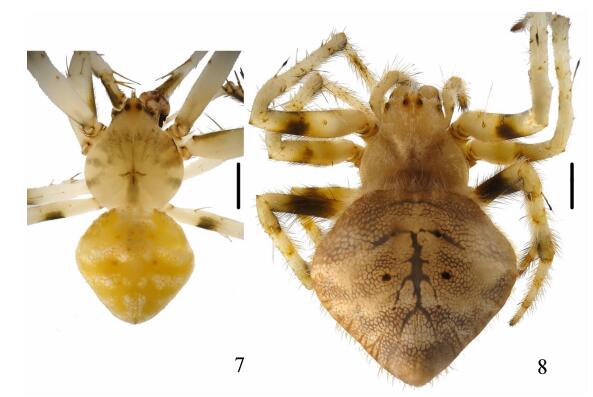
Diagnosis: This species is resemble to E. laglaizei (Simon, 1877) in having similar appearance and genitals (see Han & Zhu, 2010: Figs. 3A~B, 10A~I, and specimens from Maolan National Nature Reserve in Guizhou province were examined), but can be separated from E. laglaizei by: 1) abdomen without a hump-like tail (Figs. 7~8), whereas with a hump-like tail in E. laglaizei (see Han & Zhu, 2010: Figs. 3A~B); 2) 2 branches of the median apophysis have the same width from prolateral view (Figs. 9~10), compared to right branch is 2 times as wide as left branch from prolateral view in E. laglaizei (see Han & Zhu, 2010: Figs. 10H~I); 3) spermathecae spherical (Fig. 12) versus oval in E. laglaizei (see Han & Zhu, 2010: Figs. 10E, G).

|
| Fig. 7 Eriovixia poonaensis (Tikader & Bal, 1981) 7. Male habitus, dorsal view; 8. Female habitus, dorsal view; scale bars=1 mm. |
| |

|
| Fig. 9~12 Eriovixia poonaensis (Tikader & Bal, 1981) 9. Left palp, lateral view; 10. Left palp, ventral view; 11. Epigynum, ventral view; 12. Vulva, posterior view; C.conductor, CD.copulatory ducts, CO.copulatory openings, E.embolus, FD.fertilization ducts, MA.median apophysis, S.spermatheca, TA.terminal apophysis; scale bars=0.1 mm. |
| |
Description: Male: Carapace pear shaped, yellowish with dark radiated patches, cervical groove inconspicuous, thoracic fovea transverse. Chelicerae yellow, has 4 promarginal and 3 retromarginal teeth. Sternum yellow with dark patches, shield shaped. Legs pale with big brown patches in femurs. Abdomen pentagonal, dorsal yellowish with white scaly patches (Fig. 7). Venter pale with irregular brown patches. Spinnerets yellow brown. Male palp with a distally expanded paracymbium; median apophysis prominent, bifurcate; conductor thick, guiding the embolus; embolus fused with terminal apophysis in the base; terminal apophysis large, pointed distally (Figs. 9~10). Total length 4.90. Carapace length 2.50, width 2.10; abdomen length 2.50, width 2.50. Eye sizes and interdistances: AME 0.13, ALE 0.10, PME 0.13, PLE 0.10, AME-AME 0.15, AME-ALE 0.28, PME-PME 0.15, PME-PLE 0.35, MOA length 0.38 with front width 0.40 and back width 0.35. Leg measurements: Ⅰ 9.40 (3.20, 3.40, 2.10, 0.70), Ⅱ 6.90 (2.40, 2.20, 1.60, 0.70), Ⅲ 4.70 (1.80, 1.50, 0.90, 0.50), Ⅳ 6.80 (2.40, 2.20, 1.60, 0.60).
Female: Coloration and body shape as in male (Fig. 8). Epigynum with a lateral rimmed scape; copulatory openings situated on posterior surface; copulatory ducts twisted near the copulatory openings; spermathecae spherical (Figs. 11~12). Total length 6.40. Carapace length 2.50, width 2.20; abdomen length 4.30, width 4.30. Eye sizes and interdistances: AME 0.14, ALE 0.10, PME 0.14, PLE 0.10, AME-AME 0.18, AME-ALE 0.40, PME-PME 0.20, PME-PLE 0.48, MOA length 0.43 with front width 0.40 and back width 0.43. Leg measurements: Ⅰ 10.10 (3.60, 3.70, 2.00, 0.80), Ⅱ 7.90 (2.70, 2.80, 1.70, 0.70), Ⅲ 4.80 (1.70, 1.60, 0.90, 0.60), Ⅳ 7.40 (2.60, 2.50, 1.60, 0.70).
Variation: Females, total length 5.50~7.70 (n=7).
Distribution: China (Hainan, Guangxi, Yunnan), India.
Ecology: The specimens were collected by beating shrubs.
Acknowledgements: We are grateful to Jingdong Administration Bureau of Yunnan Ailaoshan National Nature Rerseve and Wuliangshan National Nature Rerseve for the kind help in the field work. We also thank an anonymous reviewer for helpful comments on this paper.| Archer AF. 1951. Studies in the orb-weaving spiders (Argiopidae). 1[J]. American Museum Novitates , 1487 : 1–52. |
| Chen ZF, Zhang ZH. 1991. Fauna of Zhejiang: Araneida[M]. Hangzhou: Zhejiang Science and Technology Publishing House: 85 -86. |
| Feng ZQ. 1990. Spiders of China in colour[M]. Changsha: Hunan Science and Technology Publishing House: 53 . |
| Han GX, Zhu MS. 2010. Taxonomy and biogeography of the spider genus Eriovixia(Araneae: Araneidae) from Hainan Island, China[J]. Journal of Natural History , 44 : 2609–2635. DOI:10.1080/00222933.2010.507315 |
| Mi XQ, Peng XJ, Yin CM. 2010. The orb-weaving spider genus Eriovixia(Araneae: Araneidae) in the Gaoligong Mountains, China[J]. Zootaxa , 2488 : 39–51. |
| Schenkel E. 1963. Ostasiatische Spinnen aus dem Muséum d'Histoire naturelle de Paris[J]. Mémoires du Muséum National d'Histoire Naturelle de Paris (A, Zool.) , 25 : 1–481. |
| Song DX, Zhu MS, Chen J. 1999. The spiders of China[M]. Shijiazhuang: Hebei University of Science and Techology Publishing House: 281, 287 -288. |
| Song DX, Zhu MS, Chen J. 2001. The fauna of Hebei, China: Araneae[M]. Shijiazhuang: Hebei University of Science and Techology Publishing House: 202 -204. |
| Song DX. 1987. Spiders from agricultural regions of China (Arachnida: Araneae)[M]. Beijing: Agriculture Publishing House: 160 -161. |
| Tikader BK, Bal A. 1981. Studies on some orb-weaving spiders of the genera Neoscona Simon and Araneus Clerck of the family Araneidae (=Argiopidae) from India[J]. Records of the Zoological Survey of India, Occasional Paper , 24 : 1–60. |
| World Spider Catalog. 2016. World Spider Catalog. Natural History Museum Bern[EB/OL].[2016-04-21]. http://wsc.nmbe.ch.version17.0. |
| Yin CM, Peng XJ, Yan HM, et al. 2012. Fauna of Hunan: Araneae in Hunan, China[M]. Changsha: Hunan Science and Technology Press: 685 -688. |
| Yin CM, Wang JF, Xie LP, et al. 1990. New and newly recorded species of the spiders of family Araneidae from China (Arachnida: Araneae) [M]//Yin CM, Wang JF. Spiders in China: one hundred new and newly recorded species of the families Araneidae and Agelenidae. Changsha: Hunan Normal University Press: 1-171.(in Chinese) |
| Yin CM, Wang JF, Zhu MS, et al. 1997. Fauna Sinica: Arachnida: Araneae: Araneidae[M]. Beijing: Science Press: 294 -302. |
| Yin CM, Zhao JZ. 1994. Some new species of family Araneidae from China (Arachnida: Araneae)[J]. Acta Arachnologica Sinica , 3(1) : 1–7. |
| Zhu MS, Song DX, Zhang YQ, et al. 1994. On some new species and new records of spiders of the family Araneidae from China[J]. Journal of Hebei Normal University (Natural Science Edition), (supplement) (supplement) : 25–52. |
 2016, Vol. 35


