 2024, Vol. 35


b School of Pharmaceutical Sciences, Guangzhou University of Chinese Medicine, Guangzhou 510006, China;
c Artemisinin Research Center, Guangzhou University of Chinese Medicine, Guangzhou 510006, China;
d State Key Laboratory of Respiratory Disease, the First Affiliated Hospital of Guangzhou Medical University, Guangzhou Medical University, Guangzhou 510120, China;
e Guangzhou Laboratory, Bio-Island, Guangzhou 510030, China;
f Nanshan Pharmaceutical Innovation Research Institute of Guangdong Province, Foshan 528225, China
The reaction between horseradish peroxidase (HRP) and a proper chemical substrate has been widely applied for quantitative measurements in vitro diagnostics and biochemical analyses [1–5]. Among all HRP techniques, the fluorescence assays have attracted increasing interests due to their excellent sensitivity and reasonably large linear detection ranges [6,7]. A dihydroxyphenoxazine (or hydroresorufin) analogue 10-acetyl-3,7-dihydroxyphenoxazine, also known as ADHP or Amplex red, is the most popular fluorescence substrate of HRP, which is supplied as the reporter in most fluorescent enzyme-linked immunosorbent assay (ELISA) kits in current market [8–12]. Additionally, HRP/ADHP has been extensively used to form multi-enzyme detection systems for monitoring H2O2-generating biochemical reactions, such as the reaction of glucose (GLU) with glucose oxidase (GOx) and the reaction of uric acid (UA) with urate oxidase (UAO) [13–17]. The reaction between ADHP (non-fluorescent), HRP and H2O2 generates fluorescent resorufin that provides the basis for detection. Up to now, the preparation and application of ADHP-based assays have led to more than 1100 published papers since its first development at 1994 (Fig. S1 in Supporting information). However, ADHP is suffering from some drawbacks that can greatly affect the accuracy of detection, especially for the detection in complex biological samples [18–22]. For example, it has been found that ADHP can react with carboxylesterases (CES) to produce resorufin, which is regardless of HRP and H2O2 [20]. The reaction could be explained as follow: First, CES recognized and removed the acetyl group, forming a hydroresorufin intermediate; Second, the unstable hydroresorufin was oxidized by molecular oxygen (O2) in air. With these features, the H2O2 levels in biological samples with CES expression will be over-valued when using ADHP/HRP for analysis. In addition, it has been noted that the fluorescent product resorufin is easily subject to further oxidation in the presence of high levels of H2O2 or peroxidase, which leads to fluorescence unstable [23]. For these reasons, developing a new generation of fluorescence substrates for HRP is thus attracting great attentions. Until now, several types of HRP fluorescence substrates have been reported, and some of them have been evaluated in fluorescence immunoassays [24]. A table to summarize the recent developments in this field can be found in the supporting information (Table S1 in Supporting information). For example, Yoo et al. reported a naphthalene aldehyde-based N,N-dimethylhydrazone molecule as a substrate for detecting HRP/H2O2 through fluorescence turn-on manners [25]. However, the fluorescence of the reaction product peaked at 375 nm within ultraviolet-to-blue range, which was subject to interferences from biological molecules. A recent study found that the reaction between polyethyleneimine (PEI) and p-phenylenediamine (PPD) could generate green fluorescent copolymers upon catalysis by HRP/H2O2. It thus offers a way to develop a new fluorescent ELISA, which has been successfully applied for determining cardiac troponin I and severe acute respiratory syndrome coronavirus 2 (SARS-CoV-2) N protein [26]. Although some of recent HRP/fluorescence substrate systems have shown higher sensitivity than typical colorimetric assays, there is still lack of direct comparisons between the new developments and ADHP. Discovering novel alternatives with improved performances over the most common commercial substrates is highly challenged.
Modification on hydroresorufin scaffold is another possible way to offer HRP substrates with improved properties for sensing (Scheme S1 in Supporting information), however, a systematic study is still lacking. The fact that ADHP has undesirable reactivity to CES has been pointed out several years ago, but there have no new developments to overcome it. Herein, several hydroresorufin analogues (AR-1 to AR-5) have been prepared and tested as fluorescence substrates to HRP (Scheme 1). The most promising one, 10-cyclopropylcarbonyl-dichloro-dihydroxyphenoxazine (AR-2), exhibited excellent sensitivity to HRP/H2O2 reaction and showed superiorities over the commercial ADHP in real applications, including the sensing of UA/UAO reaction and the detection of IgG to SARS-CoV-2 in serum samples. The experiments have been described in details and the results have been discussed as well.

|
Download:
|
| Scheme 1. Schematic graph shows the design principles of hydroresorufin analogues. | |
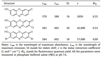
Halogenation on the aromatic rings of a dye often leads to significant influences on photophysical and photochemical properties. The most common cases are xanthene fluorophores and their halogenated analogues [27–29]. For example, 2′,7′-dichloride modification on fluorescein can lower the phenolic pKa, making these analogues less sensitive to biologically relevant pH fluctuations [30]. A similar effect also exists on coumarin dyes [31]. Moreover, halogenation sometimes can improve the photo-stability of a dye [31,32]. With these considerations, this study began with the preparation of a dichloro-resorufin (resorufin-Cl) and a dibromo-resorufin (resorufin-Br). Both resorufins were easily prepared through a simple two-step synthetic procedure from the commercial materials with reasonable isolated yields (general procedure A, Supporting information). The measurements indicated that chloride and bromide modification could result in slight red-shifts to the absorbance and fluorescence spectra (Table 1, Fig. S2 in Supporting information). Density-functional theory (DFT) calculations of the highest occupied molecular orbital-lowest unoccupied molecular orbital (HOMO-LUMO) gap were in consistence with the experimental observation (Fig. S3 in Supporting information). The photophysical properties of all three resorufins were then measured. As shown in Table 1, the fluorescence quantum yield of resorufin-Cl (0.33) is close to resorufin (0.35), but that of resorufin-Br (0.09) is significantly lower than resorufin-Cl and resorufin (Table 1 and Table S2 in Supporting information). It is worth noting that the molar extinction coefficient (ε) of resorufin-Cl (42,000 L mol−1 cm−1) is much higher than that of resorufin (5050 L mol−1 cm−1) when measured at corresponding wavelength of their maximum absorbance. It is known that the molecular brightness can be defined as the product of the molar extinction coefficient and the quantum yield [33]. Therefore, it is not hard to expect that resorufin-Cl has improved brightness than resorufin. The fluorescence under 300 nm UV light irradiation clearly shows the elevated brightness of resorufin-Cl over resorufin (Fig. S4 in Supporting information). It is certain that resorufin-Cl is a promising scaffold for fluorescence substrate design. In total, five hydroresorufin-Cl analogues with acyl substituents on N atom (AR-1/2/3/4/5) have been prepared (Fig. 1a and Supporting information). To potentially suppress the recognition and nonspecific cleavage reaction by CES, the acetyl group was replaced with biologically unusual cyclic alkanoyl groups (cyclopropylcarbonyl, cyclobutanecarbonyl, cyclopentanecarbonyl and cyclohexylcarbonyl, respectively) during the preparation.
|
|
Table 1 Photophysical properties of resorufin, resorufin-Cl and resorufin-Br. |

|
Download:
|
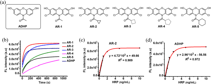
| Fig. 1. (a) The structures of synthetic substrates AR-1/2/3/4/5 and the commercial ADHP. (b) The kinetic response curves of the fluorescence substrates toward HRP/H2O2 reaction. (c) The detection curve (red line) and the linear regression (black line) of AR-2 fluorescence versus the concentration of HRP. (d) The detection curve (red line) and the linear regression (black line) of ADHP fluorescence versus the concentration of HRP. The final concentrations of AR-2 and ADHP were both at 50 µmol/L, and the concentration of H2O2 was 500 µmol/L. The concentration of HRP in (b) was 5 ng/mL. The data are expressed as mean ± standard deviation of three replicates. | |
All the synthesized substrates and ADHP were evaluated in sensing of HRP/H2O2 reaction. As shown in Fig. 1b, all probes can respond to HRP/H2O2 through fluorescence turn-on manners. However, the degrees of fluorescence enhancement from AR-1, AR-4 and AR-5 are less than that of ADHP. Among all, AR-2 shows the highest reactivity, and reaches the plateau of detection curve within 5 min. Moreover, the maximum fluorescence intensity of AR-2 reaction solution is higher than that of ADHP at the same conditions. AR-3 exhibits similar fluorescence enhancement as AR-2, however, a longer reaction time is required to reach the intensity plateau. The result indicates that HRP prefers the hydroresorufin-Cl substrate with an acyl group of three-membered ring rather than those with a smaller or a larger acyl substituent. Both Figs. 1c and d show that the fluorescence enhancement correlates well with the increase of HRP concentration from both AR-2 and ADHP reactions. There is a linear relationship between the fluorescence intensity of AR-2 and HRP concentration at pictogram-level (0.01–1.0 ng/mL). Whereas, the linear relationship for ADHP reaction system is found at the HRP concentration ranging from 0.02 ng/mL to 1.0 ng/mL, which is narrower than that of AR-2. In addition, the calibration curve's slope of AR-2 system (4.72×10³) is 1.59-fold to that of ADHP (2.96×10³). In general, a steeper calibration curve (with a larger slope) corresponds to higher sensitivity toward the changes in HRP concentration [34]. With excellent sensitivity, we then examined the reactivity of AR-2 and ADHP toward CES. Consistent with previous observation, ADHP indeed responds to CES through HRP and H2O2 independent manners. This reaction can convert ADHP to its fluorescent product resorufin (Fig. S5 in Supporting information). Inhibiting CES activity by an inhibitor phenylmethylsulfonyl fluoride (PMSF) [35] can significantly suppress the fluorescence increase. Surprisingly, AR-2 does not respond to CES as non-obvious fluorescence turn-on was observed both in the presence and in the absence of PMSF (Fig. 2a). As shown in Fig. S6 (Supporting information), the analogue with an acetyl group (AR-1) has obvious nonspecific reactivity toward CES, which is similar to ADHP. In contrast, all the ARs (AR-2 to AR-5) with cyclic alkanoyl substituents do not respond to CES. The original purpose of introducing naturally unusual acyl groups was to design substrates with suppressed unwanted responsiveness toward CES. The result is indeed of great support to the design principle.

|
Download:
|
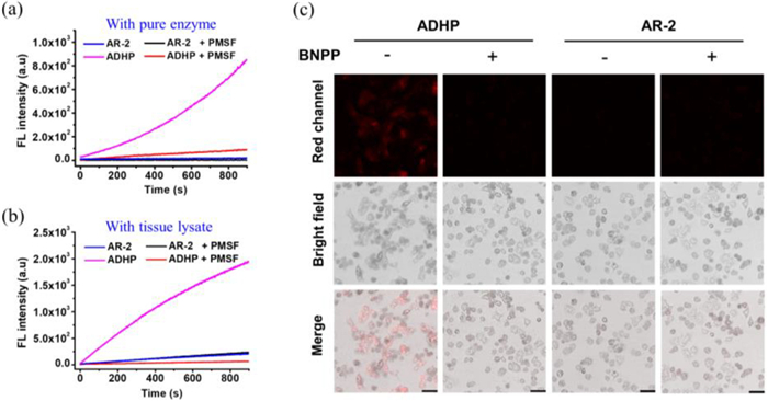
| Fig. 2. (a) Reaction kinetic curves by monitoring the fluorescence of AR-2 (50 µmol/L) or ADHP (50 µmol/L) in the presence of pure CES (0.5 U/mL). (b) Kinetic measurement of the fluorescence of AR-2 or ADHP from the incubation with fresh lysate of mouse liver tissues. (c) Fluorescence imaging of mouse primary hepatocytes by AR-2 and ADHP. Scale bar: 80 µm. The concentrations of CES inhibitor PMSF and BNPP are both at 100 µmol/L. | |
In a further study, this nonspecific reactivity has been examined by incubating ADHP and AR-2 with the lysate of fresh mouse tissues, respectively (Fig. 2b). It is known that CES are expressed in many types of tissues or cells. To measure H2O2 or H2O2-generating reaction in tissues/cells, HRP/substrate system is often mixed directly with the fresh lysates. CES activity can greatly affect the quantification accuracy if the HRP substrate itself has nonspecific responsiveness to CES. Therefore, HRP substrates without CES responsiveness are desirable. As expected, fluorescence turn-on from ADHP was observed, where 15 min incubation could lead to 80-fold increase in fluorescence intensity. Whereas, only a slight increase of fluorescence was observed from AR-2. CES inhibitor PMSF cannot suppress the fluorescence from AR-2, indicating that the slightly elevated fluorescence is not due to the CES's activity. This is mostly attributed to the reaction between AR-2 and intrinsic oxidant species in the lysates. Actually, ADHP has previously been used to image the reactive oxygen species (ROS), such as superoxide anions in living cells. We recognized that non-specific reactivity of ADHP toward CES might greatly affect the accuracy of ROS imaging. This concern has been evidenced by fluorescence imaging in mouse primary hepatocytes. As found in Fig. 2c, obvious red fluorescence in the cells was observed in the presence of ADHP. Co-incubation with bis(p-nitrophenyl)phosphate (BNPP), a specific inhibitor to CES, could dramatically suppress the fluorescence from ADHP. In contrast, only weak and background level fluorescence was observed from AR-2 treated cells either in the presence or in the absence of BNPP. To summarize, ADHP indeed responds to CES regardless of HRP and H2O2, while, AR-2 does not show responsiveness toward CES. Moreover, AR-2 is live-cell compatible at the common concentrations for cell imaging (Fig. S7 in Supporting information). Rather than only CES, in a further evaluation, some other proteins or enzymes such as human serum albumin (HSA), GOx and alkaline phosphatase (ALP) were applied to examine the selectivity and anti-interference capability of AR-2/HRP/H2O2 reaction. As illustrated in Fig. S8 (Supporting information), those interfere species did not lead to obvious influence to fluorescence readout, which indicated excellent selectivity and anti-interference capability of this reaction.
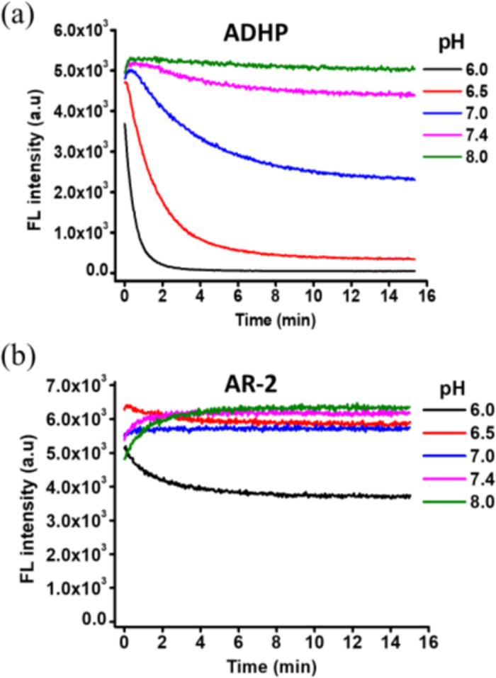
As previously described, the fluorescence product resorufin from ADHP/HRP reaction can undergo further oxidation in the presence of relatively high concentrations of H2O2 or HRP, leading to fluorescence decrease. The degree of fluorescence suppression highly depended on pH values, where low pHs could accelerate the decrease [21,36]. A similar phenomenon was observed in this study. As found in Fig. 3a, elevated HRP and H2O2 can lead to fast fluorescence turn-on from ADHP/HRP/H2O2 reaction, however, further reaction under neutral and acidic pHs can lead to rapid intensity decrease. Even under the physiological conditions (pH 7.4), 15 min reaction has resulted in decrease by 14%. In contrast, AR-2/HRP/H2O2 system works smoothly under pH ranges from 6.5 to 8.0. Further lowering pH to 6.0 can cause significant fluorescence diminution, but the degree of reduction is still much less than ADHP at the same conditions (Fig. 3b). It is assumed that the radical species generated from HRP/H2O2 might react back with resorufin, which could result in further oxidation or polymerization, however, these non-fluorescent products are hard to be identified [36]. Electron withdrawing substitution is supposedly able to decrease the electron density of the conjugation system, rendering it greater tolerance to oxidation. This could be one of the reasons to explain the improved stability of resorufin-Cl. Besides, we have noticed that pH effect might be another possible reason to explain the differences between AR-2 and ADHP in Fig. 3. As shown in Fig. S9a (Supporting information), decreasing the pH from 7.0 to 6.0 can suppress the fluorescence intensity of resorufin by 44%, while, that from resorufin-Cl is only 8%. Computational estimation has also indicated that Cl substitution on resorufin can dramatically decrease the phenolic pKa from 6.35 to 4.81 (Fig. S9b in Supporting information). Phenolic deprotonation is necessary for this sort of molecules to emit bright fluorescence in aqueous mediums [37]. Therefore, it is not hard to understand that the fluorescence of resorufin-Cl is much stable than resorufin when pH changes between neutral and weak acid. It is worth noting that, in a recent study, we have reported a dihydrodichlorofluorescein analogue (DCFH-1) with great sensitivity, selectivity and pH stability as a HRP substrate [24]. However, this substrate shows unexpected weak responsiveness toward HRP in the absence of H2O2 (Fig. S10 in Supporting information). The above results indicate that resorufin-Cl is an advanced fluorescent skeleton to construct HRP substrate and AR-2 is a promising HRP substrate for analytical practices.

|
Download:
|
| Fig. 3. Reaction kinetic curves of ADHP/HRP/H2O2 reaction (a) and AR-2/HRP/H2O2 reaction (b) by monitoring the fluorescence. The reaction was performed in PBS buffer with different pH values. The final concentrations of substrate, HRP and H2O2 were set at 50 µmol/L, 500 ng/mL and 10 mmol/L, respectively. | |
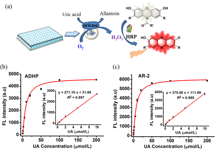
HRP/substrate is the most widely used reporter system for H2O2 and H2O2-generating biochemical reactions. H2O2 generation is a basic reaction in living organisms that can be found in numerous enzymatic reactions. For a proof of concept, UA/UAO reaction system was chosen in this study. ADHP and AR-2 were applied as HRP substrate individually to construct UA detection assays (Fig. 4a). For a typical test, HRP, substrate, and UAO were mixed firstly followed by the addition of UA. The fluorescence was measured after 15 min incubation at room temperature. As shown in Fig. 4, the fluorescence intensity of the reaction solution is UA concentration dependent. Excellent linear relationships between intensity and UA concentration (0–10 µmol/L) have been found in both ADHP and AR-2 systems. Although they have a same linear range, the standard curve's slope of AR-2 is 1.38-fold to that of ADHP, which indicating a higher sensitivity of AR-2-based assay (Figs. 4b and c).

|
Download:
|
| Fig. 4. (a) Schematic figure shows the dection of UA/UAO reaction by HRP/AR-2 or ADHP. (b) The detection curve of fluoresence intensity of ADHP/HRP in the presence of UA at different concentrations. (c) The detection curve of fluoresence intensity of AR-2/HRP in the presence of UA at different concentrations. The insets in b and c represent the linear calibration curves. The concentration of UAO was set at 5 U/mL, the fluorescence substrates and HRP were set at 50 µmol/L and 5 ng/mL, respectively. The data are expressed as mean ± standard deviation of three replicates. | |
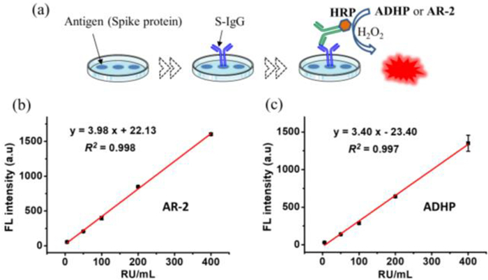
Besides, HRP is a well-known enzyme label for optical immunoassays, such as ELISAs. ADHP, as a substrate to HRP, has been widely used to construct fluorescent ELISA kits. With excellent HRP-sensing ability of AR-2, we then look into its application potency in fluorescent ELISAs. In this study, the serum immunoglobulin G (IgG) to SARS-CoV-2 spike protein was chosen as the target. Anti-virus IgG antibodies are known to play important roles against virus infection, monitoring IgG serum levels is thus important for diagnosis and guidance of therapy, which has been extensively studied in the fight against coronavirus disease 2019 (COVID-19) [38,39]. HRP substrate in the commercial ELISA kit for SARS-CoV-2 IgG was replaced with ADHP and AR-2, respectively, to form modified ELISA kits (Fig. 5a). Before IgG detection from human serum samples, the calibration curves were established according the instruction of kit. As found from Fig. 5b, an excellent linear correlation between fluorescence intensity and relative unit (RU) values of IgG is found when IgG concentration ranges from 5 RU/mL to 400 RU/mL. The standard curve's slope of AR-2 assay is 1.17 times to that of ADHP, which indicating a higher sensitivity of AR-2-based detection (Fig. 5c). In addition, the fluorescence signals from AR-2-based assays have relatively higher signal to noise ratios (SNRs) than ADHP-based assays along the linear range. It is known that the higher the SNR, the better the signal quality (Table S3 in Supporting information).

|
Download:
|
| Fig. 5. (a) Schematic figure shows the detection of S-IgG (the IgG against SARS-CoV-2 spike protein) using the modified ELISAs. (b) The standard detection curve of fluorescence intensity from AR-2 versus RU of IgG. (c) The standard detection curve of fluorescence intensity from ADHP versus RU of IgG. The data are expressed as mean ± standard deviation of three replicates. | |
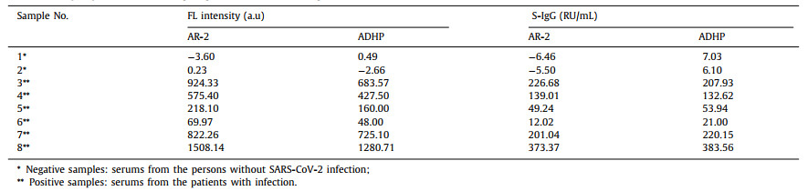
Based on the calibration curves, the IgG antibody against SARS-CoV-2 spike protein (S-IgG) from serum samples (samples 1 and 2 are from the persons without infection, samples 3 to 8 are from the patients who have just recovered from SARS-CoV-2 infection) have been quantified by the modified ELISA kits. The fluorescence readout and the calculated RU values of IgG are listed in Table 2, where the signals from blank wells have been subtracted from the measurements. Increased fluorescence intensity has been observed from all the serum samples with infection (positive samples), while, there are no meaningful fluorescence increase from the negative samples. Both AR-2 and ADHP based assays can successfully distinguish positive and negative samples according to the fluorescence changes. RU values of IgG were then calculated according to the functions in Fig. 5, which indicated a similar trend as the fluorescence observation. The RU values of all positive samples were further analyzed by Deming regression, in which the RU values from AR-2 and ADHP assays were set as x-values and y-values, respectively (Fig. S11 in Supporting information). Correlation coefficient (r) was found to be 0.995 (> 0.975), which indicated that the results from both methods exhibited excellent consistency [40]. In a word, AR-2 is undoubtedly a promising HRP substrate for setting up fluorescent ELISAs. AR-2/ELISA will become a promising alternative to replace ADHP/ELISA, especially for those requiring relatively higher sensitivity and SNR.
|
|
Table 2 Detection of IgG against SARS-CoV-2 spike protein from serum samples. |
To overcome the drawbacks of the most commonly used HRP fluorescent substrate ADHP, several new chloride hydroresorufin analogues have been designed and prepared. Among all synthetic substrates and ADHP, the analogue with a cyclopropylcarbonyl group on N atom shows the most promising sensitivity toward HRP detection. More importantly, AR-2 has improved anti-interference ability in practical applications when compared with ADHP. Furthermore, AR-2/HRP/H2O2 system has enhanced fluorescence stability than ADHP/HRP/H2O2, especially upon sensing of high concentrations of HRP or H2O2. AR-2/HRP has been used as the reporter in fluorescence analysis of UA/UAO reaction, which shows greater sensitivity than ADHP-based assays. In addition, AR-2 has been used to construct a fluorescence ELISA for successfully detecting the IgG against SARS-CoV-2 virus from human serum samples. In summary, AR-2 provides a great opportunity to improve HRP-based fluorescence biochemical analysis, which will become one of the most attractive alternatives to ADHP in real diagnostic applications.
Ethical statementThe use of human serum was reviewed and approved by the First Affiliated Hospital of Guangzhou Medical University Ethics Committee (No. 2020–77). The protocols for use of mouse tissue were approved by the Animal Ethics Committee of Guangzhou University of Chinese Medicine, in accordance with the national regulatory principles.
Declaration of competing interestThe authors declare that they have no known competing financial interests or personal relationships that could have appeared to influence the work reported in this paper.
AcknowledgmentsWe greatly acknowledge the funding support from Key-Area Research and Development Program of Guangdong Province (No. 2022B1111020003), Guangzhou Talents Program for Innovation and Entrepreneurship (No. 2021-L010) and the Foshan "Blue Ocean Talent Program" for Innovation and Entrepreneurship (No. 2230032002063). We also acknowledge the Lingnan Medical Research Center of Guangzhou University of Chinese Medicine and GIRM Biosafety (Guangzhou) Co., Ltd. for the support on facilities.
Supplementary materialsSupplementary material associated with this article can be found, in the online version, at doi:10.1016/j.cclet.2023.108970.
| [1] |
F. Chen, X. Xia, D. Ye, et al., Anal. Chem. 95 (2023) 5773-5779. DOI:10.1021/acs.analchem.3c00073 |
| [2] |
J. Wu, Z. Fu, F. Yan, H. Ju, TrAC Trends Anal. Chem. 26 (2007) 679-688. DOI:10.1016/j.trac.2007.05.007 |
| [3] |
N.C. Veitch, Phytochemistry 65 (2004) 249-259. DOI:10.1016/j.phytochem.2003.10.022 |
| [4] |
A. Jones, L. Dhanapala, R.N.T. Kankanamage, C.V. Kumar, J.F. Rusling, Anal. Chem. 92 (2020) 345-362. DOI:10.1021/acs.analchem.9b05080 |
| [5] |
F. Chen, Y. Zhang, T. Li, et al., Chin. Chem. Lett. 34 (2023) 107496. DOI:10.1016/j.cclet.2022.05.010 |
| [6] |
J. Sun, J. Zhao, L. Wang, et al., ACS Sens. 3 (2018) 183-190. DOI:10.1021/acssensors.7b00830 |
| [7] |
X. Xiao, S. Hu, X. Lai, J. Peng, W. Lai, Trends Food Sci. Tech. 111 (2021) 68-88. DOI:10.1016/j.tifs.2021.02.045 |
| [8] |
M. Zhou, Z. Diwu, N. Panchuk-Voloshina, R.P. Haugland, Anal. Biochem. 253 (1997) 162-168. DOI:10.1006/abio.1997.2391 |
| [9] |
K. Sadani, P. Nag, X.Y. Thian, S. Mukherji, Biosens. Bioelectron. 12 (2022) 100278. |
| [10] |
S. Khosrozadeh, M. Dorodnikov, T. Reitz, E. Blagodatskaya, Eur. J. Soil Sci. 73 (2022) e13225. DOI:10.1111/ejss.13225 |
| [11] |
M. Kameya, M. Himi, Y. Asano, Anal. Biochem. 447 (2014) 33-38. DOI:10.1016/j.ab.2013.11.002 |
| [12] |
H. Lee, K. Kim, C.M. Kang, et al., Anal. Chem. 95 (2023) 1038-1046. |
| [13] |
J.M. Lü, P.H. Lin, Q. Yao, C. Chen, J. Cell Mol. Med. 14 (2010) 840-860. DOI:10.1111/j.1582-4934.2009.00897.x |
| [14] |
S. Jiang, X. Chen, J. Lin, P. Huang, Adv. Mater. 35 (2023) 2207951. DOI:10.1002/adma.202207951 |
| [15] |
C. Gong, Y. Gong, M.K. Khaing Oo, et al., Biosens. Bioelectron. 96 (2017) 351-357. DOI:10.1016/j.bios.2017.05.024 |
| [16] |
A. Wojtala, M. Bonora, D. Malinska, et al., Methods Enzymol. 542 (2014) 243-262. |
| [17] |
Y. Sun, L. Wen, H. Ma, et al., Chin. Chem. Lett. 34 (2023) 107654. DOI:10.1016/j.cclet.2022.06.077 |
| [18] |
N. Wang, C.J. Miller, P. Wang, T.D. Waite, Anal. Chim. Acta 963 (2017) 61-67. DOI:10.1016/j.aca.2017.02.033 |
| [19] |
J. Serrano, M. Jové, J. Boada, et al., Biochem. Biophys. Res. Commun. 388 (2009) 443-449. DOI:10.1016/j.bbrc.2009.08.041 |
| [20] |
S. Miwa, A. Treumann, A. Bell, et al., Free Radic. Bio. Med. 90 (2016) 173-183. DOI:10.1016/j.freeradbiomed.2015.11.011 |
| [21] |
T. Wang, Y. Xiang, X. Liu, W. Chen, Y. Hu, Talanta 162 (2017) 143-150. |
| [22] |
B. Zhao, K. Ranguelova, J. Jiang, R.P. Mason, Free Radic. Bio. Med. 51 (2011) 153-159. DOI:10.1016/j.freeradbiomed.2011.03.016 |
| [23] |
C.S. Karamitros, J. Lim, M. Konrad, Anal. Biochem. 445 (2014) 20-23. DOI:10.1016/j.ab.2013.09.028 |
| [24] |
J. Zhu, T. Li, S. Zhang, et al., Chemosensors 11 (2023) 152. DOI:10.3390/chemosensors11020152 |
| [25] |
S. Yoo, S. Kim, S. Jeon, M.S. Han, RSC Adv. 12 (2022) 8668-8673. DOI:10.1039/d2ra00087c |
| [26] |
J. Liu, G. Ruan, W. Ma, et al., Biosens. Bioelectron. 198 (2022) 113823. DOI:10.1016/j.bios.2021.113823 |
| [27] |
L. Wang, M. Tran, E. D'Este, et al., Nat. Chem. 12 (2020) 165-172. DOI:10.1038/s41557-019-0371-1 |
| [28] |
X. Chen, T. Pradhan, F. Wang, J.S. Kim, J. Yoon, Chem. Rev. 112 (2012) 1910-1956. DOI:10.1021/cr200201z |
| [29] |
F. Chen, W. Liu, H. Li, et al., Anal. Sens. 2 (2022) e202100066. |
| [30] |
J.L. Turnbull, B.R. Benlian, R.P. Golden, E.W. Miller, J. Am. Chem. Soc. 143 (2021) 6194-6201. DOI:10.1021/jacs.1c01139 |
| [31] |
E. Pǎunescu, L. Louise, L. Jean, A. Romieu, P.Y. Renard, Dyes Pigments 91 (2011) 427-434. DOI:10.1016/j.dyepig.2011.05.008 |
| [32] |
W. Chyan, H.R. Kilgore, B. Gold, R.T. Raines, J. Org. Chem. 82 (2017) 4297-4304. DOI:10.1021/acs.joc.7b00285 |
| [33] |
S.J. Lim, M.U. Zahid, P. Le, et al., Nat. Commun. 6 (2015) 8210. DOI:10.1038/ncomms9210 |
| [34] |
S. Ye, H. Zhang, J. Fei, et al., Angew. Chem. Int. Ed. 60 (2021) 1339-1346. DOI:10.1002/anie.202011108 |
| [35] |
I. Mangas, E. Vilanova, J. Estévez, Chem. Res. Toxicol. 25 (2012) 2393-2401. DOI:10.1021/tx300257p |
| [36] |
V. Towne, M. Will, B. Oswald, Q. Zhao, Anal. Biochem. 334 (2004) 290-296. DOI:10.1016/j.ab.2004.07.037 |
| [37] |
X. Liu, L. He, X. Gong, et al., CCS Chem. 4 (2022) 356-368. DOI:10.31635/ccschem.021.202000679 |
| [38] |
W.N. Voss, Y.J. Hou, N.V. Johnson, et al., Science 372 (2021) 1108-1112. DOI:10.1126/science.abg5268 |
| [39] |
Q.X. Long, B.Z. Liu, H.J. Deng, et al., Nat. Med. 26 (2020) 845-848. DOI:10.1038/s41591-020-0897-1 |
| [40] |
A.L. Jensen, M. Kjelgaard-Hansen, Vet. Clin. Pathol. 35 (2006) 276-286. DOI:10.1111/j.1939-165X.2006.tb00131.x |