 2023, Vol. 34


Bone is invariably remodeled and renovated, as well as can regenerate in the whole life [1,2]. Unfortunately, the impact of inflammation, trauma, tumor and other injury factors may cause bone tissue defects and nonunion [3,4], while traditional osteogenesis materials have some disadvantages [5-9]. Therefore, it is imperative to develop novel materials to promote bone healing. Synthetic bone regeneration materials, having the superiority in solving the limitation of traditional materials, have received increasing attention in recent years. At present, synthetic bone regeneration materials, including ceramics, collagen, platelet-rich fibrin (PRF), metal-organic frameworks (MOFs) and so on [6,8,10,11], have been widely used in the clinic for their good bioactivity and bioavailability. Among them, MOFs are especially attractive due to their diversity of species and functions.
Metal-organic frameworks (MOFs), a class of hybrid materials, consist of organic linkers and bridging metal ions or clusters. Theoretically, according to the difference of organic linkers and cations or preparation methods, countless MOFs can be synthesized with tunable pore sizes and large surface areas. And these MOFs have multiple functions to adapt to special applications [12-16]. For example, MOFs can adsorb nitric oxide (NO), which participates in many processes in the body including wound repair, vasodilatation and so on. Morris et al. [17] had reviewed the applications of NO-releasing materials in biomedicine, and discussed that MOFs had high adsorption and deliverable capacities of NO. Some research [18] had found that MOFs could carry several enzymes as an immobilization support. And MOFs can also be heterogeneous catalysts [19]. Meanwhile, MOFs with luminescence, magnetism and chemical sensing can be obtained by introducing different molecules or ions [20-22]. Recently, MOFs have led to more and more applications in biomedicine for their good biocompatibility, good antibacterial ability, and abundant applicable properties [23-26]. The excellent roles of MOFs in this field have aroused widespread interest.
The porous structure and mechanical properties of MOFs are similar to substitute tissue, which are in the favor of cell adhesion and growth, nutrient transferring, metabolic waste discharging and cell differentiation. Based on this, many people have researched and confirmed the osteogenic effects of MOFs [27]. For example, our group has examined the effects of ZIF-8@AHT (nanoscale zeolitic imidazolate framework-8 (ZIF-8)-modified alkali and heat-treated titanium) in the field of osteogenesis systematically and quantitatively [28], mainly from cell behavior in vitro and osteogenesis assessment in vivo. It is indicated that ZIF-8@AHT can enhance cell bioactivity, increase extracellular matrix (ECM) mineralization, and secrete more collagen and OPG, as well as upregulate the expression of osteogenic genes and osteogenesis-related proteins. Among them, OPG could inhibit the differentiation of osteoclasts. Moreover, ZIF-8@AHT-1/8 also exhibited good osteogenic properties at the bone-implant interface. In addition to the osteogenic effects of MOFs themselves, their porous structures allow the load of osteogenic substances to promote osteogenesis [29]. Meanwhile, MOFs also have angiogenic, antibacterial and hemostatic abilities [23,27,30], which can vastly promote osseous healing. As a result, there are more and more reports about the application of MOFs in bioengineering [29]. Furthermore, MOFs can not only be used for bone tissue engineering, but also can be applied to treat bone diseases for their osteogenic, antibacterial and drug-delivering properties, which broaden the application of MOFs in bone-related applications. For instance, our group verified that nanoscale ZIF-8 can activate canonical MAPK signaling for bone repair. It is indicated that nano ZIF-8 can enter the rBMSC cytoplasm probably via caveolae-mediated endocytosis and macropinocytosis, and then, nano ZIF-8 primarily phosphorylated ERK, then activating the canonical mitogen-activated protein kinase pathway and promoting the osteogenesis of rBMSCs [31].
There have been published many excellent articles about MOFs in other fields [32-34] , and MOFs in the field of osteogenesis have been widely studied and already shown great potential, but they have not yet been systematically discussed. Therefore, it is of great importance to have a comprehensive understanding of MOFs in the field of osteogenesis. Herein, we aim to provide a review on the biological characteristics of MOFs, and their abundant applications in bone tissue engineering, as well as in bone diseases. Furthermore, the role of MOFs themselves, the role of MOFs-loading substances, and the synergy between MOFs and MOFs-loading substances are discussed.
2. Synthesis of metal-organic frameworksWith the increasing study of MOFs, many synthetic methods are exploited to synthesize MOFs for bone tissue engineering and bone diseases, including solvothermal method [35,36], non-solvothermal method [37,38], one-pot synthesis [31,39], hydrothermal method [40], reverse microemulsion [41] and ultrasonic synthesis (Fig. 1) [42,43]. By using different methods, MOFs with different properties can be synthesized, and these MOFs will induce some differences in osteogenesis of MOFs. Therefore, it is significant to discuss the synthetic methods.

|
Download:
|
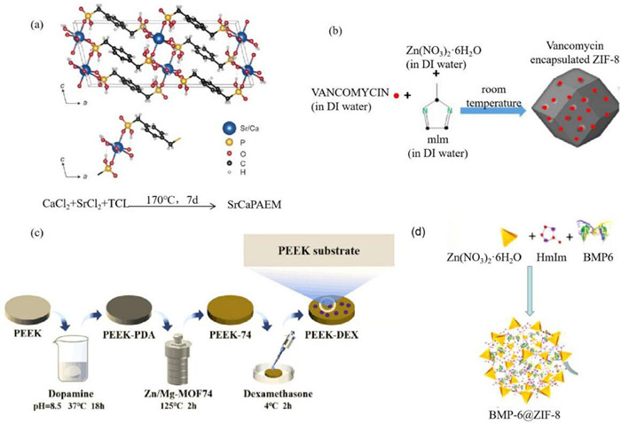
| Fig. 1. (a) Solvothermal synthesis: crystal structure and asymmetric unit of SrCaPAEM. Reproduced with permission [48]. Copyright 2019, American Chemical Society. (b) Non-solvothermal synthesis: Synthesize ZIF-8 by non-solvothermal method. Reproduced with permission [37]. Copyright 2019, Elsevier B.V. (c) Hydrothermal synthesis: the way to synthesize Zn/Mg-MOF74. Reproduced with permission [52]. Copyright 2021, American Chemical Society. (d) One-pot synthesis: crystal structure of BMP-6@ZIF-8. Reproduced with permission [44]. Copyright 2020, Elsevier B.V. | |
Non-solvothermal synthesis is one of the conventional methods. Its reaction temperature is usually below the boiling point, and the reaction condition is under ambient pressure, which immensely simplifies synthetic requirements. What is more, MOFs synthesized by this synthesis have good crystal texture and biological properties. Toprak et al. [44] synthesized ZIF-8 to carry BMP-6, and embedded it in bioresorbable electrospun fibers. Two syntheses were mentioned in this study, and one of them is non-solvothermal synthesis. 2-Methylimidazole dissolved in deionized water was added to zinc nitrate solution under continuous agitation. After stirring at room temperature for half an hour, ZIF-8 was synthesized. And the experimentation results showed that ZIF-8 had a typical dodecahedron structure and good embedding property.
Although this method has some advantages, it generally takes a long reaction time and the yield is low [45]. Joseph et al. [46] synthesized CaSr-MOFs for biodegradable orthopedic applications. The products did have uniform crystal sizes and clear boundaries. Meanwhile, it was verified that these MOFs can promote MC3T3 pre-osteoblastic cells to proliferate. However, it took 3 days to synthesize.
2.1.2. Solvothermal synthesisSolvothermal synthesis is another conventional synthesis. It refers to a closed system such as an autoclave, with organic or non-aqueous solvent as the solvent, at a certain temperature and the solution of the spontaneous pressure, the original mixture of reaction of a synthesis method. Similarly, MOFs synthesized by this synthesis have good biological properties, and it also takes a long time. The difference is this method shows higher yield, smaller and more homogeneous crystals as compared to non-solvothermal synthesis. For instance, Telgerd et al. [47] synthesized Zn–Cu imidazole MOF particles by this method: they put CuO and ZnO in acetic acid at 90 ℃. After they dissolved, added imidazole to the solution and heated to boil. And then, Zn–Cu imidazole MOF coated PLLA scaffold (PLLA@MOF). This composite scaffold can promote the osteogenic differentiation of mesenchymal cells. Maria et al. [48] synthesized SrCaPAEM with Sr/Ca ratio of 1:1 by using CaCl2, SrCl2 and tetraethyl p-xylylenebisphosphonate (TCI) as raw materials. SrCaPAEM had the ability to support biomineralization. But it needed to heat at 170 ℃ for 7 days in a sealed autoclave. Moreover, Khaled et al. [49] synthesized MIL-125-NH2 by solvothermal synthesis. According to the FE-SEM images, the particles showed a uniform and dispersed structure, and were mostly characterized by hexagonal and cubic shapes.
This method can do help to get nanocrystals of the same size. Nevertheless, sometimes it is possible to get nanocrystals with larger particle sizes by solvothermal method, which is harmful to targeted drug delivery by post-synthesis modification. Therefore, when using solvothermal method to prepare MOFs, it is essential to properly control the reaction condition and the ratio of organic ligands to metal ions so that to control the size of the particles [50].
2.2. One-pot synthesisOne-pot synthesis is also a common method in the preparation of MOFs. Compared with other methods, one-pot synthesis is a more economical and efficient way of synthesizing MOFs by reacting all the raw materials under certain conditions in the systems. As a result, the productive rate increases for the reduction of steps, and the amounts of raw materials and wastes will decrease correspondingly, which is friendly to the environment.
Among them, ZIF-8 is the most common MOFs prepared by this method. Besides synthesizing ZIF-8 by non-solvothermal synthesis, Toprak et al. [44] also synthesized BMP-6@ZIF-8 by one-pot synthesis. They just put all raw materials in the reaction system together for one hour, and BMP-6@ZIF-8 was synthesized finally. By comparison, the crystal structure of ZIF-8 did not change, and the encapsulation rate of BMP-6 was up to 98%. Then, BMP-6@ZIF-8 can slowly release BMP-6, which can enhance bone regeneration.
In many articles about ZIF-8, this method is usually used for synthesis. At the same time, based on the simplicity and convenience of this method and products with good properties, it is also widely used to synthesize other MOFs [51].
2.3. Hydrothermal synthesisHydrothermal synthesis is similar to solvothermal synthesis, but the solvent of hydrothermal synthesis is water, while the solvent of solvothermal synthesis is an organic solvent. High purity, good dispersity and easy control of particle size are the advantages of hydrothermal synthesis as compared to solvothermal synthesis.
Xiao et al. [52] synthesized Zn-Mg-MOF74 in this way. As a solvent, water was added to the reaction system. After reacting at 125 ℃ for 2 h, Zn-Mg-MOF74 crystal was obtained in the end. Meanwhile, they utilized Zn-Mg-MOF74 to carry dexamethasone. This material showed better osteogenic binding to the bone, because of the good dispersity and suitable surface roughness. Moreover, MOFs synthesized by hydrothermal method have precise particle size and dispersity, which are of great significance for the synthesis of MOFs requiring strict purity and pore size, such as being used in drug delivery system.
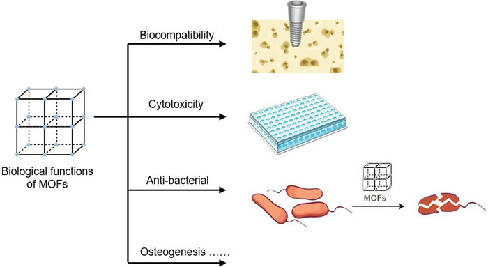
3. Biological functions of metal-organic frameworksIn recent years, quite a few studies have suggested that MOFs have led to more and more applications in biomedicine for their abundant applicable properties, especially in drug delivery, biomedical imaging, and antibacterial materials. In this section, some biological functions of MOFs relating to bone tissue engineering and bone diseases will be discussed. The major osteogenic and vasogenic effects will be detailed in the next section (Fig. 2).

|
Download:
|
| Fig. 2. The sketch map of biological functions of MOFs: Biocompatibility, cytotoxicity, and antibacterial action. | |
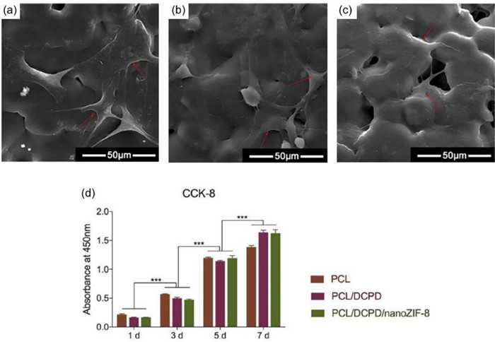
The biocompatibility of MOFs is very important for their application in vivo. The metal ions and linkers used to construct MOFs are important determinants of biocompatibility. Firstly, Ca, Mg, Zn, Fe, Ti, and Zr are suitable metals for constructing biocompatible MOFs [53]. For example, nanoscale ZIF-8 scaffolds showed biocompatibility with bone mesenchymal stem cells (BMSCs) (Fig. 3) [54]. NanoZIF-8 films applied to the surface modification of titanium also showed good biocompatibility [35]. Then, a variety of organic linkers can be used to construct biocompatible MOFs, which can be classified as exogenous and endogenous ligands. Exogenous linkers are synthetic linkers whose excretion or metabolism after in vivo application are important to ensure biocompatibility [55]. On the other hand, linkers that are naturally present in the body are called endogenous linkers. Their application enhances biocompatibility because they can be safely absorbed by the body [56]. When MOFs are used for drug delivery, they should have good drug delivery capabilities and biocompatibility. For example, electrospun pectin/modified copper-based MOF nanofibers as a drug delivery system have good biocompatibility while being biologically active [57]. However, some MOFs have poor stability in aqueous solutions, which leads to premature release of the drugs. Coating functional polymers on MOFs is a commonly used method. Chitosan, PVP, PSS, and heparin, which have good water solubility, can be wrapped around MOFs surfaces, thus improving the biocompatibility of MOFs [58-61].

|
Download:
|
| Fig. 3. Biocompatibility of the 3D printed scaffolds seeded with BMSCs. Morphology of the cells seeded on the surface of the scaffolds observed by SEM: (a) PCL, (b) PCL/DCPD, (c) PCL/DCPD/nanoZIF-8. Arrows point to the BMSCs adhering to the surface of scaffolds. (d) Cell viability measured by CCK-8 assays on day 1, 3, 5 and 7: Significant differences were marked among a series of points for each group, respectively. Data were shown as mean ± SD, n = 4, ***P < 0.001. Reproduced with permission [54]. Copyright 2020, The Royal Society of Chemistry. | |
The cytotoxicity of MOFs comes from the metal ions they release as well as the drugs they carry. Usually, it is the fast releasing of metal ions of MOFs that cause cytotoxicity. For example, the cytotoxicity of ZIF-8 may be due to the competition of Zn2+ with Fe2+ and Ca2+ through ion channels and DNA damage when the concentration of Zn2+ is too high [62]. To control cytotoxicity, the options that can be adopted are: (1) Enhance linker interaction. MOFs are composed of metal ions and organic linkers. Even though certain metal ions and organic linkers have some cytotoxicity, the two are tightly bound together by chemical bonds and their release rate is not sufficient to cause cytotoxicity [56]. (2) Control the release rate. One of the Cu-MOF NPs, called HKUST-1, is modified to slowly release Cu, thereby reducing cytotoxicity [63]. The unstable MOFs can also slowly release antibacterial metal ions from their own structure [64-68]. (3) Reduce cytotoxicity by modifying MOFs. Hf-MOFs can be modified by β-targeted peptide LPFFD, which can effectively reduce the cytotoxicity of Cu(Ⅱ)-induced a β aggregation can be reduced by chelating Cu(Ⅱ) [69]. In many papers about MOFs, the researchers have explored maximum optimum concentration of MOFs to guarantee better effects. At present, we have been able to control the concentration of MOFs so that they can play a good biological role without causing cytotoxicity. Testing the cytotoxicity is necessary for studies on the preparation of MOFs.
3.3. Antibacterial and anti-inflammatory action of MOFsMOFs have many special biological effects, including antibacterial and anti-inflammatory effects [70]. Many experiments have studied the possible antibacterial mechanisms of MOFs, and which mainly include the structural degradation of MOFs, along with the release of metal ions and the reaction of organisms with active metals on their surface [23]. The structural degradation of MOFs is due to the slow release of their internal metal ions, therefore, an unstable MOFs structure is needed to achieve the ideal antibacterial effect. Unstable MOFs can slowly release antibacterial metal ions, such as Ag+ [64], Cu2+ [65-67] , Zn2+ [68], from their own structure, making MOFs also have antibacterial effects. In addition, MOFs can also have a direct reaction with organisms the metal active sites on their surface [71]. What is more, MOFs can achieve the antibacterial effect by carrying antibiotics such as ciprofloxacin [72], vancomycin [37], too. In addition, Chen et al. [73] integrated the photosensitized porphyrin and boronic acid ligand into one single MOF, which showed a better effect to eradicate multi-drug-resistant bacteria.
Some studies also indicate that MOFs have anti-inflammatory effect. Li et al. [74] reported that mg/HCOOH MOF can promote the release of anti-inflammatory factors, and inhibit the expression of inflammatory genes, and thus play an anti-inflammatory role. Besides, MOFs can also get the anti-inflammatory effect by carrying non-steroidal anti-inflammatory drugs, such as flurbiprofen (FBP) [75], ketoprofen [76], diclofenac sodium (DFNA) [77].
Sometimes, wound healing needs the synergistic action of antibacterial and anti-inflammatory. Chen et al. [78] synthesized Zn-BTC with the ability of slowly releasing Zn2+. And the experimental results indicated that Zn-BTC had good bactericidal and anti-inflammatory effect, and can promote fibroblasts migration and proliferation, as well as promote the expression of wound healing genes.
4. Application of MOFs in bone tissue engineeringBone tissue engineering refers to the cultivation and amplification of bone marrow stromal stem cells, and then put them into a natural or synthetic scaffold or extracellular matrix with good biocompatibility [79]. These biomaterial scaffolds can provide three-dimensional space for cells to survive, which is beneficial for cells to conduct gas exchange, obtain enough nutrients, and remove waste materials, so that cells can grow on the prefabricated three-dimensional scaffold [79]. Ultimately, the hybrid material is implanted into the bone defect site. At the same time of the gradual degradation of the biomaterial, the implanted bone cells proliferated continuously, so as to achieve the purpose of repairing bone tissue defects.
Compared with traditional materials, excellent structure and better biocompatibility of MOFs are beneficial to cell growth and differentiation, which lead to better osteogenesis [80]. As mentioned above, MOFs consist of organic linkers and bridging metal ions or clusters. Meanwhile, it is verified that many kinds of MOFs have osteogenic effects, mainly including MOFs with Zn, Zr, Ca as metal ions. And different metal ions promote osteogenesis by affecting different proteins. That is why there are more and more kinds of MOFs are being applied in bone tissue engineering [81], and have gained quite a few achievements (Table S1 in Supporting information). In this section, the application of MOFs in bone tissue engineering mainly from MOFs themselves contributing to osteogenesis, as a drug delivery system, and the combination of porous scaffolds promoting osteogenesis are discussed.
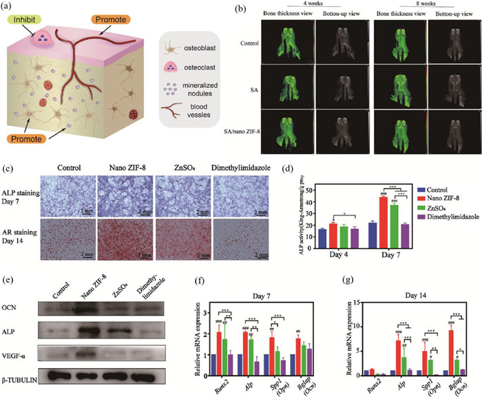
4.1. MOFs themselves contribute to osteogenesis and related applicationsThe porous structure and similar mechanical properties and good biocompatibility to those of substitute tissue of MOFs are in the favor of cell adhesion and growth, nutrient transferring, metabolic waste discharging and cell differentiation. This provides basic conditions for MOFs themselves to promote osteogenesis. And then, according to the research about MOFs contributing to osteogenesis, MOFs take effects mainly through three aspects: promoting osteoblasts, inhibiting osteoclasts, and promoting angiogenesis (Fig. 4).

|
Download:
|
| Fig. 4. (a) The sketch map of MOFs themselves promoting osteogenesis. (b) In vivo bone regeneration capability of SA/nano ZIF-8 scaffolds in rat premaxillary critical-size defect models. 3D-reconstructed micro-CT images of rat premaxillae implanted with different scaffolds. Osteogenic differentiation of rBMSCs caused by nano ZIF-8, ZnSO4, and dimethylimidazole. (c) ALP and AR staining with the materials stated above. (d) ALP activity after 4 and 7 days. (e) Western blot results after 14 days. PCR results at day 7 (f) and day 14 (g). *P < 0.05, **P < 0.01, and ***P < 0.001 between each group. #, ##, and ### indicate significant differences in comparison with the control group. #P < 0.05, ##P < 0.01, and ###P < 0.001. Copied with permission [39]. Copyright 2021, American Chemical Society. | |
Osteoblasts play a vital role in the osteogenic process. And the condition of bone repair is a positive correlation with the proliferation and differentiation of osteoblasts. In recent years, research show that MOFs have the ability to promote osteoblasts, and substantiate relevant mechanisms. In brief, MOFs can promote osteogenic genes expression of osteoblasts. After transcription and translation, the level of relevant proteins increases. Finally, the quantity of mineralized nodules becomes more.
During early osteogenesis, MOFs promote the osteogenic process mainly by regulating Runx2 and Osx. Runx2 is a central control gene of the osteoblast phenotype and plays an important role in osteoblast differentiation by binding to the osteoblast-specific cis-acting element OSE2 [82]. A variety of MOFs promote Runx2 expression [15,28,35,38-40,83-85]. For example, calcium(Ⅱ)-carboxylate MOFs (Ca2+ as metal ions) increase the mRNA expression level of Runx2 [38], and SA@ZIF-8 (Zn2+ as metal ions) upregulates Runx2 mRNA and protein levels in BMSCs [39]. Osx (Osterix, also known as Sp7), a zinc finger-containing osteoblast-specific transcription factor, in the osteoblast differentiation signaling pathway is a downstream gene of Runx2 [86]. Poly(ε-caprolactone) (PCL)@DCPD@nanoZIF-8 was shown to promote Osx mRNA expression [54]. In mid-osteogenesis, MOFs promote osteogenesis by promoting alkaline phosphatase (ALP) expression and upregulating ALP activity. ALP is the typical protein product of osteoblast phenotyping and differentiation [87]. It hydrolyzes various phosphate compounds to produce phosphate [88]. Its products, particularly inorganic phosphate (Pi), are released into the extracellular matrix, initiating the nucleation of extracellular matrix calcium deposits, leading to the formation of hydroxyapatite [89]. Several studies have shown that MOFs can increase the expression of ALP [15,28,39,46,83-85] and promote osteogenesis in MSCs [47,84], BMSCs [83,85], pre-osteoblasts [84], and osteoblasts [15]. MOFs also promote the osteogenic activity of cells by upregulating the activity of ALP [35,37,39,47,90]. In late osteogenesis, OCN (osteocalcin, also known as BGLAP) is a determinant of bone formation [91] and plays a role in the growth of apatite crystals in bone by increasing the degree of carbonate substitution [92]. MOFs such as PCL@DCPD@nanoZIF-8 [54], SA@ZIF-8 [39], PCL@Col@ZIF-8 [15], Cu-TCPP-TCP [83], and Col/MOF/NG [84] can promote osteogenesis by facilitating the expression of OCN.
MOFs upregulate the expression of osteogenesis-related genes, and transcribed and translated proteins promote osteogenesis. Type Ⅰ collagen (COL I), an extracellular matrix protein, stimulates osteoblast adhesion and differentiation [93]. Elevated expression of Col1 was detected in the induction of osteogenesis by various MOFs, including PCL@Col@ZIF-8 [15], Mg/Zn-MOF74-AT [90], Col/MOF/NG [84], MAO@MOF/ZIF-8 [85], and ZIF-8@AHT [28]. BMP-2 is the most important protein that promotes bone formation and induces BMP-2 is one of the most important extracellular signaling molecules for promoting osteoblast differentiation and inducing osteogenesis in vitro, and the results of both in vivo and in vitro assays demonstrate the ability of BMP-2 to promote osteoblast differentiation and induce osteogenesis in vitro [94]. BMP-2 promotes osteogenesis by upregulating the expression of 13 related transcription factors, including Smad6, Smad7, and Msx2 [95], and also by decreasing intracellular collagenase 11 mRNA expression thereby decreasing collagenase expression by osteoblasts and maintaining the bone matrix [95,96]. Cu-TCPP-TCP 2 [83] promotes the expression of BMP-2 and thus osteogenesis.
These MOFs can well upregulate osteogenic genes, increase intracellular calcium deposition [37], accelerate extracellular matrix mineralization [15], and increase the formation of mineralized nodes [38], showing good potential as bone tissue engineering materials.
4.1.2. Inhibit osteoclastsBesides osteoblasts, osteoclasts are another important cell that deserves to focus. The difference is osteoclasts will damage the bone structure, while osteoblasts can promote osteogenesis. Therefore, inhibiting osteoclasts is also an orientation.
Fortunately, MOFs do have the property to inhibit osteoclasts. In a study researched by our group [28], ZIF-8@AHT was verified that could increase secretion of OPG and decrease secretion of TRAP. OPG has an effect on inhibiting the function and differentiation of osteoclasts, while TRAP allows osteoclasts to transfer from bone surface to a new resorptive place. As a result, bone resorption is restrained. Pang et al. [97] found that MOFs inhibit osteoclasts cytosine-phosphate-guanosine (CpG)-loaded MOFs-treated bone marrow-derived macrophages (BMMs) reduce osteoclast formation and completely abolish bone resorption; treatment of BMMs with MOFs nanoparticles alone did not affect the number of osteoclasts, but the size of osteoclasts and bone resorption function was significantly reduced.
4.1.3. Promote angiogenesisThe osteogenic effect of MOFs is certainly important, but nutrients for cell growth and differentiation mainly come from blood vessels. Some MOFs have also been shown to promote angiogenesis [98], which guarantees an adequate supply of nutrients. The corresponding mechanism is as follows.
MOFs have a pro-angiogenic effect. Microencapsulated Cu-MOFs, M-Cu-MOFs, and M-Cu&Zn-MOFs with alginate shells and copper/zinc-nicotinic acid framework cores encapsulated in nicotinic acid MOFs significantly increased vascular density in wound beds of mice treated with M-Cu&Zn-MOFs [41]. NO-loaded MOFs encapsulated in PCL/Gel aligned coaxial scaffolds (NMPGA) can promote tendon regeneration through angiogenesis within a shorter healing period, improve the tube-forming capacity of endothelial cells in vitro, and significantly increase blood perfusion near the injured tendon in vivo [99].
MOFs promote the expression of angiogenesis-related genes. Folic acid-modified copper-based metal-organic framework nanoparticles F-HKUST-1 significantly accelerated wound healing by increasing neointimal formation through upregulation of vascular endothelial growth factor-α (VEGF-α) [63]. VEGF-α expression was upregulated in nanoSA@ZIF-8 [39], PCL/Col/ZIF-8 composite membrane [15], Cu-TCPP-TCP scaffolds [83]. Cu-TCPP-TCP (Cu2+ as metal ions) scaffolds also promoted the expression of the angiogenic differentiation-related genes VE-cad and eNOS in HUVECs [83]. The Mg2+ released after degradation of Zn-Mg-MOF74 coating flowed into cells via magnesium transporter 1 (MagT1), which activated hypoxia-inducible factor (HIF)-1α, which stimulates the transcription of vascular endothelial growth factor (VEGF) and induces angiogenesis [52]. MOFs have a core-shell structure of NO/copper-based metal-organic framework HKUST-1 sustained release system (NO@HKUST-1) and poly(ε-caprolactone) + gelatin (Gel) composed of NO@HKUST-1. The expression levels of angiogenesis-related genes (HIF-1α, eNOS and VEGF) in HUVECs were elevated by the action of PCL/Gel (NO@HPG) [100].
MOFs act on endothelial cells to promote angiogenesis. Copper ions released by NO@HPG act synergistically with NO to promote endothelial cell growth and significantly improve angiogenesis. With NO@HPG, HUVECs exhibited a significant cytoskeleton and improved cell spreading, increased cell proliferation efficiency and cell migration rate; wider capillary-like network structures were formed [100]. PCL/Col/ZIF-8 composite membranes significantly increased the number of migrating cells of human umbilical vein endothelial cells (HUVECs), and the number of branching points of tubular structures, the number of meshwork and the length of tubules of HUVECs were significantly increased [15].
4.1.4. Promote the growth of new boneThree aspects mentioned above are of the molecular level. Although the mechanisms are different, the final manifestation is the same. In the animal studies, it is realistic to find newly formed bones directly. Insertion of ZIF-8@AHT implants into healed first molars of mice promoted osseointegration at the bone-implant interface [28]. Additionally, some studies indicate that MOFs combines with PCL and osteogenic drugs show better effects to promote bone regeneration and new bone formation as compared to single MOFs. This provides potential directions for MOF-related osteogenic materials. Zhong et al. [54] found that PCL/DCPD/nanoZIF-8 scaffolds showed more new bone growth than controls, with significantly higher bone tissue volume/total tissue volume, bone mineral density, trabecular number and trabecular thickness, while trabecular separation showed the opposite trend. Moreover, Toprak et al. [44] embedded BMP-6@ZIF-8 in bioresorbable electrospun fibers (PCL/BMP-6@ZIF-8). It was the continuous release of BMP-6 that promoted the proliferation and differentiation of MC3T3-E1 pre-osteoblasts. And in vivo studies showed that continued slow release of BMP-6 is responsible for the formation of well-mineralized new bone in rat skull defects. Meanwhile, compared with PCL/ZIF-8, PCL/BMP-6@ZIF-8 exhibited more efficient and extensive newly formed bone.
4.2. MOFs as a drug delivery system contribute to osteogenesis and related applicationsIn bone tissue engineering, targeted drugs are often needed in clinical practice, and the dosage should be controlled as needed. As mentioned earlier, it is known that MOFs are widely used in drug delivery systems for their good drug load, controlled release rate, adjustable pore size, large surface area and good biocompatibility [101-105]. According to these properties of MOFs, it is feasible for MOFs to carry osteogenic drugs, which confers a great potential in bone tissue engineering.
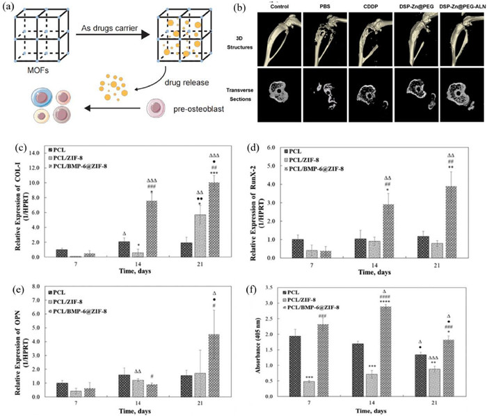
First of all, the osteogenic effects of MOFs loading drugs are embodied in promoting the expression of osteogenic genes and promoting calcium deposition. BMP-6-coated ZIF-8 nanocrystals embedded with PCL (PCL/BMP-6@ZIF-8) promote proliferation of pre-osteoblasts (MC3T3-E1 cells), expression of Col 1 gene, Opn and other osteogenic genes, upregulate ALP activity and promote calcium deposition (Fig. 5). In an in vivo bone defect model, the PCL/BMP-6@ZIF-8 fibrous membrane group exhibited the most extensive mature and effective bone regeneration and newly formed bone, confirming the stimulatory effect of BMP-6 on osteogenesis [44]. A stable, effective and sustained dexamethasone delivery platform SF-DEX@ZIF-8-Ti disk was constructed by immobilizing dexamethasone@zeolitic imidazolate framework-8 nanoparticles in micrometer-scale artificially etched pits on a titanium substrate, encapsulated using methanol-induced regenerative silk fibrillar membranes on titanium discs. acting on mouse pre osteoblasts (MC3T3-E1 cells), exhibited more mineralized nodules and increased mRNA transcript levels of Runx2, Opn and Ocn [106]. DEX@Zn-Mg-MOF74/PDA-PEEK resulted in elevated ALP, increased mineralized/calcified nodules and increased expression of osteogenic genes (Runx2, Col 1, Opn and Ocn). In vivo, PEEK-DEX samples showed significant osseointegration with the host bone and formed new bone bridges on the surface; both new bone volume and bone density were increased [50].

|
Download:
|
| Fig. 5. (a) The sketch map of MOFs as a drug delivery system contributing to osteogenesis. (b) Micro-CT 3D reconstruction image, the 2D transverse sections of tibia. Reproduced with permission [107]. Copyright 2017, Elsevier B.V. Expression of osteogenic-related genes in MC3T3-E1 cells cultured on membranes measured by quantitative RT-PCR. COL-I (c), RunX2 (d) and OPN (e). Quantitative ALP activity of cells cultured on membranes (f). n = 3, *P < 0.05, **P < 0.01, ***P < 0.005 when the control group is PCL, #P < 0.05, ##P < 0.01, ###P < 0.005 when the control is PCL/ZIF-8, ΔP < 0.05, ΔΔP < 0.01, ΔΔΔP < 0.005 when the control group is day 7 and ●P < 0.05, ●●P < 0.01, ●●●P < 0.005 when the control is day 14. The colorimetric quantitative results showing the matrix mineralization of cells cultured on PCL, PCL/ZIF-8 and PCL/BMP-6@ZIF-8 membranes on days 14 and 21 by ARS staining. Reproduced with permission [44]. Copyright 2020, Elsevier B.V. | |
On the base of the osteogenic effects of MOFs themselves, researchers explored other properties further, such as controlling drug release, targeted delivery, biodegradability and antibacterial property. The chitosan scaffold loaded with ZIF-8 [37] can not only promote the differentiation of MC3T3-E1 pre-osteoblasts, but also deliver vancomycin to treat severe bone infections. More importantly, this material could control drug release according to pH, which satisfied the purpose of precise administration. He et al. [107] synthesized Zn@PEG-ALN NPs, and utilized them to carry and deliver cisplatin prodrug (DSP) to bone metastases to treat bone metastatic breast cancer. Through adjusting the volume ratio of the oil phase to the water phase in the microemulsion, the particle size of DSP-Zn@PEG-ALN NPs could be adjusted. Studies of biological distribution in vivo had shown that DSP-Zn@PEG-ALN NPs delivered four times as much cisplatin to bone metastases as healthy bone, which indicated that these nano-particles reduced the toxicity of cisplatin and decreased bone destruction. Maria et al. [48] proposed the concept of biocompatible MOFs (bioMOFs), and had done some research about them. BioMOFs can carry and convey a variety of ingredients, such as bisphosphonates molecules and alkaline earth cations. They verified there was nontoxicity, high bioavailability and good biosafety of bioMOFs. Based on these peculiarities of bioMOFs, they could support biological mineralization to treat osteoporosis. This material provided new ideas in creating a biofunctional and safe platform for target delivery to confront osteoporosis. Yu et al. [84] designed a multifunctional mineralized collagen coating on titanium surface, and utilized MOF nanocrystals to control naringin release. Naringin had the ability to prevent bacterial infection and promote osteogenesis. The study found that mesenchymal stem cells adhered and proliferated well on the coating, and their mineralization and differentiation were significantly enhanced. Meanwhile, the proliferation of Staphylococcus aureus on this coating was affected, indicating the promotion of antibacterial ability.
In a word, drug-loaded MOFs are good materials for bone tissue engineering. That is because the drugs themselves can promote bone formation and fight bacteria. And MOFs can precisely deliver drugs to specific sites and control their release. It is believed that more such materials will be used for bone tissue engineering in the future.
4.3. MOFs combined with porous scaffolds contribute to osteogenesisIn addition to exploring more variety MOFs used for bone tissue engineering, we can find not a few reports that MOFs combined with biological scaffolds are used to promote osteogenesis. Similarly, porous scaffolds have interconnected porous structures and good biocompatibility, which can also promote cell growth, attachment, and differentiation [108]. However, the osteogenic effect of porous scaffolds is not enough, which limits the practical use in bone tissue engineering. Besides porous structures and good biocompatibility, MOFs also have a prominently osteogenic effect. Some research indicates that MOFs combining scaffolds provide good prospects for bone tissue engineering.
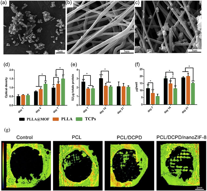
Combining with ordinary scaffolds, MOFs show better osteogenesis and bioactivity. Telgerd et al. [47] synthesized Zn-Cu imidazole MOF-coated PLLA scaffold (PLLA@MOF). They did water contact assay, tensile strength assay, isolation and expansion of adipose tissue-derived MSCs, cell seeding and osteogenic differentiation, cell proliferation, ALP activity, calcium content assay. These results reflected the change of scaffold (Fig. 6). In addition to the great biocompatibility and hydrophilicity of the scaffold have been mentioned, compared with pure PLLA scaffold, PLLA@MOF had better osteogenesis of MCSs derived from human adipose tissue. Generally speaking, Zn-Cu Imidazole MOF particles were coated on the surface of PLLA nanofiber scaffolds, which improved the bioactivity of PLLA scaffolds and enabled them to be used in bone tissue engineering. As a new type of bioactivity scaffold, PLLA@MOF showed a great prospect. Xia et al. [109] employed C-ZnO nanoparticles to embellish fibrous scaffold. This modified scaffold showed increased expression of IBSP, ALP, and vinculin, which substantiated the feasibility of bone regeneration and therapeutic therapies based on mesenchymal stem cells (MSCs). In addition, the ZnO nanoparticles allowed the slow release of Zn2+ ions, which not only activated various signaling pathways to guide osteogenic differentiation, but also prevent the potential bacterial infection during implantable applications. These represented that this material can be well used in bone tissue engineering.

|
Download:
|
| Fig. 6. FESEM image of MOF particles (a), SEM images of PLLA (b), and SEM images of PLLA@MOF (c). Rate of MSCs proliferation (d), the activity of alkaline phosphatase enzyme (e), and level of calcium deposition (f). Reproduced with permission [47]. Copyright 2019, Wiley Periodicals, Inc. New bone formation analyzed by micro-CT at 12 weeks after surgery. Representative 3D reconstructive images (g). Reproduced with permission [54]. Copyright 2020, The Royal Society of Chemistry. | |
In recent years, the use of 3D printing in medicine has attracted increasing attention. Compared with traditional materials production methods, 3D printing can produce more delicate and accurate medical materials, which is more conducive to the treatment of diseases. Similarly, some researchers have found that the MOFs produced by 3D printing also has good effects in promoting osteogenesis and bone regeneration, showing a good prospect in the application of bone tissue engineering. Zhong et al. [54] incorporated nanoZIF-8 into composite scaffolds composed of dicalcium phosphate dihydrate (DCPD) and polycaprolactone (PCL) by 3D printing. Research [28,110] have shown that nanoZIF-8 has minimal cytotoxicity, antibacterial and anti-inflammation effects, and can also promote osteogenesis in vitro and in vivo. Then, they studied the properties of nanoZIF-8-loaded 3D printed scaffolds. In vitro studies showed that the scaffolds had good biocompatibility with bone mesenchymal stem cells (BMSCs). The scaffolds also significantly up-regulated the expression of osteogenic proteins and genes, and promote extracellular matrix mineralization at the same time. In addition, in vivo studies showed that the scaffolds promoted bone formation in pivotal bone defect sites of rabbits. These indicate that nanoZIF-8-loaded 3D printed scaffolds have the ability to accelerate the re-establishment of bone defect. Dang et al. [83] successfully synthesized MOF Cu-TCPP nanosheets interface-structured β-tricalcium phosphate (TCP) scaffold (Cu-TCPP-TCP) by integrating 3D printing with in situ growth method in a solvothermal system. Research showed that this scaffold supported adhesion of HBMSCs and HUVECs. It also significantly stimulated the expression of osteogenic and angiogenic differentiation-related genes in HBMSCs and HUVEC. Besides, Cu-TCPP-TCP scaffold can promote osteoanagenesis after implanting into deficient bones of rabbits.
These two bioactive scaffolds synthesized by 3D print both manifest great osteogenic abilities. This provides theoretical feasibility for other new 3D-printed bioactive scaffolds to be used in bone tissue engineering.
5. Application of MOFs in bone diseases treatmentAs we all know, bone diseases occur in all ages of human beings. Major bone diseases include osteoarthritis, osteosarcoma, osteoporosis, osteomyelitis, and so on. These diseases may lead to limited movement, joint deformity, bone fracture and harm to other systems, such as the cardiovascular system, and digestive system. In addition, osteosarcoma may even lead to death.
The applications of MOFs in the treatment of bone diseases are mainly divided into two categories: one is modifying the structure of MOFs to change their internal biological activity and their own structural characteristics to treat diseases. The other is as carriers releasing the loaded drugs to treat diseases. According to different types of diseases and different therapeutic purposes, MOFs can play different roles (Fig. 7 and Table S2 in Supporting information).

|
Download:
|
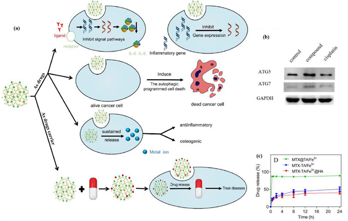
| Fig. 7. (a) Two pathways of MOFs treating bone diseases. As drugs: Inhibit intracellular signal pathways. Induce cancer cells apoptosis. Inhibit the expression of inflammatory genes. Anti-inflammatory and osteogenic effects of MOFs by releasing metal ion steadily. (b) Increased level of autophagic programmed cell death in human osteosarcoma Saos2 cells after compound treatment. The relative expression of the ATG5 and ATG7 was detected with Western blot. Copied with permission [125]. Copyright 2020, Elsevier B.V. (c) In vitro MTX release from MTX@TA/Fe3+ MOFs, MTX-TA/Fe3+ MOFs, and MTX-TA/Fe3+@HA MOFs in PBS. Copied with permission [149]. Copyright 2020, Elsevier B.V. | |
Osteoarthritis (OA) is a common degenerative joint disease. In recent years, the incidence rate of this disease has increased worldwide. The life quality of osteoarthritis patients, especially elderly patients, has been seriously affected [111]. At present, the idea of applying MOFs to the OA treatment is still under exploration.
The new modified MOFs mostly take zinc (Zn) as the metal center and can inhibit some intracellular signal transduction pathways and reducing the level of inflammatory cytokines, thus treating OA. As we all know, PI3K/Akt signaling pathway is an intracellular signaling pathway, which can accept extracellular signals and transmit signals into cells to promote metabolism, proliferation, cell survival, growth, and angiogenesis. After continuous research and exploration, scholars found that there was an abnormally activated PI3K/Akt signal pathway in the synovium of OA patients [112]. In the synovial cells, the pathway plays a role in regulating the production of downstream inflammatory cytokines, such as IL-6 and IL-18. Cai et al. [113] prepared a new metal-organic framework in the solvothermal conditions with Zn as the metal center. This MOF has a microporous Zn(Ⅱ)-MOF with rich water-coordinated metal centers and a high density of free N-donor groups, which can significantly inhibit the PI3K/Akt signaling pathway and reduce the IL-18 and IL-6 levels in cells. Among the related diseases of osteoarthritis, postmenopausal osteoarthritis deserves our attention. As the name suggests, postmenopausal osteoarthritis is more common in postmenopausal women over 50 years old. The main cause is the significant reduction of estrogen in postmenopausal women and the abnormal level of related cytokines [114]. Wei et al. [115] also took Zn as the metal center to prepare a new MOFs with multiple functional active sites. The compound can inhibit the inflammatory signaling pathway, reduce the NF-κb level in bone tissue in postmenopausal osteoarthritis animals, and delay the progress of postmenopausal osteoarthritis by increasing estrogen levels.
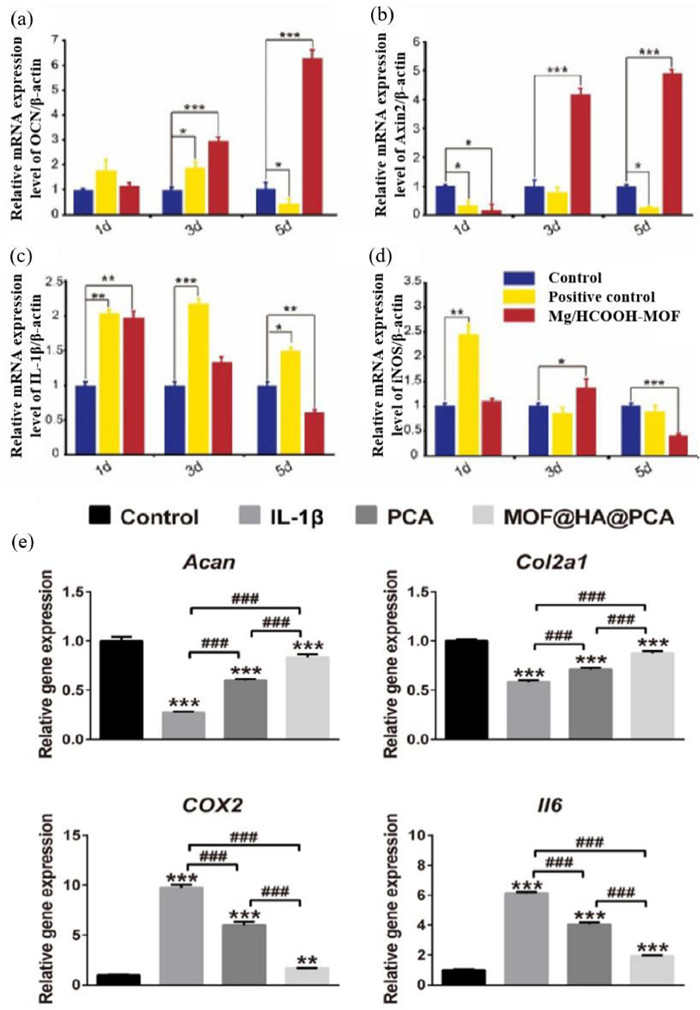
For the application of MOFs in OA treatment, in addition to regulating the activation level of the signal pathway, promoting bone formation is another way to treat OA with MOFs. In OA pathological development, cartilage loss progressively and abnormal bone tissue form. As a mineral, magnesium can promote bone formation. At the same time, some studies have found that magnesium has a strong anti-inflammatory effect [116]. Li et al. [74] synthesized Mg/HCOOH MOF and found Mg/HCOOH MOF a potential drug to treat OA due to the anti-inflammatory and bone-promoting properties of magnesium and the stability of MOFs (Fig. 8). They further showed that Mg/HCOOH MOF can stably and slowly release Mg2+ for a long time and up-regulate the pro-inflammatory factor IL-1β while inhibiting the expression of inflammatory genes.

|
Download:
|
| Fig. 8. Treat osteoarthritis. The expression levels of osteogenesis-related genes and inflammation-related genes of MG63 cells cultured with Mg/HCOOH−MOF detected by qPCR. (a) OCN, (b) Axin2, (c) IL-1β, (d) iNOS. Copied with permission [74]. Copyright 2019, Wiley‐VCH Verlag GmbH & Co. KGaA, Weinheim. (e) Relative mRNA levels of chondrogenic markers (Acan, Col2a1) and OA-relative genes (COX2, Il6). Data was presented as the mean ± SD (n = 3). *, #: P < 0.05; **, ##: P < 0.01; ***, ###: P < 0.001. *,**,*** indicate significant differences in comparison with the control group. Reproduced with permission [131]. Copyright 2020, The Author(s). | |
Osteoporosis is a systemic bone disease in which the fracture can happen easily because of the declining bone mineral density, the bone microstructure is destructed, increasing bone fragility [117,118]. Therefore, Calcium supplementation is one of the treatment principles for patients with osteoporosis. However, the blood calcium fluctuation may eventually lead to problems such as hypercalcemia and urinary stones due to the great impact on the calcium level in the body after oral or intravenous infusion. Since MOFs can stably release metal ions for a long time, Hou et al. [38] used calcium as the metal center, synthesized five calcium carboxylate compounds that exist stably in water. These compounds can be used as calcium agents that stably release calcium ions to supplement the calcium content in patients with osteoporosis and alleviate symptoms, along with preventing fracture.
For fractures caused by osteoporosis, one treatment method is implanting magnesium-based metal. However, magnesium can corrode in the body easily, and the bone formation-promoting ability needs to be improved. Zhang et al. [119] added a layer of metal-organic complex coating with magnesium combined phytic acid (PA) and zoledronic acid (ZA) on the surface of magnesium-based metal implants, which greatly enhanced the corrosion resistance of metal implants, inhibited osteoclasts and promoted the proliferation of pre-osteoblasts, promoting osteogenesis.
Another traditional treatment for osteoporosis is bisphosphate (BP). Bisphosphate can selectively combine with hydroxyapatite in the bone to inhibit osteoclast, thus inhibiting bone absorption. It has a good effect on osteoporosis treatment [120]. However, bisphosphate also has undeniable side effects, such as jaw osteonecrosis [121], hypocalcemia, and atrial fibrillation [122]. At present, the risk of side effects increases according to the increasing diphosphate dosage due to the low oral bioavailability of BP, to solve this problem, researchers found that MOFs can store and release active drugs stably for a long time, which may provide a way to solve this problem. Konstantinos et al. [123] developed metal bisphosphonate compounds/coordination polymers. Among them, the bisphosphonate ligand/linker is an active drug, which can control the stable release of bisphosphonate, improving the prognosis of osteoporosis treatment and improving the quality of life of patients. Maria et al. [124] reported that using Ca2+ and Mg2+ as metal centers and several anti-osteoporosis bisphosphate drugs as organic linkers, the new MOFs can release bisphosphate drugs at a specific rate for a long time. What is more, the drug releasing rate and releasing percentage of MOFs obtained after the combination of different drugs and different metal ions are different, the reason may be the different internal structure of MOFs.
5.1.3. OsteosarcomaOsteosarcoma is a common primary malignant tumor in bone tissue. Because the pathogenesis is still unclear, and the pathological progress is rapid, which poses a great threat to the life quality of patients and greatly increases the treatment difficulty.
At present, the new MOFs developed for the osteosarcoma treatment mainly take Zn as the metal center, and the main anti-cancer mechanism is regulating the autophagy program of tumor cells to eliminate cancer cells. Song et al. [125] prepared a new MOF containing Zn(Ⅱ). The compound contains a one-dimensional chain network based on Zn2(CO2)4, which can induce cells to start an autophagy program (Fig. 7), resulting in cell death, inhibiting the growth of human osteosarcoma cell line Saos2. Peng et al. [126] also prepared a new MOF containing Zn. The structure of the compound contains high-density open metal sites and one-dimensional channels rich in O donor sites, which can promote autophagy of MG63 cancer cells, kill cancer cells and achieve the treatment purpose. Previously, Chen et al. [127] prepared a porous MOF containing π electron-rich ligand through solvothermal method, inhibiting human osteosarcoma cells of 293T, 3T3, B16, and 769-p cell lines.
In addition, scholars found that many complexes with copper as the metal center also have high anticancer activity. Inspired by this, scientists have developed a variety of copper-based MOFs for various cancer treatments, including colon cancer [128], breast cancer, and so on. At the same time, copper-based MOFs have also been used in the treatment of osteosarcoma. Luo et al. [129] synthesized a new MOF with high-density pores and one-dimensional hexagonal channels under the condition of hot solvent. Through relevant biological experiments, it was found that the compound can induce a large accumulation of ROS in osteosarcoma cells, significantly inhibiting the OS-732 osteosarcoma cells.
5.2. As a drug delivery systemIn general, MOFs have the following characteristics supporting them to be as drug delivery systems: (1) The pore network is orderly and the pore size is very uniform so that developers can accurately control the drug loading and release rate. (2) The number of pores is large, which can meet the required drug load. (3) Large surface area can provide large drug adsorption capacity. Furthermore, drugs for bone diseases nowadays usually have some problems such as short half-life, strong anticoagulation and obvious side effects [76,130-133], which can damage the gastrointestinal liver function [134].
According to the types and characteristics of drugs, MOFs can load drugs in different ways, mainly including making drugs as organic ligands and direct physical packaging [105]. For non-neoplastic bone diseases, MOFs mainly load traditional clinical drugs or treatment-related substances directly to the focus for accurate treatment, avoid waste, reduce the harm of side effects and optimize the treatment effect. For neoplastic bone diseases, due to the complexity of tumor treatment, MOFs can play a role in each process of treatment. The following mainly introduces several major bone diseases.
5.2.1. OsteoarthritisIn recent years, the commonly used methods for the treatment of osteoarthritis are drug treatment and surgical treatment. The drugs include oral drugs and intra-articular injection drugs, oral drugs such as ketoprofen, glycosaminoglycan (GAG), intra-articular injection drugs such as anti-inflammatory agents protocatechuic acid (PCA), antioxidants. However, due to the limitations mentioned above, MOFs have attracted considerable attention in the therapy of osteoarthritis.
Aiming at improving oral drug delivery, Zhen et al. [76] successfully loaded ketoprofen with uio-66 metal organic framework (MOF). At the same time, they found that the loading amount of −NH2 functional group was higher than that of −NO2 functional group, which may be due to the high hydrogen bond capacity and −NH2 is an alkaline functional group. Velasquez-Hernandez et al. [130] designed three pH responsive frameworks based on azo metals (ZIF-8, ZIF-90 and MAF-7) and showed that different MOFs can be selected according to the different required loading amounts and release rates due to the different chemical properties and matrix, achieving different therapeutic purposes.
As for improving intra-articular drug injection, to avoid poor water stability and short drug half-life of MOFs, Xiong et al. [131] modified the MIL-100 (Fe) surface with HA to improve its water stability, and HA also has the anti-inflammatory effects and lubricating cartilage, and finally synthesized MOF@HA-NPs, which is used as a drug delivery carrier, and successfully loaded with the anti-inflammatory agent protocatechuic acid (PCA). MOF@HA@PCA can continuously release HA and PCA in the acidic inflammatory microenvironment, prevent the OA progression and protect cartilage. In addition to anti-inflammatory agents, using antioxidants to scavenge active free radicals in Osteoarthritis Chondrocytes is also a therapeutic method. However, antioxidants have some cytotoxicity, Song et al. [132] loaded bilirubin (BR) into the MOF shell and rapamycin (RAP) into the MOF mesoporous to design a near-infrared laser responsive drug release system, which can remove oxygen free radicals in chondrocytes, reduce apoptosis and prevent cartilage lesions (Fig. 8). Moreover, Yang et al. [135] synthesized gelatin (Gel)-glucosamine hydrochloride (GH) mixed crosslinked-cyclodextrin metal-organic framework@IBU (G-GH/CL-CD-MOF@IBU). To solve poor water solubility and mechanical properties, they used polyethylene glycol diglycidyl ether as the crosslinking agent. It is indicated that G-GH/CL-CD-MOF@IBU composite hydrogel had certain mechanical properties, good biocompatibility, and sustained drug releasing behavior, which provided good prospects for OA treatment.
5.2.2. OsteoporosisBone pain is a big problem for osteoporosis, and ketoprofen is a vital choice to release the pain. However, it has some side effects that limit its practical use. To solve this problem, Ge et al. [136] prepared Mg-MOF-74 as a drug delivery system which loaded ketoprofen to treat osteoporotic pain. They synthesized Mg-MOF-74 by hydrothermal synthesis, and XDR showed it had perfect crystallinity and crystal structure. Then, ketoprofen was loaded into Mg-MOF-74, it is verified that Ket@Mg-MOF-74 had a high loading rate and good controlled release of ketoprofen. More importantly, it was indicated that this compound could enhance the osteogenic cytokines expression obviously, and reduce the secretion of COX2 and proinflammatory factors. It is the osteogenic and anti-inflammatory effects of Ket@Mg-MOF-74 that make it be an ideal drug system to treat osteoporosis.
5.2.3. OsteosarcomaThere are many cancer treatment strategies, and chemotherapy drugs are of great significance in cancer treatment. However, their toxicity cannot be ignored and limited bioavailability needs to be improved due to their side effects and rapid elimination in vivo [137,138]. Recently, the use of carriers to provide anticancer drugs has shown promising potential to overcome these difficulties. For osteosarcoma treatment, besides developing new MOF and regulating autophagy program to kill cancer cells, MOF can also change the genes of cancer cells, load anticancer drugs and improve the radiotherapy sensitivity.
It has been reported that 5-fluorouracil, one of the commonly used anticancer drugs, has been successfully loaded into MOF. Zhao et al. [139] prepared a new porous MOF with V-shaped Poly carboxylic acid organic linker under solvothermal conditions. It has a high-density o donor site, which can load the anticancer drug 5-fluorouracil (5-FU), and release the drug depending on the pH in the environment. This new MOF has an obvious anticancer effect and a low toxicity to human normal cells. Guo et al. [140] also synthesized a new porous metal organic skeleton based on Dy(III) in a porous environment using a curved Poly carboxylic acid linker under solvothermal conditions. The MOF has a functionalized 1D channel with polar atoms, which can load anticancer drug 5-FU, and has cytotoxicity to human osteosarcoma cell line MG63. In another study, Wu et al. [141] combined MOFs with erythrocyte membrane nano vesicles modified by a VEGF ligand to form a bilayer drug delivery system (V-RZCD). The core MOF was synthesized by ZA and Ca2+ and loaded Adriamycin (DOX), and the shell was an erythrocyte membrane containing nanovesicles VEGF. According to their experiments, the drug delivery system has good biocompatibility and biodegradability. It can release the ZA and Ca2+ to the tumor site accurately with the help of VEGF, so as to inhibit the growth of osteosarcoma cells and resist osteolysis at the same time.
In addition to chemotherapy, radiotherapy is also one of the main treatments for osteosarcoma. By irradiating the tumor sites with X-rays, γ-rays, etc., it can eliminate tumor cells in the active dividing phase. However, it has great side effects on patients due to poor sensitivity and high-dose radiation. Du et al. [142] developed D-arginine loaded MOF nanoparticles to improve the radiosensitivity of osteosarcoma and reduce side effects, so as to provide a safe and effective way for radiotherapy of osteosarcoma. Undoubtedly, radiotherapy in cooperation with MOFs has significant advances over radiotherapy alone, but the corresponding theory is not mature and correlative research reports are few.
Besides, MOFs has also emerged as a candidate for the treatment of bone metastases. In a recent work, Nafiseh et al. [143] developed a new bone searching agent based on the structure of MOF. In their experiments, the bone searching agent was closely combined with 177Lu, which had good therapeutic characteristics for bone metastasis and fully alleviated the pain caused by bone metastasis. In another work, Ma et al. [144] designed a double shell nano therapeutic system with gold nanoparticle core, MOF and mesoporous silica (MS). The system can encapsulate chemotherapeutic drug cisplatin (CIS) and inhibitor alpelisib simultaneously to achieve pH responsive drug release in acidic tumor microenvironment and combined with photothermal effect in the treatment of metastatic spinal tumors.
5.2.4. Rheumatoid arthritisRheumatoid arthritis (RA) is a chronic, systemic disease dominated by inflammatory synovitis. The main clinical manifestation is a joint deformity, which has a great adverse impact on the patients’ life quality [145,146]. At present, the main disease-modifying antirheumatic drugs (DMARDs) used in clinic include leflunomide, methotrexate and cyclosporine, inhibiting the inflammatory cytokines production, delaying the rheumatoid arthritis progress and alleviating symptoms [147]. However, these drugs have some non-ignorable side effects mentioned above.
Therefore, MOFs can be used for drug loading to accurately deliver drugs to arthritis joints, reducing side effects, improving prognosis and avoiding drug waste. In the report of Kritskiy et al. [148], they loaded LEF on gamma-cyclodextrin-metal organic frameworks according to its layered structure and high specific surface area, synthesized a drug delivery system which can delivery LEF efficiently. At present, several research have reported that MTX loaded MOFs have great potential in the RA targeted therapy. It is worth mentioning that Guo et al. [149] noticed that MTX is a weak metal ions affinity drug, so they covalently linked MTX to Ta/Fe3+ MOFs (Fig. 7), thus solved the defect that the drugs are released prematurely after direct coordination or physical encapsulation.
5.2.5. OsteomyelitisOsteomyelitis is a bacterial inflammation. It often occurs repeatedly, and serious affects the physical and mental health and living ability of patients. Because osteomyelitis is mainly caused by bacterial infection, the antibiotics application is essential. However, due to the drug-resistant bacteria and side effects of antibiotics, it is necessary to use MOF to deliver antibiotics for accurately killing the bacteria. In fact, some studies have shown that chitosan can be used as a bone tissue scaffold to enhance cell adhesion and promote osteoblast differentiation. Karakecili et al. [37] loaded vancomycin in ZIF-8 and embedded it into chitosan fiber scaffold, so that the scaffold has two important properties: inhibiting the growth of bacterial and promoting osteogenesis. Moreover, Kundu et al. [150] synthesized copper sericin (Cu-SER) MOFs. The sericin has antibacterial and osteogenic properties, and plays the part of organic template for Cu-SER MOFs deposition. Importantly, Cu-SER MOFs provide potential approaches for minimizing antibiotic dependence.
The major limitation of antibiotics is drug-resistance, and scientists have found microwaves to solve this problem. However, microwave is harmful to the normal tissue around the lesion. Thus, developing a material to absorb microwaves is of great significance. MOFs has shown great potential in absorbing microwaves due to its structure that can realize multiple reflection and refraction from microwave. Wei et al. [151] developed a Na+ inserted Pb microwave response system based on the characteristics of MOF. The system can induce synergy between microwave, microwave thermal effect and ROS, and can be used as microwave absorbing material and iron ion carrier to treat bacterial infected osteomyelitis.
6. Conclusions and prospectsIn view of quite a few reports on MOFs in the field of bone tissue engineering and bone diseases, we attempt to summarize the effects of MOFs on bone tissue engineering and bone diseases in this section.
For a start, all MOFs consist of organic linkers and bridging metal ions or clusters, however, different synthesis will get MOFs with different properties. For example, MOFs synthesized by non-solvothermal synthesis can have uniform crystal size and clear boundary, while MOFs have high purity and good dispersity if using hydrothermal synthesis. Therefore, it is necessary to choose an appropriate synthesis. Secondly, many organic linkers and metals can be used to construct MOFs. Using suitable raw materials can synthesize MOFs with good biocompatibility [53]. Meanwhile, organic linkers and metals are tightly bound together by chemical bonds and their release rate is not sufficient to cause cytotoxicity [56]. The two advantages provide a foundation for being utilized in biosome. Nowadays, how to promote utilization rate of raw materials, which is friendly to environment, is an issue deserved researching.
In bone tissue engineering, whether MOFs themselves or carrying drugs, MOFs have attracted much attention. The porous structure and similar mechanical properties of MOFs are in favor of cell adhesion and growth. For osteoblasts, MOFs can promote the expression of osteogenic genes [15,39]. After transcribing and translating, the content of homologous proteins increases [93,94]. As signaling molecules, these proteins promote osteoblast differentiation. Ultimately, MOFs increase intracellular calcium deposition [37], and accelerate extracellular matrix mineralization [15]. When it comes to osteoclasts, MOFs inhibit their size and function. What is more, it is confirmed that MOFs can promote angiogenesis. And blood vessels are vital structures to provide nutrients for cells, which guarantees an adequate energy supply for cell differentiation. Besides MOFs themselves, MOFs as a drug delivery system still play an important role. Drug-loaded MOFs can promote the expression of osteogenic genes and calcium deposition [106]. Moreover, combined with porous scaffolds, MOFs show better osteogenesis and bioactivity.
MOFs are still widely used to treat bone diseases, such as osteoarthritis, osteoporosis, osteosarcoma, osteomyelitis. Similarly, MOFs work by themselves or carrying drugs. For a start, MOFs with zinc as the metal center can inhibit some intracellular signal transduction pathways and reduce the level of inflammatory cytokines to treat osteoarthritis. As a drug delivery system, MOFs are better than drugs for bone diseases nowadays, owing to their orderly pore network and uniformed pore size, they own accurate and stable drug loading and release rate; in addition, the large number of pores make them meet the required drug load easily; last, they can provide large drug adsorption capacity due to their large surface area. Moreover, targeted agent is a hot research direction in recent years, therefore, whether MOFs can be used in the drug targeted is a direction worthy researching.
In summary, MOFs are promising synthetic bone graft materials for the design of osteogenic drugs. There are more and more MOFs having been applied to bone tissue engineering and bone diseases, may serve directly as osteogenic drugs, or be used to construct drug-MOFs synergistic systems, or combine with the biological scaffolds. For the clinical application of MOFs, it is necessary to make a breakthrough in the biosafety of new MOFs in the future. Metal ions and organic linkers released during the dissociation of MOFs may cause potential biosafety problems. The potential solution to this problem is to use biocompatible ligands to obtain nontoxic MOFs. In addition, when MOFs are used as drug delivery systems, the drugs delivered may have toxic and side effects on the healthy tissues around the lesion site. How to reduce the long-term accumulation of the delivered drugs in the healthy sites will also be a major challenge for the clinical application of MOFs in the future.
Although the development of clinical drugs is still at early stages and some challenges remain to be solved, there is no doubt that MOF-based osteogenic drugs could innovate clinical synthetic bone graft materials under the corporate efforts of researchers.
Declaration of competing interestWe declare that we have no financial and personal relationships with other people or organizations that can inappropriately influence our work, there is no professional or other personal interest of any nature or kind in any product, service and/or company that could be construed as influencing the position presented in, or the review of, the manuscript entitled.
AcknowledgmentsThis material is based upon work supported by the National Natural Science Foundation of China (Nos. 82071164 and 82271016) and Key Research Program of Sichuan Science and Technology Department (No. 2021YFS0052).
Supplementary materialsSupplementary material associated with this article can be found, in the online version, at doi:10.1016/j.cclet.2022.107986.
| [1] |
F. Donnaloja, E. Jacchetti, M. Soncini, M.T. Raimondi, Polymers (Basel) 12 (2020) 905. DOI:10.3390/polym12040905 |
| [2] |
E.R. Oliveira, L. Nie, D. Podstawczyk, et al., Int. J. Mol. Sci. 22 (2021) 903. DOI:10.3390/ijms22020903 |
| [3] |
J.A. McGovern, M. Griffin, D.W. Hutmacher, Dis. Model Mech. 11 (2018) dmm033084. DOI:10.1242/dmm.033084 |
| [4] |
J. Zhang, Y. Jiang, Z. Shang, et al., Bioact. Mater. 6 (2021) 4027-4052. DOI:10.1016/j.bioactmat.2021.03.035 |
| [5] |
S. Wei, J.X. Ma, L. Xu, et al., Mil. Med. Res. 7 (2020) 54. |
| [6] |
M. Gerressen, D. Riediger, R.D. Hilgers, et al., J. Oral Implantol. 41 (2015) 276-283. DOI:10.1563/AAID-JOI-D-13-00246 |
| [7] |
Y. Ma, S. Gu, Q. Yin, et al., BMC Musculoskelet. Disord. 20 (2019) 346. DOI:10.1186/s12891-019-2713-y |
| [8] |
A. Singh, M. Kohli, N. Gupta, J. Maxillofac. Oral Surg. 11 (2012) 430-434. DOI:10.1007/s12663-012-0351-0 |
| [9] |
R. Zhao, R. Yang, P.R. Cooper, et al., Molecules 26 (2021) 3007. DOI:10.3390/molecules26103007 |
| [10] |
I. El Bialy, W. Jiskoot, M. Reza Nejadnik, Pharm. Res. 34 (2017) 1152-1170. DOI:10.1007/s11095-017-2147-x |
| [11] |
K.Q. Huang, J. Huang, J.M. Zhao, Z.P. Gu, J. Wu, Chin. Chem. Lett. 33 (2022) 1941-1945. DOI:10.1016/j.cclet.2021.10.073 |
| [12] |
W. Zhu, J. Zhao, Q. Chen, Z. Liu, Coord. Chem. Rev. 398 (2019) 113009. DOI:10.1016/j.ccr.2019.07.006 |
| [13] |
J.Y. Kim, R. Balderas-Xicohtencatl, L. Zhang, et al., J. Am. Chem. Soc. 139 (2017) 15135-15141. DOI:10.1021/jacs.7b07925 |
| [14] |
T. Simon-Yarza, A. Mielcarek, P. Couvreur, C. Serre, Adv. Mater. 30 (2018) e1707365. DOI:10.1002/adma.201707365 |
| [15] |
Y. Xue, Z. Zhu, X. Zhang, et al., Adv. Healthc. Mater. 10 (2021) e2001369. DOI:10.1002/adhm.202001369 |
| [16] |
M. Cai, G. Chen, L. Qin, et al., Pharmaceutics 12 (2020) 232. DOI:10.3390/pharmaceutics12030232 |
| [17] |
R.E. Morris, P.S. Wheatley, Angew. Chem. Int. Ed. 47 (2008) 4966-4981. DOI:10.1002/anie.200703934 |
| [18] |
B. Ogunbadejo, S. Al-Zuhair, Molecules 26 (2021) 680. DOI:10.3390/molecules26030680 |
| [19] |
F.G. Cirujano, R. Luque, A. Dhakshinamoorthy, Molecules 26 (2021) 1445. DOI:10.3390/molecules26051445 |
| [20] |
M. Mon, A. Pascual-Alvarez, T. Grancha, et al., Chem. Eur. J. 22 (2016) 539-545. DOI:10.1002/chem.201504176 |
| [21] |
S.N. Zhao, X.Z. Song, M. Zhu, et al., Chem. Eur. J. 21 (2015) 9748-9752. DOI:10.1002/chem.201500562 |
| [22] |
M. Zhu, X.Z. Song, S.Y. Song, et al., Adv. Sci. 2 (2015) 1500012. DOI:10.1002/advs.201500012 |
| [23] |
M. Shen, F. Forghani, X. Kong, et al., Compr. Rev. Food Sci. Food Saf. 19 (2020) 1397-1419. DOI:10.1111/1541-4337.12515 |
| [24] |
J. Della Rocca, D. Liu, W. Lin, et al., Acc. Chem. Res. 44 (2011) 957-968. DOI:10.1021/ar200028a |
| [25] |
J.E. Cun, X. Fan, Q. Pan, et al., Adv. Colloid Interface Sci. 305 (2022) 102686. DOI:10.1016/j.cis.2022.102686 |
| [26] |
X. Ge, R. Wong, A. Anisa, S. Ma, Biomaterials 281 (2022) 121322. DOI:10.1016/j.biomaterials.2021.121322 |
| [27] |
M. Shyngys, J. Ren, X. Liang, et al., Front. Bioeng. Biotechnol. 9 (2021) 603608. DOI:10.3389/fbioe.2021.603608 |
| [28] |
X. Zhang, J. Chen, X. Pei, et al., ACS Appl. Mater. Interfaces 9 (2017) 25171-25183. DOI:10.1021/acsami.7b07800 |
| [29] |
Y. Liu, Y. Zhao, X. Chen, Theranostics 9 (2019) 3122-3133. DOI:10.7150/thno.31918 |
| [30] |
E. Lamei, M. Hasanzadeh, Int. J. Biol. Macromol. 208 (2022) 409-420. DOI:10.1016/j.ijbiomac.2022.03.117 |
| [31] |
L. Yin, D. Wang, X. Li, et al., Sci. Total Environ. 815 (2022) 151962. DOI:10.1016/j.scitotenv.2021.151962 |
| [32] |
H. Zhao, F. Wang, L. Cui, et al., Nanomicro Lett. 13 (2021) 208. |
| [33] |
S.N. Zhao, G. Wang, D. Poelman, P.V. Voort, Materials (Basel) 11 (2018) 572. DOI:10.3390/ma11040572 |
| [34] |
P. Gao, Y. Chen, W. Pan, et al., Angew. Chem. Int. Ed. 60 (2021) 16763-16776. DOI:10.1002/anie.202102574 |
| [35] |
J. Chen, X. Zhang, C. Huang, et al., J. Biomed. Mater. Res. A 105 (2017) 834-846. DOI:10.1002/jbm.a.35960 |
| [36] |
M.A. Khalili, E. Tamjid, Sci. Rep. 11 (2021) 8645. DOI:10.1038/s41598-021-87783-x |
| [37] |
A. Karakecili, B. Topuz, S. Korpayev, M. Erdek, Mater. Sci. Eng. C 105 (2019) 110098. DOI:10.1016/j.msec.2019.110098 |
| [38] |
Y. Hou, C.Z. Luo, D.H. Xie, et al., Int. J. Pharm. 608 (2021) 121083. DOI:10.1016/j.ijpharm.2021.121083 |
| [39] |
X. Gao, Y. Xue, Z. Zhu, et al., ACS Appl. Mater. Interfaces 13 (2021) 97-111. DOI:10.1021/acsami.0c15945 |
| [40] |
F.N. Shi, J.C. Almeida, L.A. Helguero, et al., Inorg. Chem. 54 (2015) 9929-9935. DOI:10.1021/acs.inorgchem.5b01634 |
| [41] |
G. Chen, Y. Yu, X. Wu, et al., Research 2019 (2019) 6175398. |
| [42] |
L.L. Tan, N. Song, S.X. Zhang, et al., J. Mater. Chem. B 4 (2016) 135-140. DOI:10.1039/C5TB01789K |
| [43] |
N. Liu, Z. Zou, J. Liu, et al., Analyst 144 (2019) 6254-6261. DOI:10.1039/c9an01671f |
| [44] |
O. Toprak, B. Topuz, Y.A. Monsef, et al., Mater. Sci. Eng. C 120 (2021) 111738. DOI:10.1016/j.msec.2020.111738 |
| [45] |
S. He, L. Wu, X. Li, et al., Acta Pharm. Sin. B 11 (2021) 2362-2395. DOI:10.1016/j.apsb.2021.03.019 |
| [46] |
N. Joseph, H.D. Lawson, K.J. Overholt, et al., Sci. Rep. 9 (2019) 13024. DOI:10.1038/s41598-019-49536-9 |
| [47] |
M.D. Telgerd, M. Sadeghinia, G. Birhanu, et al., J. Biomed. Mater. Res. A 107 (2019) 1841-1848. |
| [48] |
M.A. Matlinska, M. Ha, B. Hughton, et al., ACS Appl. Mater. Interfaces 11 (2019) 32739-32745. DOI:10.1021/acsami.9b11004 |
| [49] |
K. AbouAitah, I.M. Higazy, A. Swiderska-Sroda, et al., Drug Deliv. 28 (2021) 1478-1495. DOI:10.1080/10717544.2021.1949073 |
| [50] |
H. Lu, X. Yang, S. Li, et al., Inorg. Chem. Commun. 61 (2015) 48-52. DOI:10.1016/j.inoche.2015.08.015 |
| [51] |
J. Li, Y.C. Wang, Y. Yu, Q.W. Li, Chin. Chem. Lett. 29 (2018) 837-841. DOI:10.5194/isprs-archives-xlii-3-837-2018 |
| [52] |
T. Xiao, L. Fan, R. Liu, et al., ACS Appl. Mater. Interfaces 13 (2021) 50836-50850. DOI:10.1021/acsami.1c18088 |
| [53] |
P. Horcajada, R. Gref, T. Baati, et al., Chem. Rev. 112 (2012) 1232-1268. DOI:10.1021/cr200256v |
| [54] |
L. Zhong, J. Chen, Z. Ma, et al., Nanoscale 12 (2020) 24437-24449. DOI:10.1039/d0nr06297a |
| [55] |
N. Singh, S. Qutub, N.M. Khashab, J. Mater. Chem. B 9 (2021) 5925-5934. DOI:10.1039/d1tb01044a |
| [56] |
I. Imaz, R.M. Marta, J. An, et al., Chem. Commun. 47 (2011) 7287-7302. DOI:10.1039/c1cc11202c |
| [57] |
S. Zirak Hassan Kiadeh, A. Ghaee, M. Farokhi, et al., Int. J. Biol. Macromol. 173 (2021) 351-365. DOI:10.1016/j.ijbiomac.2021.01.058 |
| [58] |
T. Hidalgo, M. Gimenez-Marques, E. Bellido, et al., Sci. Rep. 7 (2017) 43099. DOI:10.1038/srep43099 |
| [59] |
E. Bellido, T. Hidalgo, M.V. Lozano, et al., Adv. Healthc. Mater. 4 (2015) 1246-1257. DOI:10.1002/adhm.201400755 |
| [60] |
L.E. Smith, S. Rimmer, S. MacNeil, Biomaterials 27 (2006) 2806-2812. DOI:10.1016/j.biomaterials.2005.12.018 |
| [61] |
Y. Long, S. Song, J. Li, et al., ACS Catal. 8 (2018) 8506-8512. DOI:10.1021/acscatal.8b01851 |
| [62] |
C. Tamames-Tabar, D. Cunha, E. Imbuluzqueta, et al., J. Mater. Chem. B 2 (2014) 262-271. DOI:10.1039/C3TB20832J |
| [63] |
J. Xiao, Y. Zhu, S. Huddleston, et al., ACS Nano 12 (2018) 1023-1032. DOI:10.1021/acsnano.7b01850 |
| [64] |
N.A. Travlou, M. Algarra, C. Alcoholado, et al., ACS Appl. Bio. Mater. 1 (2018) 693-707. DOI:10.1021/acsabm.8b00166 |
| [65] |
M. Askarinia, M. Ghaedi, L. Manzouri, et al., Jundishapur J. Microb. 11 (2018) e60680. |
| [66] |
V. Celis-Arias, S. Loera-Serna, H.I. Beltrán, et al., New J. Chem. 42 (2018) 5570-5579. DOI:10.1039/C8NJ00120K |
| [67] |
A. Rauf, J. Ye, S. Zhang, et al., Polyhedron 166 (2019) 130-136. DOI:10.1016/j.poly.2019.03.039 |
| [68] |
X. Li, M. Qi, C. Li, et al., J. Mater. Chem. B 7 (2019) 6955-6971. DOI:10.1039/c9tb01743g |
| [69] |
D. Yu, Y. Guan, F. Bai, et al., Chem. Eur. J. 25 (2019) 3489-3495. DOI:10.1002/chem.201805835 |
| [70] |
B.C. Yan, J. Tan, H.F. Zhang, et al., Biomater. Adv. 134 (2022) 10. |
| [71] |
R. Karimi Alavijeh, S. Beheshti, K. Akhbari, A. Morsali, Polyhedron 156 (2018) 257-278. DOI:10.1016/j.poly.2018.09.028 |
| [72] |
M. Nasrabadi, M.A. Ghasemzadeh, M.R. Zand Monfared, New J. Chem. 43 (2019) 16033-16040. DOI:10.1039/c9nj03216a |
| [73] |
M. Chen, J. Zhang, J. Qi, et al., ACS Nano 16 (2022) 7732-7744. DOI:10.1021/acsnano.1c11613 |
| [74] |
Z. Li, Y. Peng, X. Pang, B. Tang, ChemMedChem 15 (2020) 13-16. DOI:10.1002/cmdc.201900546 |
| [75] |
M. Al Haydar, H.R. Abid, B. Sunderland, S. Wang, Drug Des. Dev. Ther. 11 (2017) 2685-2695. DOI:10.2147/DDDT.S145716 |
| [76] |
Z. Li, S. Zhao, H. Wang, et al., Colloids Surf. B 178 (2019) 1-7. DOI:10.1016/j.colsurfb.2019.02.027 |
| [77] |
M.P. Abucafy, B.L. Caetano, B.G. Chiari-Andreo, et al., Eur. J. Pharm. Biopharm. 127 (2018) 112-119. DOI:10.1016/j.ejpb.2018.02.009 |
| [78] |
Y. Chen, J. Cai, D. Liu, et al., Regen. Biomater. 9 (2022) rbac019. DOI:10.1093/rb/rbac019 |
| [79] |
X. Hu, W. Zhao, Z. Zhang, et al., Chin. Chem. Lett. 34 (2023) 107451. DOI:10.1016/j.cclet.2022.04.049 |
| [80] |
M. Li, S. Yin, M. Lin, et al., J. Mater. Chem. B 46 (2022) 13818. |
| [81] |
C. Liu, X. Xu, W. Cui, H. Zhang, Eng. Regen. 2 (2021) 105-108. |
| [82] |
P. Ducy, R. Zhang, V. Geoffroy, A.L. Ridall, G. Karsenty, Cell 89 (1997) 747-754. DOI:10.1016/S0092-8674(00)80257-3 |
| [83] |
W. Dang, B. Ma, B. Li, et al., Biofabrication 12 (2020) 025005. DOI:10.1088/1758-5090/ab5ae3 |
| [84] |
M. Yu, D. You, J. Zhuang, et al., ACS Appl. Mater. Interfaces 9 (2017) 19698-19705. DOI:10.1021/acsami.7b05296 |
| [85] |
W. Teng, Z. Zhang, Y. Wang, et al., Small 17 (2021) e2102315. DOI:10.1002/smll.202102315 |
| [86] |
K. Nakashima, X. Zhou, G. Kunkel, et al., Cell 108 (2002) 17-29. DOI:10.1016/S0092-8674(01)00622-5 |
| [87] |
J. An, H. Yang, Q. Zhang, et al., Life Sci. 147 (2016) 46-58. DOI:10.1016/j.lfs.2016.01.024 |
| [88] |
J.R. Farley, S.L. Hall, M.A. Tanner, J.E. Wergedal, J. Bone Miner. Res. 9 (1994) 497-508. |
| [89] |
M. Balcerzak, E. Hamade, L. Zhang, et al., Acta Biochim. Pol. 50 (2003) 1019-1038. DOI:10.18388/abp.2003_3629 |
| [90] |
X. Shen, Y. Zhang, P. Ma, et al., Biomaterials 212 (2019) 1-16. DOI:10.1155/2019/7602427 |
| [91] |
P. Ducy, C. Desbois, B. Boyce, et al., Nature 382 (1996) 448-452. DOI:10.1038/382448a0 |
| [92] |
N.B. Kavukcuoglu, P. Patterson-Buckendahl, A.B. Mann, J. Mech. Behav. Biomed. Mater. 2 (2009) 348-354. DOI:10.1016/j.jmbbm.2008.10.010 |
| [93] |
M. Mizuno, Y. Kuboki, J. Biochem. 129 (2001) 133-138. DOI:10.1093/oxfordjournals.jbchem.a002824 |
| [94] |
D. Chen, M.A. Harris, G. Rossini, et al., Calcif. Tissue Int. 60 (1997) 283-290. DOI:10.1007/s002239900230 |
| [95] |
T. Liu, Y. Gao, K. Sakamoto, et al., J. Cell. Physiol. 211 (2007) 728-735. DOI:10.1002/jcp.20988 |
| [96] |
S. Varghese, S. Rydziel, E. Canalis, J. Cell. Physiol. 202 (2005) 391-399. DOI:10.1002/jcp.20130 |
| [97] |
Y. Pang, Y. Fu, C. Li, et al., Nano Lett. 20 (2020) 829-840. DOI:10.1021/acs.nanolett.9b02916 |
| [98] |
C. Xu, Y. Kang, X. Dong, D. Jiang, M. Qi, Chin. Chem. Lett. 34 (2023) 107528. DOI:10.1016/j.cclet.2022.05.042 |
| [99] |
J. Chen, D. Sheng, T. Ying, et al., Nanomicro Lett. 13 (2020) 23. |
| [100] |
P. Zhang, Y. Li, Y. Tang, et al., ACS Appl. Mater. Interfaces 12 (2020) 18319-18331. DOI:10.1021/acsami.0c01792 |
| [101] |
T. Baheri, Y. Yamini, M. Shamsayei, M. Tabibpour, J. Sep. Sci. 44 (2021) 2814-2823. DOI:10.1002/jssc.202100070 |
| [102] |
M. Vallet-Regi, F. Balas, D. Arcos, Angew. Chem. Int. Ed. 46 (2007) 7548-7558. DOI:10.1002/anie.200604488 |
| [103] |
A. Hamedi, A. Anceschi, A. Patrucco, M. Hasanzadeh, J. Drug Target. 30 (2022) 381-393. DOI:10.1080/1061186x.2021.2012683 |
| [104] |
L. Li, Z. Qi, S. Han, et al., Mini Rev. Med. Chem. 22 (2022) 2564-2580. DOI:10.2174/1389557522666220330152145 |
| [105] |
B. Maranescu, A. Visa, Int. J. Mol. Sci. 23 (2022) 4458. DOI:10.3390/ijms23084458 |
| [106] |
J. Ran, H. Zeng, J. Cai, et al., Chem. Eng. J. 333 (2018) 20-33. DOI:10.1016/j.cej.2017.09.145 |
| [107] |
Y. He, Y. Huang, Z. Huang, et al., J. Control. Release 264 (2017) 76-88. DOI:10.1016/j.jconrel.2017.08.024 |
| [108] |
Y. Lv, B. Wang, G. Liu, et al., Front. Bioeng. Biotechnol. 9 (2021) 641130. DOI:10.3389/fbioe.2021.641130 |
| [109] |
Y. Xia, X. Fan, H. Yang, et al., Small 16 (2020) e2003010. DOI:10.1002/smll.202003010 |
| [110] |
Y. Liu, Z. Zhu, X. Pei, et al., ACS Appl. Mater. Interfaces 12 (2020) 36978-36995. DOI:10.1021/acsami.0c12090 |
| [111] |
G.B.D. Disease, I. Injury, C. Prevalence, Lancet 390 (2017) 1211-1259. DOI:10.1016/S0140-6736(17)32154-2 |
| [112] |
K. Sun, J. Luo, J. Guo, et al., Osteoarthr. Cartil. 28 (2020) 400-409. DOI:10.1016/j.joca.2020.02.027 |
| [113] |
Z. Cai, J. Qian, Y. Zhao, Y. Cheng, J. Solid State Chem. 283 (2020) 121169. DOI:10.1016/j.jssc.2019.121169 |
| [114] |
R. Kolhe, M. Hunter, S. Liu, et al., Sci. Rep. 7 (2017) 2029. DOI:10.1038/s41598-017-01905-y |
| [115] |
W. Wu, Y. Yuan, Y. Chen, D. Yan, B. Wang, J. Mol. Struct. 1242 (2021) 130827. DOI:10.1016/j.molstruc.2021.130827 |
| [116] |
L. Sun, X. Li, M. Xu, et al., Regen. Biomater. 7 (2020) 391-401. DOI:10.1093/rb/rbaa010 |
| [117] |
J.A. Kanis, C. Cooper, R. Rizzoli, J.Y. Reginster, C. Scientific, Osteoporos. Int. 30 (2019) 3-44. DOI:10.1007/s00198-018-4704-5 |
| [118] |
P. Pisani, M.D. Renna, F. Conversano, et al., World J. Orthop. 7 (2016) 171-181. |
| [119] |
W. Zhang, S. Zhao, X. Mo, et al., Surf. Coat. Technol. 413 (2021) 127075. DOI:10.1016/j.surfcoat.2021.127075 |
| [120] |
Z. Mbese, B.A. Aderibigbe, Int. J. Mol. Sci. 22 (2021) 6869. DOI:10.3390/ijms22136869 |
| [121] |
Z.B. Gonen, H.A. Colpak, M.E. Onger, J. Stomatol. Oral Maxillofac. Surg. 120 (2019) 513-516. DOI:10.1016/j.jormas.2019.04.003 |
| [122] |
G.A. Schmidt, K.E. Horner, D.L. McDanel, M.B. Ross, K.G. Moores, Am. J. Health Syst. Pharm. 67 (2010) 994-1001. DOI:10.2146/ajhp090506 |
| [123] |
K.E. Papathanasiou, M. Vassaki, A. Spinthaki, et al., Pure Appl. Chem. 91 (2019) 421-441. DOI:10.1515/pac-2018-1012 |
| [124] |
M. Vassaki, K.E. Papathanasiou, C. Hadjicharalambous, et al., Chem. Commun. 56 (2020) 5166-5169. DOI:10.1039/d0cc00439a |
| [125] |
K. Song, N. Liu, Inorg. Chim. Acta 510 (2020) 119784. DOI:10.1016/j.ica.2020.119784 |
| [126] |
T. Peng, P. Jia, R. Wang, H.Y. Feng, X.M. Han, J. Coord. Chem. 73 (2020) 2235-2249. DOI:10.1080/00958972.2020.1819538 |
| [127] |
G. Chen, L. Gao, Biomed. Res. 27 (2016) S330-S332. |
| [128] |
V. Gandin, M. Pellei, F. Tisato, et al., J. Cell. Mol. Med. 16 (2012) 142-151. DOI:10.1111/j.1582-4934.2011.01292.x |
| [129] |
J. Luo, L.F. Ying, F. Zhang, Z. Zhou, Y.G. Zhang, ACS Omega 6 (2021) 5856-5864. DOI:10.1021/acsomega.0c06270 |
| [130] |
M.J. Velasquez-Hernandez, E. Astria, S. Winkler, et al., Chem. Sci. 11 (2020) 10835-10843. DOI:10.1039/d0sc01204a |
| [131] |
F. Xiong, Z. Qin, H. Chen, et al., J. Nanobiotechnol. 18 (2020) 139. DOI:10.1186/s12951-020-00694-3 |
| [132] |
S. Xue, X. Zhou, W. Sang, et al., Bioact. Mater. 6 (2021) 2372-2389. DOI:10.1016/j.bioactmat.2021.01.017 |
| [133] |
X. You, L. Wang, L. Wang, J. Wu, Adv. Funct. Mater. 31 (2021) 2100805. DOI:10.1002/adfm.202100805 |
| [134] |
R.U. Pliquett, C. Lubbert, C. Schafer, M. Girndt, J. Med. Case Rep. 14 (2020) 26. DOI:10.1186/s13256-020-2349-4 |
| [135] |
H. Yang, Y. Hu, M. Kang, et al., Biomed. Mater. 17 (2022) 035003. DOI:10.1088/1748-605x/ac61fa |
| [136] |
Y. Ge, K. Wang, H. Li, et al., Mater. Sci. Eng. C 129 (2021) 112386. DOI:10.1016/j.msec.2021.112386 |
| [137] |
X. Zhang, X. Duan, Y. Hu, et al., Nano Today 37 (2021) 101098. DOI:10.1016/j.nantod.2021.101098 |
| [138] |
C. Li, X. You, X. Xu, et al., Adv. Sci. 9 (2022) 2104134. DOI:10.1002/advs.202104134 |
| [139] |
L.C. Zhao, M. Tang, Q.H. Zhang, et al., J. Chem. 2018 (2018) 1523154. |
| [140] |
Y. Guo, B. Yan, Y. Cheng, L. Mu, J. Coord. Chem. 72 (2019) 262-271. DOI:10.1080/00958972.2018.1546850 |
| [141] |
X. Wu, X. Zhang, W. Feng, et al., ACS Appl. Mater. Interfaces 13 (2021) 27920-27933. DOI:10.1021/acsami.1c06059 |
| [142] |
C. Du, M. Zhou, F. Jia, et al., Biomaterials 269 (2021) 120642. DOI:10.1016/j.biomaterials.2020.120642 |
| [143] |
N. Salek, A. Bahrami-Samani, S. Shirvani-Arani, et al., IEEE Trans. Nanobiosci. 17 (2018) 361-371. DOI:10.1109/tnb.2018.2830812 |
| [144] |
Y. Ma, L. Chen, X. Li, et al., Biomaterials 275 (2021) 120917. DOI:10.1016/j.biomaterials.2021.120917 |
| [145] |
J.S. Smolen, D. Aletaha, A. Barton, et al., Nat. Rev. Dis. Primers 4 (2018) 18001. DOI:10.1038/nrdp.2018.1 |
| [146] |
H.U. Scherer, T. Haupl, G.R. Burmester, J. Autoimmun. 110 (2020) 102400. DOI:10.1016/j.jaut.2019.102400 |
| [147] |
M.K. Demoruelle, K.D. Deane, Curr. Rheumatol. Rep. 14 (2012) 472-480. DOI:10.1007/s11926-012-0275-1 |
| [148] |
I. Kritskiy, T. Volkova, A. Surov, I. Terekhova, Carbohydr. Polym. 216 (2019) 224-230. DOI:10.1016/j.carbpol.2019.04.037 |
| [149] |
L. Guo, Y. Chen, T. Wang, et al., J. Control. Release 330 (2021) 119-131. DOI:10.1016/j.jconrel.2020.10.069 |
| [150] |
B. Kundu, R.L. Reis, S.C. Kundu, Biomimetics 7 (2022) 64. DOI:10.3390/biomimetics7020064 |
| [151] |
S.B. Wei, Y.Q. Qiao, Z.C. Wu, et al., Nano Today 37 (2021) 101090. DOI:10.1016/j.nantod.2021.101090 |