 2020, Vol. 31


b Center of Materials Science and Optoelectronics Engineering, University of Chinese Academy of Science, Beijing 100049, China
Since the successful exfoliation of graphene from graphite in 2004, the research interest of various scientific communities has been dramatically focused on ultrathin two-dimensional (2D) materials due to their unique planar nanostructure and unprecedented physicochemical properties, such as large specific surface area, high Young's modulus and superior carrier mobility [1]. Encouraged by the success of graphene, the research of 2D nanomaterials has achieved significant progress in recent years and they have been applied to various property-dependent applications such as energy conversion and storage [2-7], environmental protection [8-12], catalysis [13-17], sensor [18-24], biomedicine [25-35], electromagnetic interference shielding [36-38] and other applications based on the accelerated global development in nanotechnology. With the revolutionary advance in material science and the intrinsic synthetic chemistry, the family of 2D nanomaterials has been enriched in the past decade. In addition to graphene, the emerging 2D materials mainly comprise hexagonal boron nitride (hBN) [39-42], transition metal carbides and/or nitrides (MXenes) [43-46], layered double hydroxides (LDHs) [47-50], transition metal dichalcogenides (TMDCs) [9, 51-55], monoelemental materials (Xenes) [56-62], graphitic carbon nitride (g-C3N4) [9, 52, 63, 64], transition metal oxides (TMOs) [65-67], covalent-organic frameworks (COFs) [15, 68, 69] and metal-organic frameworks (MOFs) [70-72]. The typical graphene-like 2D topological structure endows their versatile properties, but their compositions are significantly different from that of graphene.
Among diverse 2D materials, the emerging MXenes, as a novel type of graphene-like 2D material, have rapidly developed since the initial discovery of 2D titanium carbide (Ti3C2) nanosheets in 2011 [73]. MXenes are generally fabricated by selective removing of A-element from their corresponding layered MAX phases, which are layered ternary metal carbides and/or nitrides with a versatile formula termed as Mn+1AXn, where M stands for an early transition metal element (e.g., Ti, Zr, V, Nb, Ta, Mo, Sc or W), A refers to an element from group 13 or 14 (e.g., Al, In, Si, Ga or Ge), and Xrepresents C and/or N (Fig. 1a). MAX phases are usually classified as three types, including 211 (M2AX), 312 (M3AX2) and 413 (M4AX3) structures (Fig. 1b). Up to now, nearly thirty MXenes with different compositions have been triumphantly synthesized in laboratory, and more than seventy different compositions of MXenes are predicted to exist by theoretical calculation (Fig. 1c) [74]. Therefore, based on the tunable chemical composition, intrinsic physiochemical properties and biological effects, it is highly expected to explore the biomedical applications of 2D MXenes.

|
Download:
|
| Fig. 1. Compositions and structures of MAX phases and MXenes. (a) 12 M transition metals (red), 2 X elements (gray) and 12 A elements (blue). (b) 3D ball-and-stick models of MAX (211, 312, and 413) phases and selective etching of A layer to fabricate 2D MXenes (M2X, M3X2 and M4X3). (c) Thirty MXenes as synthesized in laboratory (green) and many more predicted theoretically (black). Adapted with permission [74]. Copyright 2018, Wiley-VCH. | |
Benefit from their planar nanostructure and intriguing physiochemical properties, 2D MXenes have shown broad applications in theranostic nanomedicine. For instance, (Ⅰ) some MXenes are featured with the intensive absorption in the near-infrared (NIR) region (including 7501000 nm for the first NIR (NIR I) biological window and 10001350 nm for the second NIR (NIR II) biological window) and high photothermal-conversion performance, showing the high potential in both photoacoustic (PA) imaging and photothermal therapy (PTT). (Ⅱ) MXenes exhibit the high specific surface area due to the planar structure, providing the opportunities for loading therapeutic drugs for chemotherapy, which can also integrate with PTT to achieve highly efficient synergistic disease treatment. (Ⅲ) Several transition metal atoms in MXenes have high atomic number (e.g., Ta and W), exhibiting strong X-ray attenuation ability for computed tomography (CT) imaging and radiation sensitization. (Ⅳ) Based on the presence of paramagnetic transition metal components (e.g., Cr and V), MXenes could be potentially employed as contrast agents for magnetic resonance (MR) imaging.
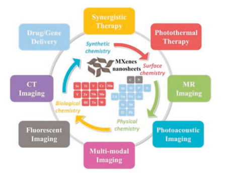
In this review, we briefly summarize the recent advances of 2D MXenes in theranostic nanomedicine on the basis of four sub-disciplines of chemistry for satisfying desirable theranostic outcomes, including synthetic chemistry, surface chemistry, physiochemistry and biological chemistry (Fig. 2). The emerging miscellaneous theranostic applications of MXenes, including therapeutic applications (e.g., drug delivery and PTT) and diagnostic imaging (e.g., CT, PA, MR and photoluminescence (PL) imaging), provide alternative material nanoplatforms for disease theransotics. By gaining a deeper insight into the chemistry of 2D MXenes based on the existing and emerging therapeutic modalities, we provide a perspective on future chances and possibilities to promote the multiple functionalization and clinical translation of MXenes in cancer theranostics and beyond.

|
Download:
|
| Fig. 2. Summary of 2D MXenes used in oncological applications and corresponding fundamental principles of intrinsic chemistry for prospective anti-tumor applications. | |
2. Rational construction of 2D MXenes by synthetic chemistry
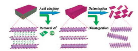
To date, the synthesis of MXenes can be typically classified into two categories: top-down approach and bottom-up method. As a typical top-down approach, MXenes are synthesized mainly using liquid exfoliation by selective removal of A elements from their layer-structured precursors of MAX phases, as shown for Mo2CTx MXene in Fig. 3 [75]. This top-down method to fabricate MXenes from parent MAX phases undergoes two steps, including etching and delamination process, which could be accomplished separately. Hydrofluoric acid (HF) is a frequently-used selective etching acids for corroding A element. As an example, the multilayerstacked Ti3C2 MXene was synthesized at room temperature using HF aqueous solution as the etchant (reaction (1)). In addition, as shown in reactions (2) and (3), the method based on HF treatment results in different surface terminations with O, OH and/or F terminal groups [2].

|
Download:
|
| Fig. 3. Schematic diagram of the synthesis and delamination process of 2D Mo2C MXenes. Adapted with permission [75]. Copyright 2019, Wiley-VCH. | |

|
(1) |
|
(2) |

|
(3) |
To reduce the risk of concentrated HF, MXenes can be prepared by using non-HF-etching approaches, which make use of the insitu production of HF via the reaction between the acid and fluoride salts (commonly LiF/NaF and HCl) [76]. However, the etching conditions vary from one kind of parent MAX phases to another, largely depending on the atomic bond of the materials. Therefore, it is required to explore the detailed etching conditions for different MXenes. Very recently, Li and coworkers reported a fluorine-free method to fabricate Ti3C2 MXene by employing the alkali-assisted hydrothermal method. They selectively removed Al element from Ti3AlC2 using NaOH solution. The experimental results exhibited that the high-quality Ti3C2 powder with 92 wt% purity was efficiently fabricated in a 27.5 mol/L NaOH solution at 270 ℃ [77].
In the delamination process, polar organic solvents (e.g., isopropylamine and dimethyl sulfoxide (DMSO)), organic base molecules (e.g., tetrabutylammonium hydroxide (TBAOH) and tetrapropylammonium hydroxide (TPAOH)) and metal cations (e.g., halide salts) have been employed as the intercalant. Subsequently, to achieve the desired size and high yield, the hand-shaking or sonication of the intercalated multilayered MXenes are utilized as supplementary means to obtain few- or single-layered nanosheets. Based on these synthetic methodologies, a series of ultrathin 2D MXenes have been synthesized for versatile biomedical applications, including Ti3C2, Nb2C, Mo2C and Ta4C3 [75, 78-80].
In addition to the top-down strategy, there are some reports on exploring bottom-up methods to synthesize MXenes. Unlike the top-down strategy that could be applied to fabricate 2D materials from layered bulky crystals, the bottom-up strategy is an alternative approach to synthesize 2D materials via atomic level control, which may create new possibilities for the broad range applications of MXenes. Compared to traditional 2D materials, the current bottom-up approaches to fabricate MXenes are not suitable for biomedical applications because of the low yield and nonsupport of surface engineering. For instance, 2D ultrathin α-Mo2C crystals were manufactured by Ren and co-workers via chemical vapor deposition (CVD) growth in 2015 [81]. The α-Mo2C nanosheets with large area and high quality were fabricated by using methane as the carbon source and Cu/Mo foils as the substrate to grow crystals at temperatures above 1085 ℃. It is worth noting that the maximal lateral size of the 2D ultrathin α-Mo2C is over 100 μm and α-Mo2C with the lateral size under 100 μm could be acquired flexibly by controlling the process of CVD [81]. Obviously, high-quality α-Mo2C synthesized by CVD with few defects and few surface groups is difficult to support surface engineering for biomedical applications, not to mention the low production yield and difficulty in controlling the key parameters for satisfying the biomedical application requirements. Xu et al. fabricated high-quality graphene/2D α-Mo2C crystal vertical heterostructures by CVD, which showed no defect both in graphene and 2D α-Mo2C crystal and uniformly well-aligned lattice orientation. Compared to the traditional stacking method, CVD endows strong interface coupling between two different phases in heterostructures. The heterostructures contribute to the implementation of highly transparent Josephson junction devices [82]. In addition, other bottom-up synthetic methods (such as template method [83, 84] and plasma enhanced pulsed laser deposition (PEPLD) [85, 86]) have been explored to synthesize MXenes, especially for some MXenes which cannot be obtained by top-down methods such as the aforementioned heterostructures [82, 87]. Xu Xiao and coworkers reported a scalable salt-templated synthesis by using reduction of 2D hexagonal oxides in ammonia to produce 2D metallic MoN, W2N and V2N efficiently [84]. Zhang et al. synthesized continuous single-crystalline and face-centered cubic structure Mo2C films on sapphire at 700 ℃ by PEPLD. This method combined the advantages of plasma enhanced CVD and pulsed laser deposition, which endowed the film with much lower crystalline quality, more stacking faults and large upper critical magnetic fields [85]. These methods provide an efficient way to expand the family of MXene materials and add many members with attractive properties for further applications.
3. Surface engineering of 2D MXenes for guaranteeing biomedical applicationsPlanar topography, nanoscale size and abundant compositions have gifted MXenes with unique surface chemistry for various biomedical applications. Although MXenes are hydrophilic, the bare MXenes are not stable and easily aggregate in physiological environments, similar to other inorganic nanosheets. In order to improve their biomedical performance (e.g., biocompatibility, blood circulation, targeting capability and loading capacity) in nanomedicine, 2D MXenes should be equipped with appropriate surface modification/functionalization. The fabricated 2D MXenes are typically featured with terminating functional groups (e.g., -F, -Cl, -OH and -O) through most of the current fabrication methods [44, 88]. Together with the high surface charge and abundant surface functional groups, the physical modification of organic moieties or loaded with functional agents onto the surface of MXenes is able to broaden their potential biomedical applications. Especially, the surface modification of MXene nanosheets has achieved some successes via physical routes (e.g., physical absorption and electrostatic attraction). Afterwards, the surface functionalized MXenes exhibit essentially enhanced dispersion stability, tunable surface reactivity, and desirable in vivo degradation kinetics, achieving maximal biological outcomes and minimal physiological toxic responses. In this section, we introduce some paradigms on surface modification and functionalization of 2D MXenes, mainly including molecules or polymers coating, targeting ligands decorating and therapeutic agents loading.
Regarding the biomedical applications of 2D MXenes, the excellent physiological stability and high biocompatibility are the basic principles for in vivo studies. Under physiological conditions, biocompatible organic molecules or polymers coating, including polyethylene glycol (PEG), poly(vinylpyrrolidone) (PVP), polyvinyl alcohol (PVA) and soybean phospholipid (SP), is one of the most common and economical strategies to improve their colloidal stability in physiological conditions. For instance, we provided a facile organic modification of Mo2C MXenes with PVA [75]. Compared to bare Mo2C, after PVA modification, the obtained Mo2C-PVA nanosheets exhibited remarkably enhanced stability and dispersibility in physiological environment, which was employed as an efficient nanoplatform for cancer PTT over both NIR I and NIR II biowindows [75]. In addition, researchers have modified the surface of other MXene nanosheets, such as Ti3C2 and Ta4C3 nanosheets with SP, Nb2C with PVP, and V2C with PEG to ensure the long-period blood circulation for in vivo photonic hyperthermia [78-80, 89].
Analogous to other 2D materials, surface modification with targeting ligands enables MXenes with active-targeting response for cancer therapy, which could effectively enhance the therapeutic effect and reduce damage to normal organs or tissues. For instance, Cao and coworkers reported an efficient strategy of nucleus-targeted PTT in the NIR II region to achieve effective thermal ablation of tumor (Fig. 4a) [89]. RGD, referring to three amino acid peptide arginine-glycine-aspartic acid, was used as a functionalized protein to target tumor cells. V2C quantum dots (QDs) with high photothermal-conversion performance in the NIR II region were modified with cell nucleus-target TAT peptides and packaged into vector with RGD modification (designated as V2C-TAT@Ex-RGD) [89]. It has been proved that these targeted delivery platforms exhibited significantly enhanced oncoma gathering and improved therapeutic efficacy compared to their corresponding non-targeted modalities. Therefore, it is highly expected that on basis of the adequate surface chemistry, the precise regulation and control of the surface modification would endow MXene-based nanoplatforms with multiple functions for versatile biomedical applications.

|
Download:
|
| Fig. 4. (a) Schematic representation of the preparation of dual-target V2C-TAT@Ex-RGD nanoagents. Adapted with permission [89]. Copyright 2019, American Chemical Society. (b) Synthetic process of PEGylated Ti3C2@Au and the therapeutic function with PA/CT imaging guidance and monitoring. Adapted with permission [91]. Copyright 2019, American Chemical Society. | |
In addition, the surface of 2D nanosheets could be processed with some other functional agents to endow multiple theranostic functions for biomedical applications. The large surface area and abundant terminal chemical groups of ultrathin MXenes with single-atomic thickness provide large amounts of anchoring points for guest therapeutic agents such as photosensitizers and chemotherapeutics. Therefore, they can act as the carrier systems for photodynamic therapy or chemotherapy. On the one hand, MXenes could be decorated with multifunctional inorganic nanoparticles to broaden their potential applications by inorganic nanoparticles-based surface chemistry. For example, biocompatible GdW10-based POMs/Ti3C2 composite nanosheets were the typical paradigm of integrating therapeutic platforms with CT and MR imaging guidance functionality [90]. Tang et al. synthesized 2D core–shell nanocomposites (Ti3C2@Au) employing a seedgrowth method for imaging-guided cancer therapy (Fig. 4b) [91]. With the high atomic number of Au element, the Ti3C2@Au nanosheets was used in photothermal-enhanced radiotherapy, as guided by the PA and CT dual-modality imaging, which endowed Ti3C2@Au with an excellent synergistic effect in cancer theranostics. Another typical paradigm is that, modified with calcium peroxide, the Nb2C MXene was employed as a photothermal agent and a carrier for photothermal-promoted Fenton reaction as triggered by self-supplied H2O2. This multifunctional nanoparticle, characterized with significantly enhanced reactive oxygen species (ROS) production and high photothermal effect, provided an efficient cancer-therapeutic modality (Figs. 5a and b) [92]. On the other hand, surface engineering endows MXenes with additional function as a drug or gene delivery vehicle, through direct growth of mesoporous silica layer or other porous material onto the surface of MXenes. As a typical paradigm, we introduced a surfacenanopore engineering strategy for the MXenes' surface functionalization, which achieved the homogeneous coating of a thin mesoporous-silica layer onto the surface of 2D Nb2C MXene (Nb2C@mMSNs) [93]. Under the alkaline synthetic condition, the surface of Nb2C was coated with a thin layer of mesoporous silica (Nb2C@mMSNs) using cetyltrimethylammonium chloride (CTAC) as the mesopore-directing agent and tetraethylorthosilicate (TEOS) as the silica precursor. 2, 20-Azobis[2-(2-imidazolin-2-yl) propane] dihydrochloride (AIPH) molecules, as the free-radicals supplier, were encapsulated into the mesopores of Nb2C@mSiO2 nanoparticles to promote cancer-cell apoptosis in both normoxic and hypoxic microenvironment by photothermal-activated ROS production as enabled by Nb2C MXene core under the NIR irradiation (Figs. 5c–e), initiating a promising thermodynamic therapeutic modality [93].

|
Download:
|
| Fig. 5. (a) Schematic illustration of the preparation of H2O2 self-supplied Nb2C–IO–CaO2–PVP composite nanocatalysts and (b) photothermal-enhanced sequential catalytic reaction as induced by Nb2C–IO–CaO2–PVP for synergistic cancer treatment. Adapted with permission [92]. Copyright 2019, Royal Society of Chemistry. Schematic illustration of (c) the preparation of AIPH@Nb2C@mSiO2 NPs, (d) thermal-responsive decomposition of AIPH to produce radicals, and (e) systematic delivery of AIPH@Nb2C@mSiO2 nanoparticles as a photo-sensitive nanoplatform for imaging-guided collaborative photothermal cancer ablation and photonic thermodynamic cancer therapy. Adapted with permission [93]. Copyright 2019, American Chemical Society. | |
4. Intrinsic physiochemical property of 2D MXenes for versatile biomedical applications
Compared to other 2D materials, MXenes exhibit their intrinsic physicochemical properties. Nevertheless, the underlying relationship between the intrinsic physicochemical properties of MXenes and their versatile biomedical functions for disease theranostics has not been fully clarified yet. Additionally, it is highly important to excavate the optimal physicochemical parameters based on the unique physical chemistry of MXenes during disease theranostics. Therefore, based on our currently limited understanding of the potential fundamental mechanisms of physical-biological interactions, we mainly focus on the physicochemical properties of MXenes in cancer theranostics (i.e., photothermal-conversion, photoacoustic effect, X-ray attenuation, magnetic responsiveness, PL property and electron transference) in this section, with additional paradigm of radiation protection by MXenes.
The optical properties (e.g., light absorption and emission) of MXenes are decisive and important for their oncological applications. Combined with external light stimulus, MXenes could act as photo-therapy and photo-imaging nanoagents, which provided considerable superiorities in the efficacy of cancer theranostics [94, 95]. Especially, some MXenes exhibit strong absorption in a broad range of spectrum from UV–vis to NIR, which has high potential to be used in PTT and PA imaging. As a compelling approach with low cost, site-specific action, mitigant side effect and high therapeutic efficiency, PTT is recommended for targeted heating of photothermally active nanomaterials. The lightabsorbing agents with desirable photothermal-conversion property are employed to transfer the absorbed NIR optical energy into heat. MXenes exhibit excellent photothermal-conversion performance in cancer photonic hyperthermia, which has been extensively demonstrated very recently. For instance, we synthesized 2D ultrathin Ti3C2 and Ta4C3 (MXene) nanosheets for efficient photothermal tumor elimination in NIR I biowindow [78, 80].
However, the photothermal-conversion performance of 2D MXenes for effective tumor hyperthermia ablation is strongly influenced by tissue absorption and scattering of laser. Compared to the traditional NIR I biowindow, the NIR II biowindow exhibits higher penetration depth and larger maximum permissible exposure (MPE), which displays prominent advantages in phototherapy. On this ground, we and other groups have achieved significant breakthroughs on developing MXene-based photothermal nanoagents in NIR II biowindow. Recently, we successfully constructed ultrathin 2D Nb2C and Mo2C MXenes for highly efficient photothermal ablation of the tumor in NIR II biowindow [75, 79]. The extinction coefficient (ε) of Nb2C was 37.6 L g-1 cm-1 of 808 nm and 35.4 L g-1 cm-1 of 1064 nm. Meanwhile, the laser photothermal-conversion efficiency (η) of Nb2C nanosheets of 808 nm was 36.4% and of 1064 nm was 45.65%. Due to the ambiguous guideline in present experimental trial-and-error method and predicting the photonic performance by theoretical simulation, the desirable photothermal-conversion efficiency (24.5% for NIR I and 43.3% for NIR II) of Mo2C MXenes was subsequently validated by the experiments.
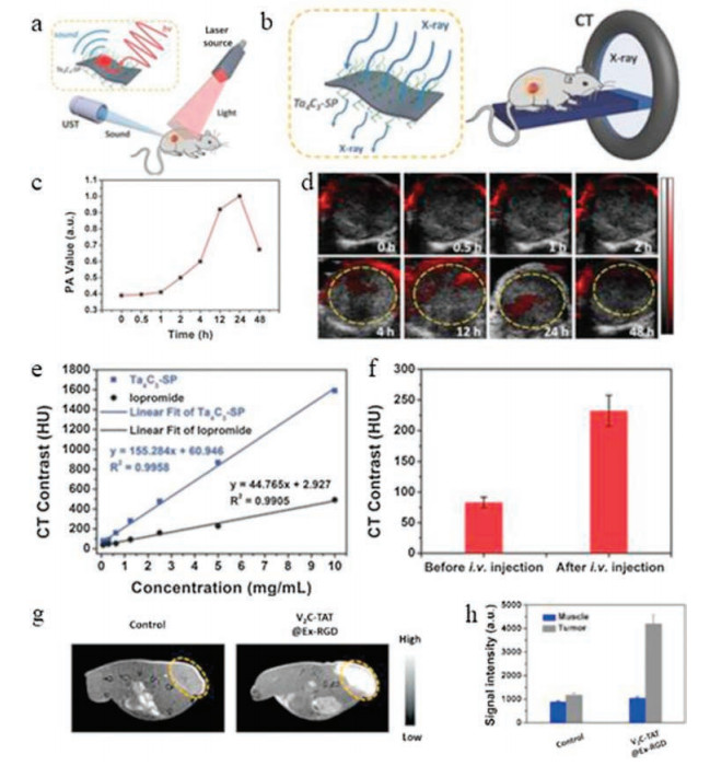
Laser can not only be used for photothermal ablation, but also act as photo-imaging tools. Based on the NIR light-absorption capability of MXenes, they could generate acoustical signal for further imaging namely PA imaging (Fig. 6a) [96-98], which is featured with remarkably enhanced imaging depth and spatial resolution compared to conventional optical imaging. For desired optical absorption performance, biocompatible Ta4C3-SP composite nanosheets acted as the contrast-enhancing nanoagents for effective PA imaging of tumor (Figs. 6c and d) [80]. It was obvious that the intensity of PA signal in the tumor site was gradually enlightened due to the accumulation of nanosheets in 24 h by the typical enhanced permeability and retention (EPR) effect. With high spatial resolution and deep tissue penetration, CT is considered as a powerful technique for in vivo bioimaging, especially for recognizing tumor and hard tissues [99-101]. Recently, MXenes such as Ta4C3 with high atomic number elements and strong X-rays attenuation, have attracted great interest in the potential application for CT imaging (Fig. 6b) [80]. Consistent with PA imaging results, the slope of the HU value for Ta4C3-SP was much higher than that of the commercial iodine-based CT contrast agents (Figs. 6e and f). Therefore, these Ta4C3-SP MXenes could act as promising contrast agents for PA and CT imaging of tumors [80].

|
Download:
|
| Fig. 6. Schematic diagram of (a) PA imaging and (b) CT imaging. (c) In vivo temporal evolution of PA value. (d) PA images of the tumor site after i.v. administration of MXenes for different time intervals. (e) Comparison between CTcontrasts of Ta4C3-SP and commercial iodine-based CTcontrast in vitro. (f) Statistical data of in vivo CTcontrasts before and after i.v. injection. Adapted with permission [80]. Copyright 2018, Wiley-VCH. (g) T1-weighted MR imaging of MCF-7 tumor-bearing mice before and after the administration of V2C. (h) Quantification of the MR signals in tumor site and muscle. Adapted with permission [89]. Copyright 2019, American Chemical Society. | |
MR imaging is another popular modern medical-imaging technology, featured with excellent contrast difference and high spatial resolution [102-104]. The V element in emerging 2D V2C endow MXenes with T1-weighted MR imaging function due to the 3d1 electronic configuration of quadrivalent vanadium (V) and the quantum mechanical confinement. Compared to normal muscle, obviously enhanced MR signals were observed in tumor sites because of the concentration by EPR effect after i.v. injection with V2C (Figs. 6g and h) [89].
In addition, the unique PL property of MXene QDs could be used in bioimaging including Ti3C2 QDs, N-doped Ti3C2 QDs, N, P-doped Ti3C2 QDs and V2C QDs as shown in Table 1 [89, 105-107]. Xue and coworkers successfully prepared luminescent Ti3C2 MXene-based QDs by a facile hydrothermal method [107]. Because of strong quantum confinement, the obtained Ti3C2 MXene QDs with quantum yields of up to 10% were used as biocompatible multicolor cellular imaging probes. This result broadens the biomedical applications of MXenes in multicolor cellular imaging and in vivo PL imaging. To expand the applications in biomedical and optical fields, Quan Xu and co-workers prepared N-doped Ti3C2 quantum dots and N, P-doped Ti3C2 quantum dots with a high PL quantum yield of 18.7% and 20.1% through a facile top-bottom hydrothermal method [105, 106]. These biocompatible Ti3C2 MXene-based QDs demonstrated outstanding PL performance, which showed high potential to act as PL imaging agents [108].
|
|
Table 1 Summary of the state of the art of MXenes in biomedical applications and crucial parameters related to their applications. |

In addition, due to the unusual electronic properties, it is worth noting that 2D MXene nanosheets exhibit the capability of eliminating reactive oxygen species (ROS), including hydroxyl (·OH), superoxide anion (O2·–) and hydrogen peroxide (H2O2), which could be applied to scavenge free radicals against ionizing radiation (IR), especially during the cancer treatment. As an intriguing radioprotectant, Nb2C MXene was demonstrated to significantly reverse the damage of ROS induced by IR through regulating superoxide dismutase (SOD) activities and intrinsic reducing property (Fig. 7) [109]. Experimental results showed that Nb2C-PVP could significantly enhance the survival rate of irradiated mice and significantly increase viability of mouse embryonic fibroblast 3T3/A31 cells after IR. To further explore the protection mechanism, ROS levels, SOD activities and malondialdehyde (MDA) levels in different organs of BALB/C mice were measured. The results demonstrated that Nb2C-PVP enhanced the SOD activities and reduced MDA levels via scavenging IR-induced ROS. Based on this result, these Nb2C MXenes could be potentially employed to mitigate the undesirable damage to normal tissues in cancer radiotherapy.

|
Download:
|
| Fig. 7. Schematic illustration of 2D Nb2C MXene acting as radiation-protection agents with reducibility and SOD-like activity. Adapted with permission [109]. Copyright 2019, American Chemical Society. | |
5. Unique biological chemistry for potential clinical translation
Because of the complex interaction between 2D ultrathin nanomaterials and physiological environment, it is difficult to maintain steady and achieve satisfactory biosafety performance in cancer theranostics, which should be taken into consideration in the practical applications. Furthermore, the desirable responsiveness to biological triggers may broaden the biomedical applications of 2D MXenes. In this section, the biological chemistry of MXenes is highlighted, including their bio-responsiveness, biodegradation, in vivo biodistribution and metabolism pathway.
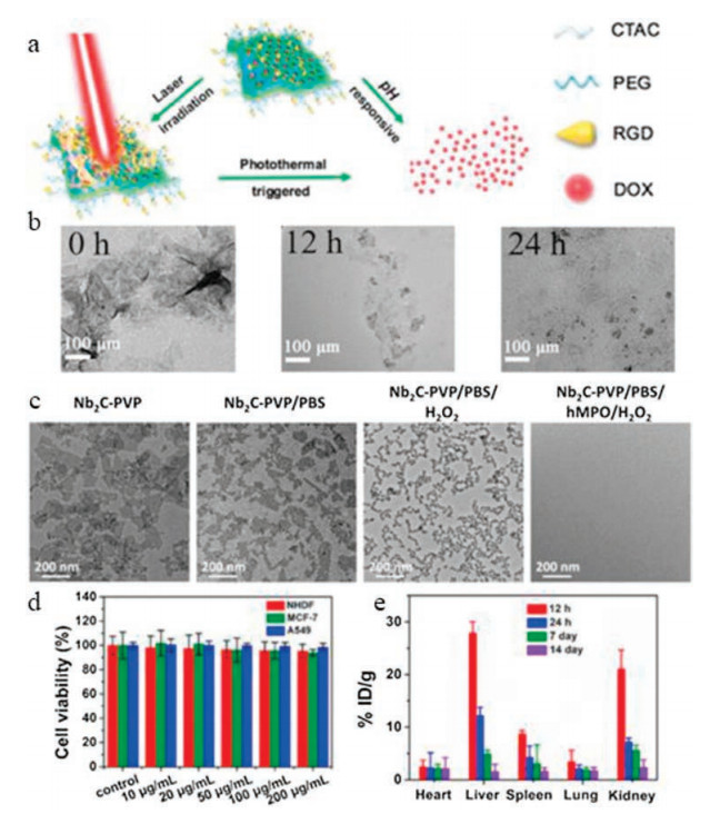
Based on the excellent responsiveness to biological triggers (e.g., temperature, pH value, enzyme), MXenes could be designed and fabricated as "smart" bioresponsive nanoreactors to optimize therapeutic outcomes. For instance, we have successfully developed intelligent 2D theranostic nanosystem based on doxorubicin (DOX)-loaded 2D Ti3C2 MXene for pH- and temperature-responsive chemotherapy and PTT-based synergetic therapy (Fig. 8a) [110]. Along with the pH-driven protonation behavior of DOX and temperature rising in the tumor site, the fast release of DOX was promoted to facilitate tumor microenvironment (TME)-specific chemotherapy. This strategy is hopefully conducive to the concurrent therapeutic outcomes with inappreciable damage to normal tissues by inducing the intelligent response and synergetic therapeutic outcome.

|
Download:
|
| Fig. 8. (a) Schematic illustration of pH-triggered and photothermal-accelerated drug release from DOX-loaded Ti3C2@mMSNs-RGD. Adapted with permission [110]. Copyright 2018, Wiley-VCH. (b) The degradation of MoC2-PVA nanosheets in PBS (pH 7.4). Adapted with permission [75]. Copyright 2019, Wiley-VCH. (c) The hMPOresponsive biodegradation of Nb2C-PVP nanoflakes were incubated in PBS for 24 h. Adapted with permission[79]. Copyright 2017, American Chemical Society. (d) Cytotoxicity of V2C after the incubation with NHDF, MCF-7 and A549 cell lines for 24 h. (e) Biodistribution of V (% ID of V per gram of tissues) in main tissues after intravenous administration of V2C QDs dispersed in PBS for varied time intervals (12 h, 24 h, 7 day and 14 day). Adapted with permission [89]. Copyright 2019, American Chemical Society. | |
Similar to the most inorganic nanosystems, poor biodegradability of 2D MXene nanosheets is the pivotal issue, which may hinder their further oncological research as well as clinical translation. Therefore, it is of high significance to systematically evaluate the biodegradation performance of MXene nanosheets under the complex physiological environment. We investigated the biodegradability of MoC2-PVA nanosheets (Fig. 8b) in various pH environment and the human myeloperoxidase (hMPO)- responsive biodegradability of Nb2C-PVP nanoflakes (Fig. 8c) [75, 79]. The characteristic 2D planar structures of MoC2-PVA and Nb2C-PVP were almost completely destroyed when MoC2-PVA nanosheets were incubated in phosphate buffered solution (PBS) (pH 7.4) and Nb2C-PVP nanoflakes were incubated with hMPOmixed PBS for 24 h. Therefore, a desirable route of biodegradation of MoC2 and enzyme-triggered biodegradation of Nb2C nanosheets was explored, which enabled their facile degradation in vivo after accomplishing the therapeutic functions.
The in vivo circulation and metabolism behaviors have a fundamental influence on the biosafety of MXenes. Therefore, systematic evaluation of in vivo biodistribution, circulating halflife, tumor accumulation and toxicology profiles is essential for the biomedical applications of MXenes. It is hopefully to identify the most promising nanoplatform with fast excretion and low toxicity. Up to now, several MXenes have been undergone individual studies and case-by-case phenomenon evaluations, including Ti3C2 [78], Ta4C3 [80], Nb2C [79], MoC2 [75], V2C [89] and TiN [111]. As a paradigm, the in vitro and in vivo results suggested that V2C had no appreciable cytotoxicity and long-term toxicity (Figs. 8d and e). After 14 days, most of the V2C nanosheets were metabolized from major tissues through the liver, spleen, and kidney due to the ultrasmall size of the V2C quantum dots, demonstrating that V2C could be cleared from the body with decreased long-term toxicity [89]. However, these biological effects of MXenes are only preliminarily assessed at cellular and animal level currently, the solid data on the long-term biological effects and biosafety are still highly lacking, which requires further systematic and in-depth evaluation and assessment.
6. Conclusions and outlookThis review summarizes the current developments of the emerging 2D MXenes in terms of their fabrication, modification and versatile biomedical applications, from the viewpoint of chemistry. Based on the insight into the fundamental mechanisms of synthetic methodology, surface functionalization, physiochemical properties and biological effect, we outline the current stateof-the-art of MXenes in the field of theranostic nanomedicine, including therapeutic modality (chemotherapy, phototherapy and synergistic therapy) and diagnostic imaging (PA, CT, PL and MR imaging). As shown in Fig. 9, we clarify the critical insights in the following discussions, aiming at increasing the potential of MXenes in clinical translation and further broadening their applications in biomedical fields. In addition, we systematically summarize the state-of-the-art of MXenes in biomedical applications in Table 1 [78-80, 89, 93, 105-109, 111-118], and crucial parameters related to their applications are also presented. Obviously, considerable efforts should be put into, because most of the MXenes have not been used in this filed yet, including the promising Cr2C, Sc2C, Zr3C2, Hf2C, Ti4N3, V4C3, etc.

|
Download:
|
| Fig. 9. Summary of the cancer theranostic of 2D MXenes from the viewpoints of chemistry, and current advances and further expectation for future biomedical applications. | |
As a new kind of 2D materials, due to the integrated metal atomic layers, copious surface terminations, adjustable element composition and unique electronic structure, MXenes display some unique electronic, optical and magnetic properties. Take the electronic properties as a paradigm, it is the most special peculiarity compared to other 2D materials. According to theoretical studies and experimental confirmations, MXenes show metallic to semiconductor-like properties because of the copious surface terminations and the innate quality of M, X. The transition metal component, preparation methods, outer layers, defects and surface terminations influences the electronic properties of MXenes to some extent. Based on the unique electronic properties, MXenes could be employed as ultrasensitive detector of several gases and other oxide/reducing molecules, as well as the biocatalyst and producer of ROS [112]. In addition, the abundant O, OH and/or F terminal groups on the surface of MXenes can combine with diverse drugs, proteins, nucleic acids, and even nanoparticles, and thus achieve desirable synergistic treatment outcomes in biomedical applications. In view of these unique properties, MXenes can combine with other 2D materials to arm them with broader applications, such as the graphene/2D Mo2C vertical heterostructures for highly transparent Josephson junction devices [82].
Actually, the biomedical applications of 2D MXenes are limited by their easy aggregation, difficult surface engineering, unsatisfactory stability and potential toxicity. The lateral size, the number of layers and the degree of oxidation all have significant influences in the degradation and stability during the in vivo circulation. The interaction between physiological environment and nanosheets, the analysis of metabolic pathway and the side effects as induced by MXene nanosheets have not been clarified yet currently. There is a remarkable contradiction that fast degradation may influence the therapeutic outcomes we desired, while their degradation meets requirements of biological safety. Therefore, appropriate degradation rate should be taken into consideration during the design and synthesis of MXenes to pursue ideal therapeutic outcomes and low toxicity concurrently.
Although many studies have demonstrated the great potential of MXenes in oncological applications, their clinical translation is still in its infancy. Therefore, the systematic investigation on the interaction between MXenes and biological microenvironments should be systematically investigated in the following fundamental researches. Additionally, the effect of surface functionalization on the biological behavior of MXenes requires further clarification. Especially, the scale-up fabrication of 2D MXenes is a significant respect for their design and engineering. A further and deeper exploration of material science and clinical translation require the cooperation among researchers of interdisciplinary and industrial sectors. It is highly expected that the development of nanotechnology and bioscience would achieve more fundamental and technological breakthroughs to afford the limitless application of MXenes in varied biomedical areas in the near future, provided the facing challenges and critical issues are adequately solved.
Declaration of competing interestThe authors declare that they have no known competing financial interests or personal relationships that could have appeared to influence the work reported in this paper.
AcknowledgmentsThis work was financially supported by the National Key R & D Program of China (No. 2016YFA0203700), Postdoctoral Science Foundation of China (No. 2018M630475), National Science Foundation for Young Scientists of China (No. 51802336), National Nature Science Foundation of China (Nos. 51672303, 51722211) and Program of Shanghai Academic Research Leader (No. 18XD1404300).
| [1] |
K.S. Novoselov, A.K. Geim, S.V. Morozov, et al., Science 306 (2004) 666-669. DOI:10.1126/science.1102896 |
| [2] |
J. Pang, R.G. Mendes, A. Bachmatiuk, et al., Chem. Soc. Rev. 48 (2019) 72-133. DOI:10.1039/C8CS00324F |
| [3] |
C. Chen, X. Xie, B. Anasori, et al., Angew. Chem. Int. Ed. 57 (2018) 1846-1850. DOI:10.1002/anie.201710616 |
| [4] |
O. Mashtalir, M.R. Lukatskaya, M.Q. Zhao, M.W. Barsoum, Y. Gogotsi, Adv. Mater. 27 (2015) 3501-3506. DOI:10.1002/adma.201500604 |
| [5] |
S. Niu, Z. Wang, M. Yu, et al., ACS Nano 12 (2018) 3928-3937. DOI:10.1021/acsnano.8b01459 |
| [6] |
Y. Wen, T.E. Rufford, X. Chen, et al., Nano Energy 38 (2017) 368-376. DOI:10.1016/j.nanoen.2017.06.009 |
| [7] |
Y. Xie, Y. Dall'Agnese, M. Naguib, et al., ACS Nano 8 (2014) 9606-9615. DOI:10.1021/nn503921j |
| [8] |
H. Xie, P. Li, J. Shao, et al., ACS Sens. 4 (2019) 2303-2310. DOI:10.1021/acssensors.9b00778 |
| [9] |
Q.H. Wang, K.K. Zadeh, A. Kis, J.N. Coleman, M.S. Strano, Nat. Nanotechnol. 7 (2012) 699-712. DOI:10.1038/nnano.2012.193 |
| [10] |
K. Ai, C. Ruan, M. Shen, L. Lu, Adv. Funct. Mater. 26 (2016) 5542-5549. DOI:10.1002/adfm.201601338 |
| [11] |
G. Liu, B. Debnath, T.R. Pope, et al., Nat. Nanotechnol. 11 (2016) 844-850. |
| [12] |
P. Wang, Q. Xu, Z. Li, et al., Adv. Mater. 30 (2018) 201801991. |
| [13] |
C. Huang, C. Li, G. Shi, Energy Environ. Sci. 5 (2012) 8848-8868. DOI:10.1039/c2ee22238h |
| [14] |
D. Deng, K.S. Novoselov, Q. Fu, et al., Nat. Nanotechnol. 11 (2016) 218-230. DOI:10.1038/nnano.2015.340 |
| [15] |
X. Feng, X. Ding, D. Jiang, Chem. Soc. Rev. 41 (2012) 6010-6022. DOI:10.1039/c2cs35157a |
| [16] |
P. Niu, L. Zhang, G. Liu, H.M. Cheng, Adv. Funct. Mater. 22 (2012) 4763-4770. DOI:10.1002/adfm.201200922 |
| [17] |
C. Tan, X. Cao, X.J. Wu, et al., Chem. Rev. 117 (2017) 6225-6331. DOI:10.1021/acs.chemrev.6b00558 |
| [18] |
M. Barzegar, B. Tudu, Surf. Innov. 6 (2018) 205-230. DOI:10.1680/jsuin.18.00013 |
| [19] |
C. Gong, K. Hu, X. Wang, et al., Adv. Funct. Mater. 28 (2018) 201706559. |
| [20] |
P.K. Kannan, D.J. Late, H. Morgan, C.S. Rout, Nanoscale 7 (2015) 13293-13312. DOI:10.1039/C5NR03633J |
| [21] |
X. Liu, T. Ma, N. Pinna, J. Zhang, Adv. Funct. Mater. 27 (2017) 1702168. DOI:10.1002/adfm.201702168 |
| [22] |
G. Ren, B.Y. Zhang, Q. Yao, et al., Small 15 (2019) 1805251. DOI:10.1002/smll.201805251 |
| [23] |
Y. Ma, N. Liu, L. Li, et al., Nat. Commun. 8 (2017) 1207. DOI:10.1038/s41467-017-01136-9 |
| [24] |
P. Panigrahi, T. Hussain, A. Karton, R. Ahuja, ACS Sens. 4 (2019) 2646-2653. DOI:10.1021/acssensors.9b01044 |
| [25] |
Y. Chen, C. Tan, H. Zhang, L. Wang, Chem. Soc. Rev. 44 (2015) 2681-2701. DOI:10.1039/C4CS00300D |
| [26] |
L. Cheng, J. Liu, X. Gu, et al., Adv. Mater. 26 (2014) 1886-1893. DOI:10.1002/adma.201304497 |
| [27] |
D. Chimene, D.L. Alge, A.K. Gaharwar, Adv. Mater. 27 (2015) 7261-7284. DOI:10.1002/adma.201502422 |
| [28] |
G.E. Christensen, R.D. Rabbitt, M.I. Miller, IEEE Trans. Image Process. 5 (1996) 1435-1447. DOI:10.1109/83.536892 |
| [29] |
W. Fan, W. Bu, B. Shen, et al., Adv. Mater. 27 (2015) 4155-4161. DOI:10.1002/adma.201405141 |
| [30] |
T. Liu, C. Wang, X. Gu, et al., Adv. Mater. 26 (2014) 3433-3440. DOI:10.1002/adma.201305256 |
| [31] |
M. Soleymaniha, M.A. Shahbazi, A.R. Rafieerad, A. Maleki, A. Amiri, Adv. Healthc. Mater. 8 (2019) 1801137. DOI:10.1002/adhm.201801137 |
| [32] |
H. Lin, Y. Chen, J. Shi, Adv. Sci. 5 (2018) 1800518. DOI:10.1002/advs.201800518 |
| [33] |
B. Yang, Y. Chen, J. Shi, Chemistry 4 (2018) 1284-1313. DOI:10.1016/j.chempr.2018.02.012 |
| [34] |
B. Yang, J. Yin, Y. Chen, et al., Adv. Mater. 30 (2018) 1705611. DOI:10.1002/adma.201705611 |
| [35] |
K. Huang, Z. Li, J. Lin, G. Han, P. Huang, Chem. Soc. Rev. 47 (2018) 5109-5124. DOI:10.1039/C7CS00838D |
| [36] |
M. Alhabeb, K. Maleski, B. Anasori, et al., Chem. Mater. 29 (2017) 7633-7644. DOI:10.1021/acs.chemmater.7b02847 |
| [37] |
J. Liu, H.B. Zhang, R. Sun, et al., Adv. Mater. 29 (2017) 1702367. DOI:10.1002/adma.201702367 |
| [38] |
F. Shahzad, M. Alhabeb, C.B. Hatter, et al., Science 353 (2016) 1137-1140. DOI:10.1126/science.aag2421 |
| [39] |
L. Ci, L. Song, C. Jin, et al., Nat. Mater. 9 (2010) 430-435. DOI:10.1038/nmat2711 |
| [40] |
C.R. Dean, A.F. Young, I. Meric, et al., Nat. Nanotechnol. 5 (2010) 722-726. DOI:10.1038/nnano.2010.172 |
| [41] |
D. Golberg, Y. Bando, Y. Huang, et al., ACS Nano 4 (2010) 2979-2993. DOI:10.1021/nn1006495 |
| [42] |
K. Watanabe, T. Taniguchi, H. Kanda, Nat. Mater. 3 (2004) 404-409. DOI:10.1038/nmat1134 |
| [43] |
M.R. Lukatskaya, O. Mashtalir, C.E. Ren, et al., Science 341 (2013) 1502-1505. DOI:10.1126/science.1241488 |
| [44] |
M. Naguib, V.N. Mochalin, M.W. Barsoum, Y. Gogotsi, Adv. Mater. 26 (2014) 992-1005. DOI:10.1002/adma.201304138 |
| [45] |
V.M.H. Ng, H. Huang, K. Zhou, et al., J. Mater. Chem. A 5 (2017) 3039-3068. DOI:10.1039/C6TA06772G |
| [46] |
D.A. Kuznetsov, Z. Chen, P.V. Kumar, et al., J. Am. Chem. Soc. 141 (2019) 17809-17816. DOI:10.1021/jacs.9b08897 |
| [47] |
G. Fan, F. Li, D.G. Evans, X. Duan, Chem. Soc. Rev. 43 (2014) 7040-7066. DOI:10.1039/C4CS00160E |
| [48] |
P.J. Sideris, U.G. Nielsen, Z. Gan, C.P. Grey, Science 321 (2008) 113-117. DOI:10.1126/science.1157581 |
| [49] |
F. Zhang, L. Zhao, H. Chen, et al., Angew. Chem. Int. Ed. 47 (2008) 2466-2469. DOI:10.1002/anie.200704694 |
| [50] |
G. Chen, J. Liu, Y. Li, et al., Nanotechnology 30 (2019) 335201.
|
| [51] |
K.F. Mak, J. Shan, Nat. Photonics 10 (2016) 216-226. DOI:10.1038/nphoton.2015.282 |
| [52] |
S. Manzeli, D. Ovchinnikov, D. Pasquier, O.V. Yazyev, A. Kis, Nat. Rev. Mater. 2 (2017) 17033. DOI:10.1038/natrevmats.2017.33 |
| [53] |
X. Zhang, X.F. Qiao, W. Shi, et al., Chem. Soc. Rev. 44 (2015) 2757-2785. DOI:10.1039/C4CS00282B |
| [54] |
T.Y. Jeong, H. Kim, S.J. Choi, et al., Nat. Commun. 10 (2019) 3825. DOI:10.1038/s41467-019-11751-3 |
| [55] |
X. Li, S. Zhang, H. Huang, et al., Nano Lett. 19 (2019) 6005-6012. DOI:10.1021/acs.nanolett.9b01812 |
| [56] |
J. Ouyang, C. Feng, X. Ji, et al., Angew. Chem. Int. Ed. 58 (2019) 13405-13410. DOI:10.1002/anie.201908377 |
| [57] |
M. Pumera, Z. Sofer, Adv. Mater. 29 (2017) 1605299. DOI:10.1002/adma.201605299 |
| [58] |
W. Tao, N. Kong, X. Ji, et al., Chem. Soc. Rev. 48 (2019) 2891-2912. DOI:10.1039/C8CS00823J |
| [59] |
L. Zhang, W. Chu, Q. Zheng, et al., J. Phys. Chem. Lett. (2019) 6151-6158.
|
| [60] |
A. Molle, J. Goldberger, M. Houssa, et al., Nat. Mater. 16 (2017) 163-169. DOI:10.1038/nmat4802 |
| [61] |
A. Molle, C. Grazianetti, L. Tao, et al., Chem. Soc. Rev. 47 (2018) 6370-6387. DOI:10.1039/C8CS00338F |
| [62] |
H. Lin, W. Qiu, J. Liu, et al., Adv. Mater. 31 (2019) 1903013. DOI:10.1002/adma.201903013 |
| [63] |
I. Niehues, A. Blob, T. Stiehm, S.M. de Vasconcellos, R. Bratschitsch, Nanoscale 11 (2019) 12788-12792. DOI:10.1039/C9NR03332G |
| [64] |
Q. Wang, Q. Zhang, X. Zhao, et al., Nano Lett. 19 (2019) 5595-5603. DOI:10.1021/acs.nanolett.9b02136 |
| [65] |
J. Meyer, S. Hamwi, M. Kroeger, et al., Adv. Mater. 24 (2012) 5408-5427. DOI:10.1002/adma.201201630 |
| [66] |
R.R. Salunkhe, Y.V. Kaneti, Y. Yamauchi, ACS Nano 11 (2017) 5293-5308. DOI:10.1021/acsnano.7b02796 |
| [67] |
J. Cuan, Y. Zhou, J. Zhang, et al., ACS Nano 13 (2019) 11665-11675.
|
| [68] |
X. Hu, Y. Long, M. Fan, et al., Appl. Catal. B-Environ. 244 (2019) 25-35. DOI:10.1016/j.apcatb.2018.11.028 |
| [69] |
F. Wang, X. Wu, X. Yuan, et al., Chem. Soc. Rev. 46 (2017) 6816-6854. DOI:10.1039/C7CS00205J |
| [70] |
R. Dong, P. Han, H. Arora, et al., Nat. Mater. 17 (2018) 1027-1032. DOI:10.1038/s41563-018-0189-z |
| [71] |
C.N.R. Rao, K. Pramoda, R. Kumar, Chem. Commun. 53 (2017) 10093-10107. DOI:10.1039/C7CC05390H |
| [72] |
D. Zhao, D.J. Timmons, D. Yuan, H.C. Zhou, Acc. Chem. Res. 44 (2011) 123-133. DOI:10.1021/ar100112y |
| [73] |
M. Naguib, M. Kurtoglu, V. Presser, et al., Adv. Mater. 23 (2011) 4248-4253. DOI:10.1002/adma.201102306 |
| [74] |
K. Hantanasirisakul, Y. Gogotsi, Adv. Mater. 30 (2018) 1804779. DOI:10.1002/adma.201804779 |
| [75] |
W. Feng, R. Wang, Y. Zhou, et al., Adv. Funct. Mater. 29 (2019) 1901942. DOI:10.1002/adfm.201901942 |
| [76] |
M. Ghidiu, M.R. Lukatskaya, M.Q. Zhao, Y. Gogotsi, M.W. Barsoum, Nature 516 (2014) 78-81. DOI:10.1038/nature13970 |
| [77] |
T. Li, L. Yao, Q. Liu, et al., Angew. Chem. Int. Ed. 57 (2018) 6115-6119. DOI:10.1002/anie.201800887 |
| [78] |
H. Lin, X. Wang, L. Yu, Y. Chen, J. Shi, Nano Lett. 17 (2017) 384-391. DOI:10.1021/acs.nanolett.6b04339 |
| [79] |
H. Lin, S. Gao, C. Dai, Y. Chen, J. Shi, J. Am. Chem. Soc. 139 (2017) 16235-16247. DOI:10.1021/jacs.7b07818 |
| [80] |
H. Lin, Y. Wang, S. Gao, Y. Chen, J. Shi, Adv. Mater. 30 (2018) 1800518. |
| [81] |
C. Xu, L. Wang, Z. Liu, et al., Nat. Mater. 14 (2015) 1135-1141. DOI:10.1038/nmat4374 |
| [82] |
C. Xu, S.A. Song, Z.B. Liu, et al., ACS Nano 11 (2017) 5906-5914. DOI:10.1021/acsnano.7b01638 |
| [83] |
S. Joshi, Q. Wang, A. Puntambekar, V. Chakrapani, ACS Energy Lett. 2 (2017) 1257-1262. DOI:10.1021/acsenergylett.7b00240 |
| [84] |
X. Xiao, H.M. Yu, H.Y. Jin, et al., ACS Nano 11 (2017) 2180-2186. DOI:10.1021/acsnano.6b08534 |
| [85] |
F. Zhang, Z. Zhang, H.C. Wang, et al., Phys. Rev. Mater. 1 (2017) 034002. DOI:10.1103/PhysRevMaterials.1.034002 |
| [86] |
Z. Zhang, F. Zhang, H.C. Wang, et al., J. Mater. Chem. C 5 (2017) 10822-10827. DOI:10.1039/C7TC03652C |
| [87] |
J.B. Qiao, Y. Gong, W.J. Zuo, et al., Phys. Rev. B 95 (2017) 201403. DOI:10.1103/PhysRevB.95.201403 |
| [88] |
Q. Zhang, J. Zhang, S. Wan, W. Wang, L. Fu, Adv. Funct. Mater. 28 (2018) 1802500. DOI:10.1002/adfm.201802500 |
| [89] |
Y. Cao, T. Wu, K. Zhang, et al., ACS Nano 13 (2019) 1499-1510. |
| [90] |
L. Zong, H. Wu, H. Lin, Y. Chen, Nano Res. 11 (2018) 4149-4168. DOI:10.1007/s12274-018-2002-3 |
| [91] |
W. Tang, Z. Dong, R. Zhang, et al., ACS Nano 13 (2019) 284-294. DOI:10.1021/acsnano.8b05982 |
| [92] |
S. Gao, X. Lu, P. Zhu, et al., J. Mat. Chem. B 7 (2019) 3599-3609. DOI:10.1039/C9TB00525K |
| [93] |
H. Xiang, H. Lin, L. Yu, Y. Chen, ACS Nano 13 (2019) 2223-2235. |
| [94] |
L. Cheng, C. Wang, L. Feng, K. Yang, Z. Liu, Chem. Rev. 114 (2014) 10869-10939. DOI:10.1021/cr400532z |
| [95] |
S.M. Park, A. Aalipour, O. Vermesh, J.H. Yu, S.S. Gambhir, Nat. Rev. Mater. 2 (2017) 17014. DOI:10.1038/natrevmats.2017.14 |
| [96] |
L.V. Wang, Nat. Photonics 3 (2009) 503-509. DOI:10.1038/nphoton.2009.157 |
| [97] |
L.V. Wang, S. Hu, Science 335 (2012) 1458-1462. DOI:10.1126/science.1216210 |
| [98] |
M.H. Xu, L.H.V. Wang, Rev. Sci. Instrum. 77 (2006) 041101. DOI:10.1063/1.2195024 |
| [99] |
S. Bipat, A.S. Glas, F.J.M. Slors, et al., Radiology 232 (2004) 773-783. DOI:10.1148/radiol.2323031368 |
| [100] |
D.J. Brenner, E.J. Hall, N. Engl. J. Med. 357 (2007) 2277-2284. DOI:10.1056/NEJMra072149 |
| [101] |
L. Wexler, B. Brundage, J. Crouse, et al., Circulation 94 (1996) 1175-1192.
|
| [102] |
J. Ashburner, K.J. Friston, Neuroimage 11 (2000) 805-821. DOI:10.1006/nimg.2000.0582 |
| [103] |
E.T. Bullmore, O. Sporns, Nat. Rev. Neurosci. 10 (2009) 186-198. DOI:10.1038/nrn2575 |
| [104] |
M.D. Fox, M.E. Raichle, Nat. Rev. Neurosci. 8 (2007) 700-711. DOI:10.1038/nrn2201 |
| [105] |
Q. Xu, L. Ding, Y.Y. Wen, et al., J. Mater. Chem. C 6 (2018) 6360-6369. DOI:10.1039/C8TC02156B |
| [106] |
Q.W. Guan, J.F. Ma, W.J. Yang, et al., Nanoscale 11 (2019) 14123-14133.
|
| [107] |
Q. Xue, H. Zhang, M. Zhu, et al., Adv. Mater. 29 (2017) 1604847. DOI:10.1002/adma.201604847 |
| [108] |
Q. Xu, W. Cai, W. Li, et al., Mater. Today Energy 10 (2018) 222-240. DOI:10.1016/j.mtener.2018.09.005 |
| [109] |
X. Ren, M. Huo, M. Wang, et al., ACS Nano 13 (2019) 6438-6454. DOI:10.1021/acsnano.8b09327 |
| [110] |
Z. Li, H. Zhang, J. Han, et al., Adv. Mater. 30 (2018) 1706981. DOI:10.1002/adma.201706981 |
| [111] |
Z. Xie, S. Chen, Y. Duo, et al., ACS Appl. Mater. Interfaces 11 (2019) 22129-22140. DOI:10.1021/acsami.9b04628 |
| [112] |
K. Huang, Z.J. Li, J. Lin, G. Han, P. Huang, Chem. Soc. Rev. 47 (2018) 5109-5124. DOI:10.1039/C7CS00838D |
| [113] |
C. Dai, H. Lin, G. Xu, et al., Chem. Mater. 29 (2017) 8637-8652. DOI:10.1021/acs.chemmater.7b02441 |
| [114] |
G.Y. Liu, J.H. Zou, Q.Y. Tang, et al., ACS Appl. Mater. Interfaces 9 (2017) 40077-40086. DOI:10.1021/acsami.7b13421 |
| [115] |
B.Z. Xu, M.S. Zhu, W.C. Zhang, et al., Adv. Mater. 28 (2016) 3333-3339. DOI:10.1002/adma.201504657 |
| [116] |
C. Dai, Y. Chen, X.X. Jing, et al., ACS Nano 11 (2017) 12696-12712.
|
| [117] |
A. Szuplewska, D. Kulpinska, A. Dybko, et al., Mat. Sci. Eng. C-Mater. 98 (2019) 874-886. DOI:10.1016/j.msec.2019.01.021 |
| [118] |
X.H. Yu, X.K. Cai, H.D. Cui, et al., Nanoscale 9 (2017) 17859-17864.
|