


 , Wan-Wan Liua,b, Shui-Ping Liua,b, Meng-Juan Lia,b, Yong-Gui Lia,b, Ming-Qiao Gea,b
b College of Textile & Clothing, Jiangnan University, Wuxi 214122, China
Electrically conductive polymers have attracted great interests because their physicochemical properties resemble organic polymers and they have electrical characteristics similar to metals [1, 2]. Further fabrication of nano/micro-structured conductive polymers has recently been demonstrated in the design and construction of various devices [3]. Although the fabrications of these nano/micro-materials are more complicated than those of flat film, such nano- or micro-structures can enhance the performance of sensors efficiently. During the past several years, electrospinning technique was widely applied to fabricate conductive polymeric nano/microfibers [4, 5, 6].
Electrospinning is currently being used to an increasing extent to produce ultra thin fibers from a wide range of polymer materials [7, 8]. This technology is highly versatile and allows the processing not only of many different polymers into polymeric microfibers but also the co-processing of polymers and low molecular weight nonvolatile materials, simply by using ternary solutions of the components for electrospinning to form a combination of fibrous functionalities [8].
In this work, the microfibers were produced by electrospinning from the DMF solution of aniline (ANi) monomers with the copolymer of poly(vinylchloride-acrylonitrile) (PVC-AN) serving as the carrier. Then, the monomers of aniline were in situ polymerized in the precursor microfibers using ammonium persulfate to obtain the conductive microfibers.
The copolymer of PVC-AN was synthesized in our lab, and the molecular weight was about 100,000. The monomer of aniline, dimethyl formamide (DMF), and ammonium persulfate were purchased from Sinopharm Chemical Reagent Co., Ltd. (Shanghai, China) and used without any further purification.
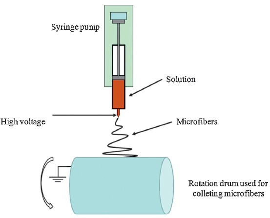
All experiments were carried out at room temperature. PVC-AN was firstly added into DMF with the concentration of 0.2 g/mL followed by magnetic stirring for 2 h to obtain a homogenous solution. Then, a suitable amount of aniline (5-20 vol% in the electrospinning solution) was added into the PVC-AN/DMF solution. As-prepared PVC-AN/ANi/DMF solution was used for electrospinning experiment. The precursor microfibers were collected on aluminum foil wrapped on a rotation drum. Electrospinning was carried out by setting the distance between spinneret and the collector at 12 cm, solution flow rate at 1.0 mL/h, voltage at 15 kV, and humidity at 55%-65%. The pure PVC-AN microfibers were electrospun for physicochemical evaluations. The scheme of electrospinning setup is shown in Fig. 1.

|
Download:
|
| Fig. 1.Experimental setup for the electrospinning process. | |
The as-spun precursor microfibrous mat was carefully peeled up from the aluminum foil; thereafter, the mat was immersed into ammonium persulfate solution (0.02 g/mL) immediately and then stored in a 4 ℃ refrigerator for 24 h.
PVC-AN-ANi precursor microfibers and PVC-AN-PANi microfibers were washed with distilled water three times and then dried in vacuum for 24 h before any tests. The morphology of the microfibers was observed using a digital vacuum scanning electron microscope (SEM, JSM-5600LV Electron Optical Laboratory, Japan) at an accelerating voltage of 5 kV. Only the PVC-AN-ANi precursor microfibers were sputter coated with gold prior to observation. Fourier-transform infrared (FTIR) spectra were obtained on a Spectrum Two IR Spectrometer (Frontier, PerkinElmer, USA) through detecting the microfibers mats directly. The scanning range was 650-4000 cm-1 and the resolution was 1 cm-1. The differential scanning calorimetry (DSC) analyses were carried out using an MDSC 2910 differential scanning calorimeter (TA Instruments Co., USA). Sealed samples were heated at 5 ℃/min from 20 ℃ to 100 ℃. The nitrogen flow rate was 40 mL/min.
Conductance was measured using a two-point method on a ZC- 90G Solid State Electrometer (Qiangjia Instruments Ltd., Shanghai). Before measurements, the fiber samples (2 cm2 × 2 cm2) were conditioned for 24 h at room temperature and 25% relative humidity. The microfibrous sample was clamped on its edges by two separated clinchers subject to the voltage of 100 V; the conductivity of the sample was obtained from the electrometer after 10 s.
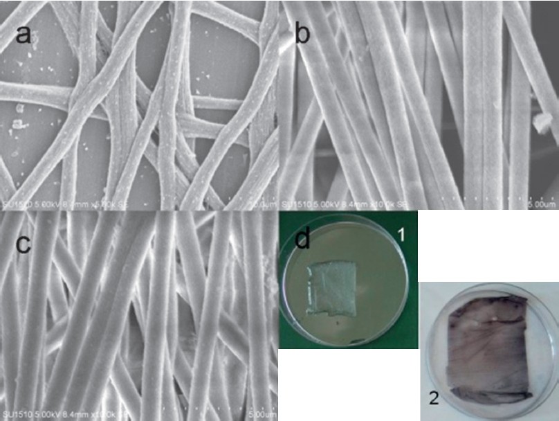
The morphology of the gold coated precursor microfibers and PVC-AN-PANi conductive microfibers were investigated by SEM, which exhibited bead-free structure (Fig. 2a-c). It should be noted that the precursor microfibers were washed with distilled water three times and then dried in vacuum for 24 h. In those processes, we believe that most of the ANi (nearly 100%) was removed from the PVC-AN microfibers by distilled water; therefore, the PVC-AN microfibers shrunk significantly and presented the rough but not the smooth surfaces (Fig. 2a). The average diameters of the microfibers with 5 wt% and 20 wt% PANi incorporated were 889 ± 71 nm and 1226 ± 119 nm, respectively. As shown in Fig. 2d, the precursor microfibers mat without treatment by ammonium persulfate solution and the conductive microfibers mat (with PANi in the fibers) were immersed in distilled water for photographing. The precursor microfibers mat was colorless and transparent, while the conductive microfibers mat was non-uniform dust blue. Following convention, the fibers were formed with evaporation of solvent during the electrospinning processes. In this study, we used DMF (boiled point of 152.8 ℃) as the solvent to prepare the electrospun solution. It is noted that the boiling point of aniline is 184 ℃. Therefore, some of the aniline would also evaporate from the microfibers matrix when microfibers formed in the process of electrospinning. Moreover, we found that the ammonium persulfate solution turned blue after treating the precursor microfibers mat, which demonstrated that some of the aniline transferred into the ammonium persulfate solution from the precursor microfibers mat and was then polymerized in the solution.

|
Download:
|
| Fig. 2.SEM microimages of electrospun nanofibrous precursors from DMF solutions of 0.2 g/mL PVC-AN as the carrier (a), conductive microfibers of PVC-AN-PANi synthesizing from the precursors with aniline contents of 5 wt% (b) and 20 wt% (c); photograph of the microfibers precursors (d-1) and conductive microfibers mats with PANi content of 5 wt% (d-2). | |
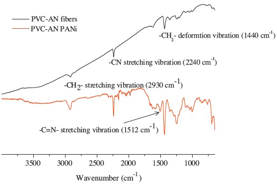
As shown in Fig. 3, the spectrum of PVC-AN fibers shows characteristic peaks at 2930 cm-1 (-CH2- stretching), 2240 cm-1 (-CN stretching) and 1440 cm-1 (-CH2- deformation). In addition to these aforementioned characteristic peaks of PVC-AN, the spectrum of PVC-AN-PANi fibers also shows more peaks at 1512 cm-1 and around 850-760 cm-1, indicating the existence of -C=N- and benzene rings in the fiber samples, respectively. This evidence demonstrated that the aniline was in situ polymerized in the PVC-AN microfibers.

|
Download:
|
| Fig. 3.FTIR spectra of PVC-AN fibers and the conductive microfibers of PVC-AN PANi. | |
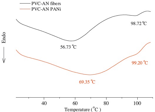
The incorporation of PANi also affected the thermal properties of PVC-AN microfibers. Fig. 4 shows the DSC thermograms of PVCAN and PVC-AN-PANi microfibers. We found that the PVC-AN microfibers exhibited an obvious endothermic band around 56.7 ℃ and a very small endothermic band at 98.7 ℃. The endothermic band of the PVC-AN-PANi fibers lies around 69.4 ℃ and the small endothermic band at 98.7 ℃ almost disappears. These differences indicated that there must be some interaction on the molecular level between PVC-AN and PANi. The aniline monomers dispersed to PVC-AN molecular chains before polymerization.

|
Download:
|
| Fig. 4.DSC curves of PVC-AN fibers and the conductive microfibers of PVC-AN PANi. | |
The electrical conductivity was approximately 2.2 × 10⁻⁸ S/cm for 20 wt% content of aniline incorporated in the microfibers; however, the electrical conductivity decreased with the decrease of aniline content. Therefore, the electrical conductivity of these microfiberous mats can be adjusted by controlling the ratio of PANi to PVC-AN. The interaction at the molecular level facilitates the formation of continuous conduction paths in PANi chains. Moreover, the stretching of fibers in the electrospinning process may orient aniline molecules along the longitudinal fiber direction, thus also potentially increasing the charge-carrier mobility after polymerization. It was mentioned that some part of the aniline was evaporated during the electrospinning and polymerization processes; therefore, the electrical conductivity would be higher than 3.0 × 10⁻⁸ S/cm when there was 20% PANi incorporated in the microfibers. It is also reasonable to believe that the conductivity can be enhanced by doping with other semi-conductive materials.
Aniline was in situ polymerized in PVC-AN precursor to prepare conductive microfibers with an electrical conductivity of about 2.2 × 10⁻⁸ S/cm for 20 wt% of aniline incorporated. The results from FTIR and DSC measurements demonstrated that the PANi was successfully polymerized in the PVC-AN microfibers matrix.
This study has been funded by the Fundamental Research Funds for the Central Universities (No. JUSRP31104), the Open Project Program of Key Laboratory of Eco-textiles (Ministry of Education, Jiangnan University, No. KLET1209), National High-tech R&D Program of China (863 Program, No. 2012AA030313), and Jiangsu Province Innovation Team in Colleges and Universities (No. Sue [2009]10).
| [1] | I.S. Chronakis, S. Grapenson, A. Jakob, Conductive polypyrrole nanofibers via electrospinning: electrical and morphological properties, Polymer 47 (2006) 1597-1603. |
| [2] | C. Schmindt, V. Shastri, J. Vacanti, R. Langer, Stimulation of neurite outgrowth using an electrically conducting polymer, Appl. Biol. Sci. 94 (1997) 8948-8953. |
| [3] | Y.S. Takahiro, T.M. Seiichi, M.Y. Koji, et al., Improvement of electrical contact reliability by conductive polymer coated elastomer structure in woven electronic textiles, Jpn. J. Appl. Phys. 51 (2012) 120204-120206. |
| [4] | S.H. Chen, P. Fu, B. Yin, et al., Immobilizing Pt nanoparticles and chitosan hybrid film on polyaniline naofibers membrane for an amperometric hydrogen peroxide biosensor, Bioprocess Biosyst. Eng. 34 (2011) 711-719. |
| [5] | A.C. Salvador, I.R. Maria, J.G. Martinez, et al., Fabrication of conductive electrospun silk fibroin scaffolds by coating with polypyrrole for biomedical applications, Bioelectrochemistry 85 (2012) 36-43. |
| [6] | L. Jin, Z.F. Feng, M.L. Zhu, et al., A novel fluffy conductive polypyrrole nano-layer coated PLLA fibrous scaffold for nerve tissue engineering, J. Biomed. Nanotechnol. 8 (2012) 779-785. |
| [7] | S.P. Liu, L.J. Tan, W.L. Hu, et al., Cellulose acetate nanofibers with photochromic property: fabrication and characterization, Mater. Lett. 64 (2010) 2427-2430. |
| [8] | X.Q. Li, M.A. Kanjwal, L. Lin, I.S. Chronakisa, Electrospun polyvinyl-alcohol nanofibers as oral fast-dissolving delivery system of caffeine and riboflavin, Colloids Surf. B Biointerfaces 103 (2013) 182-188. |