文章信息
- 黄徐建, 李敬东, 杨嘉琳, 李梦, 杨刚, 周何, 熊永福
- Huang Xujian, Li Jingdong, Yang Jialin, Li Meng, Yang Gang, Zhou He, Xiong Yongfu
- 联合长链非编码RNA Lnc-cCSC1构建结直肠癌临床预后个体化模型
- Establishment of individualized model for clinical prognosis of colorectal cancer with long non-coding RNA Lnc-cCSC1
- 实用肿瘤杂志, 2021, 36(5): 412-418
- Journal of Practical Oncology, 2021, 36(5): 412-418
基金项目
- 川北医学院附属医院博士启动基金(2020BS001);川北医学院院内课题重点项目(2020ZD001);川北医学院国家级课题预研项目(2020YY031);川北医学院院内课题基础项目(2020JC035)
-
通信作者
- 熊永福,E-mail:Xiong-Yongfu@foxmail.com
-
文章历史
- 收稿日期:2021-01-30
2. 川北医学院肝胆胰肠疾病研究所,四川 南充 637000;
3. 南充市中心医院/川北医学院第二临床医学院医学影像中心,四川 南充 637000;
4. 川北医学院附属医院胃肠外科,四川 南充 637000
2. Institute of Hepato-Biliary-Pancreatic-Intestinal Disease, North Sichuan Medical College, Nanchong 637000, China;
3. Medical Imaging Center, Nanchong Central Hospital/Second School of Clinical Medicine, North Sichuan Medical College, Nanchong 637000, China;
4. Department of Gastrointestinal Surgery, Affiliated Hospital of North Sichuan Medical College, Nanchong 637000, China
结直肠癌(colorectal cancer,CRC)是我国发病率和死亡率分别位列第3位和第5位的常见恶性肿瘤[1]。CRC患者通常在行根治性手术后能获得良好的长期生存,不过远处转移和复发仍然影响着CRC患者的长期生存[2]。准确评估患者术后生存状况对于治疗方案的制定非常重要[3]。列线图(nomogram)作为一种简洁和直观的预后评估模型,可以综合多种预后指标,被证实在多种类型的肿瘤中相较于传统的TNM分期系统具有更加可靠的预测价值[4-7]。
近年来,越来越多的证据表明,长链非编码RNA(long chain non-coding RNA,lncRNA)在调节mRNA转录和蛋白质翻译中有着重要的作用[8-9]。在既往研究中,笔者通过大样本高通量测序筛选并从中鉴定到1个未被报道的全新lncRNA,富集分析表明,其表达在CRC中具有重要的生物学作用,可能与多个microRNA构成内源性竞争参与肿瘤的侵袭与转移[10]。进一步研究证实,该lncRNA与CRC细胞干性特征维持密切相关,并能通过Hedgehog通路直接调控干性标志物表达。因此,本研究组首次将其命名为CRC细胞干性相关lncRNA 1(CRC stem cell related lncRNA 1,lncRNA-cCSC1)[11]。本研究纳入274例CRC患者,旨在验证Lnc-cCSC1在CRC中的表达及其对CRC预后的影响。此外,本研究还结合Lnc-cCSC1构建基于CRC病理特征和分子表达的nomogram预测模型,并将其与TNM分期系统进行比较。
1 资料与方法 1.1 研究对象选取2012年5月至2019年5月于川北医学院附属医院接受根治性手术治疗的CRC患者195例及川北医学院肝胆胰肠疾病研究所生物标本库保存的CRC样本79例。取术后新鲜癌组织及癌旁组织标本,组织标本采集后即刻置于液氮中,于-80℃冰箱保存。纳入标准:(1)病理组织学证实为CRC;(2)术前未接受化疗或放疗;(3)有完善临床病理资料。排除标准:(1)临床资料不完善或随访资料不完善;(2)有严重的基础疾病。本研究通过本院医学伦理委员会审批,患者及家属均签属知情同意。最终纳入274例CRC患者中,女性88例,男性186例;年龄36~82岁,(66.5±12.4)岁。
1.2 主要试剂Trizol试剂盒购于瑞士罗式公司。反转录试剂盒和实时荧光定量聚合酶链反应(quantitative polymerase chain reaction,qPCR)试剂盒购于日本Takara公司。Lnc-cCSC1和甘油醛-3磷酸脱氢酶(glyceraldehyde-3 phosphate dehydrogenase,GAPDH)的引物由上海生工生物工程有限公司合成。
1.3 qPCR检测Lnc-cCSC1表达提取组织总RNA,定量后反转录为cDNA。按照试剂盒说明书确定反应体系和反应条件,PCR反应体系为25 μL:上下游引物各0.5 μL,cDNA模板2 μL,ddH2O 10 μL,SYBR Green Mix 12 μL。扩增条件:预变性95℃5 min,1个循环;PCR扩增95℃15 s,60℃1 min,40个循环;溶解曲线:95℃5 s;60℃1 min。降温:50℃30 s,1个循环。以GAPDH为内参,采用2-ΔCt表示Lnc-cCSC1的相对表达。Lnc-cCSC1上游引物为5’-AGAGGCTCTGACCAGTGGAA-3’;下游引物为5’-ACAAACCCATGGGGACATTA-3’;GAPDH上游引物为5’-GATTTGGTCGTATTGGGCGC-3’,下游引物为5’-GCCTTCTCCATGGTGGTGAA-3’。
1.4 随访通过门诊和电话进行术后随访。术后1年内每3个月随访1次,术后2~3年每6个月随访1次,之后每年随访1次。随访内容包括肿瘤标志物[癌胚抗原(carcinoembryonic antigen,CEA)和糖类抗原(carbohydrate antigen 199,CA199)]、肠镜和影像学检查等。随访截止日期为2019年10月。术后随访1~86个月,中位随访时间为45个月。
1.5 统计学分析采用SPSS25.0软件进行数据处理。计数资料采用频数(百分比)表示,组间比较采用χ2检验。等级资料组间比较采用Mann-Whitney U检验。采用受试者工作特征(receiver operating characteristic,ROC)曲线获得计量资料的最佳截断值。采用Kaplan-Meier法和Log-rank检验绘制生存曲线。影响预后的独立危险因素采用多因素Cox回归分析;根据多因素Cox回归分析的结果在R(Version 3.5.2)中调用rms包(Version 5.1.3)绘制nomogram模型,并进行内部数据验证计算一致性指数(C-index)。以P < 0.05为差异具有统计学意义。
2 结果 2.1 患者临床特征共纳入274例CRC患者,其中于川北医学院附属医院接受根治性手术治疗的195例CRC患者作为实验组,川北医学院肝胆胰肠疾病研究所生物标本库的79例CRC患者标本作为验证组。实验组与验证组在年龄、性别、肿瘤分化、肿瘤病理类型、原发部位、TNM分期、CEA水平和CA199水平等方面比较,差异均无统计学意义(均P > 0.05,表 1)。
| 临床特征 | 所有患者(n=274) | 实验组(n=195) | 验证组(n=79) | P值 |
| 年龄(x±s,岁) | 66.5±12.4 | 66.7±11.9 | 66.1±12.3 | 0.769 |
| 性别 | 0.108 | |||
| 女性 | 88 | 57 | 31 | |
| 男性 | 186 | 138 | 48 | |
| BMI(x±s,kg/m2) | 27.5±3.2 | 27.6±2.9 | 26.9±3.6 | 0.460 |
| 肿瘤分化 | 0.914 | |||
| 高/中分化 | 145 | 110 | 35 | |
| 低/未分化 | 129 | 85 | 44 | |
| 肿瘤病理类型 | 0.306 | |||
| 无黏液腺癌 | 222 | 161 | 61 | |
| 有黏液腺癌 | 52 | 34 | 18 | |
| 肿瘤原发部位 | 0.103 | |||
| 右半结肠 | 125 | 81 | 44 | |
| 左半结肠 | 90 | 69 | 21 | |
| 直肠 | 59 | 45 | 14 | |
| TNM分期 | 0.422 | |||
| Ⅰ/Ⅱ期 | 156 | 114 | 42 | |
| Ⅲ/Ⅳ期 | 118 | 81 | 37 | |
| CEA(x±s,ng/mL) | 6.1±3.7 | 6.3±3.4 | 5.9±4.1 | 0.849 |
| CA199(x±s,ng/mL) | 54.2±21.4 | 51.9±24.7 | 55.1±23.8 | 0.751 |
| 注BMI:体质量指数(body mass index);CEA:癌胚抗原(carcinoembryonic antigen);CA199:糖类抗原199(carbohydrate antigen 199) | ||||
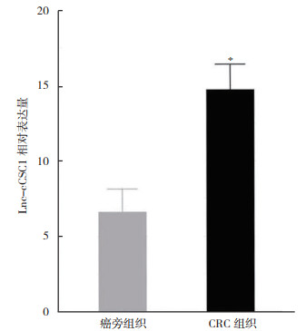
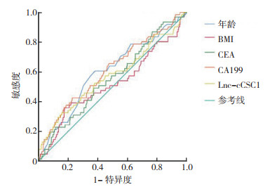
qPCR结果显示,癌组织中Lnc-cCSC1表达水平高于癌旁组织[(14.7±1.8)vs(6.5±1.6),P < 0.01;图 1)。计量资料采用ROC曲线获得最佳截断值并以此将计量资料转化为计数资料进行比较(图 2)。Lnc-cCSC1水平的曲线下面积(area under curve,AUC)为0.710(P < 0.01), 最佳截断值为14.65,敏感度和特异度分别为69.13%和64.43%。
|
| 注 * 与癌旁组织比较,P<0.01 图 1 Lnc-cCSC1在CRC组织及其癌旁组织中的相对表达水平 Fig.1 The relative expression of Lnc-cCSC1 in CRC tissues and the paired adjacent tissues |
|
| 注 BMI:体质量指数(body mass index);CEA:癌胚抗原(carcinoembryonic antigen);CA199:糖类抗原199(carbohydrate antigen 199);Lnc-cCSC1:结直肠癌细胞干性相关lncRNA 1(colorectal cancer stem cell related lncRNA 1) 图 2 临床病理因素和Lnc-cCSC1判断CRC预后的ROC曲线 Fig.2 The ROC curves of clinicopathological factors and LnccCSC1 in determining the prognosis of CRC |
根据ROC曲线,以Lnc-cCSC16的最佳截断值14.65为界,将实验组195例CRC患者分为Lnc-cCSC1高表达组(n=56)和低表达组(n=139)。Lnc-cCSC1高表达组和低表达组在肿瘤的分化程度、TNM分期及CEA水平方面比较,差异均具有统计学意义(均P < 0.05,表 2), 而在年龄、性别、BMI、肿瘤病理类型、肿瘤原发部位和CA199水平方面比较,差异均无统计学意义(均P > 0.05)。
| 临床特征 | Lnc-cCSC1低表达组(n=139) | Lnc-cCSC1高表达组(n=56) | χ2值 | P值 |
| 年龄 | 0.188 | 0.665 | ||
| ≤63岁 | 41 | 15 | ||
| > 63岁 | 98 | 41 | ||
| 性别 | 0.605 | 0.437 | ||
| 女性 | 30 | 27 | ||
| 男性 | 81 | 57 | ||
| BMI | 1.56 | 0.218 | ||
| ≤26.4 kg/m2 | 72 | 24 | ||
| > 26.4 kg/m2 | 67 | 32 | ||
| 肿瘤分化 | 21.686 | < 0.01 | ||
| 高/中分化 | 93 | 17 | ||
| 低/未分化 | 46 | 39 | ||
| 肿瘤病理类型 | 1.63 | 0.202 | ||
| 无黏液腺癌 | 117 | 44 | ||
| 有黏液腺癌 | 22 | 12 | ||
| 肿瘤原发部位 | 2.502 | 0.286 | ||
| 右半结肠 | 56 | 25 | ||
| 左半结肠 | 53 | 16 | ||
| 直肠 | 30 | 15 | ||
| TNM分期 | 8.642 | 0.003 | ||
| Ⅰ/Ⅱ期 | 88 | 26 | ||
| Ⅲ/Ⅳ期 | 51 | 30 | ||
| CEA | 7.897 | 0.005 | ||
| ≤7.5 ng/mL | 98 | 41 | ||
| > 7.5 ng/mL | 41 | 15 | ||
| CA199 | 0.733 | 0.392 | ||
| ≤32.6 ng/mL | 33 | 11 | ||
| > 32.6 ng/mL | 106 | 45 | ||
| 注Lnc-cCSC1:结直肠癌细胞干性相关lncRNA 1(colorectal cancer stem cell related lncRNA 1);BMI:体质量指数(body mass index);CEA:癌胚抗原(carcinoembryonic antigen);CA199:糖类抗原199(carbohydrate antigen 199) | ||||
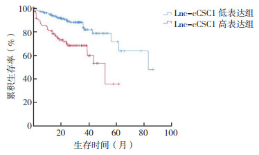
所有患者中位总生存期(overall survival,OS)为31个月,1、3和5年OS率分别为87.5%、79.2%和39.4%。生存分析结果显示,癌组织中Lnc-cCSC1高表达的患者预后较低表达患者更差(χ2=18.175,P < 0.01;图 3)。单因素回归分析表明,肿瘤的分化程度、肿瘤原发部位、TNM分期、CEA水平、CA199水平以及Lnc-cCSC1与CRC患者的预后相关(均P < 0.05)。选择单因素分析中P < 0.05的变量纳入Cox多因素回归分析,结果表明,肿瘤原发部位、TNM分期、CEA水平、CA199水平和Lnc-cCSC1是影响CRC患者预后的独立危险因素(均P < 0.05,表 3)。
|
| 图 3 实验组CRC患者术后总生存曲线 Fig.3 Postoperative overall survival curves of CRC patients in the experimental group |
| 临床特征 | 单因素分析 | 多因素分析 | |||
| HR(95%CI) | P值 | HR(95%CI) | P值 | ||
| 年龄 | |||||
| ≤63岁 | 参照 | ||||
| > 63岁 | 1.476(0.812~2.682) | 0.202 | |||
| 性别 | |||||
| 女性 | 参照 | ||||
| 男性 | 1.405(0.849~2.325) | 0.185 | 0.137 | ||
| BMI | |||||
| ≤26.4 kg/m2 | 参照 | ||||
| > 26.4 kg/m2 | 1.635(0.796~3.360) | 0.181 | 0.890 | ||
| 肿瘤分化 | |||||
| 高/中分化 | 参照 | ||||
| 低/未分化 | 2.635(1.247~5.569) | 0.011 | 1.415(0.656~3.052) | 0.376 | |
| 肿瘤病理类型 | |||||
| 无黏液腺癌 | 参照 | ||||
| 有黏液腺癌 | 1.112(0.592~2.092) | 0.741 | |||
| 肿瘤原发部位 | |||||
| 右半结肠 | 参照 | ||||
| 左半结肠 | 2.126(1.216~3.715) | 0.008 | 3.282(1.760~6.121) | < 0.01 | |
| 直肠 | 1.049(0.493~2.230) | 0.901 | 0.73 | ||
| TNM分期 | |||||
| Ⅰ/Ⅱ期 | 参照 | ||||
| Ⅲ/Ⅳ期 | 3.711(2.158~6.382) | < 0.01 | 3.360(1.036~10.895) | 0.043 | |
| CEA | |||||
| ≤7.5 ng/mL | 参照 | ||||
| > 7.5 ng/mL | 1.552(1.103~2.213) | 0.012 | 2.121(1.117~4.027) | 0.022 | |
| CA199 | |||||
| ≤32.6 ng/mL | 参照 | ||||
| > 32.6 ng/mL | 2.391(1.731~3.303) | < 0.001 | 3.040(1.123~6.453) | 0.032 | |
| Lnc-cCSC1 | |||||
| ≤14.65 | 参照 | ||||
| > 14.65 | 2.850(1.721~4.719) | < 0.01 | 2.510(1.431~4.402) | 0.001 | |
| 注BMI:体质量指数(body mass index);CEA:癌胚抗原(carcinoembryonic antigen);CA199:糖类抗原199(carbohydrate antigen 199);Lnc-cCSC1:结直肠癌细胞干性相关lncRNA 1(colorectal cancer stem cell related lncRNA 1) | |||||
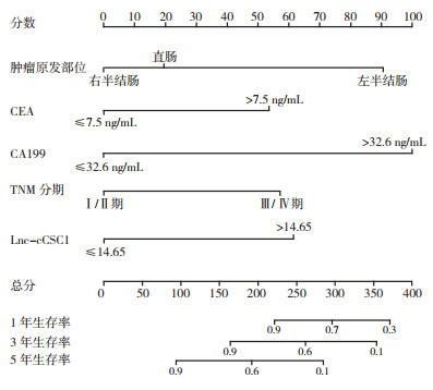
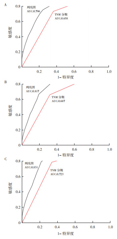
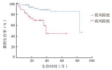
纳入CRC患者预后的独立危险因素构建nomogram预测模型(图 4)。计算生存预测的C-index为0.792。比较nomogram模型和TNM分期系统对CRC患者术后OS的预测准确性。Nomogram模型预测术后1、3和5年OS率的ROC的AUC为0.796、0.817和0.831,而TNM分期系统则分别为0.658、0.687和0.723(图 5)。结果表明,联合Lnc-cCSC1构建的nomogram模型相较于TNM分期系统在预测患者术后OS有更好的效能。利用构建的nomogram模型对验证组病例进行评估,以评分中位数为截断值将79例病例划分为高风险组和低风险组,其中高风险组40例,低风险组39例。Kaplan-Meier法生存分析结果显示,高风险组患者行根治术后OS较低风险组更差(P=0.005,图 6),这表明构建的nomogram模型能够有效评估CRC患者行根治术后临床预后效果。
|
| 注 CEA:癌胚抗原(carcinoembryonic antigen);CA199:糖类抗原199(carbohydrate antigen 199);Lnc-cCSC1:结直肠癌细胞干性相关lncRNA 1(colorectal cancer stem cell related lncRNA 1) 图 4 根治性切除术后CRC患者总生存率的nomogram模型 Fig.4 The nomogram model of overall survival rates of CRC patients after radical resection |
|
| 注 A:1年总生存ROC曲线;B:3年总生存ROC曲线;C:5年总生存ROC曲线;AUC:曲线下面积(area under curve) 图 5 列线图和TNM分期系统预后模型的总生存ROC曲线 Fig.5 ROC curves of overall survival of nomogram and TNM staging system prognostic models |
|
| 图 6 验证组 CRC 患者术后总生存曲线 Fig.6 Postoperative overall survival curves of CRC patients in the validation group |
目前研究认为CRC的发生和发展是一个多因素共同参与的复杂过程,与此同时已经有相当数量的研究表明,LncRNA在这一过程中扮演着重要的角色[12-13]。在既往研究中,本研究组通过富集分析表明,Lnc-cCSC1表达在CRC中具有重要的生物学作用,进一步的研究发现,Lnc-cCSC1与CRC细胞干性特征维持密切相关[11],并能通过Hedgehog通路直接调控干性标志物表达[10-11]。本研究进一步研究Lnc-cCSC1在CRC中的表达水平差异及其对CRC预后的影响。
本研究检测Lnc-cCSC1在CRC组织及癌旁组织中的表达水平,首次证实,CRC组织中Lnc-cCSC1的表达相较于癌旁组织更高。ROC曲线结果也表明,Lnc-cCSC1对于判断CRC患者术后总体预后有着较好的效能。根据ROC曲线将患者划分为Lnc-cCSC1高表达组与低表达组后的统计学分析表明,Lnc-cCSC1的高表达与肿瘤的分化程度、TNM分期及CEA水平密切相关,这提示Lnc-cCSC1可能在CRC中发挥着原癌基因的作用,有着促进癌细胞增殖、侵袭和转移的作用。同时Kaplan-Meier法和Log-rank分析也表明,Lnc-cCSC1高表达组患者预后较低表达组患者更差。以上结果说明Lnc-cCSC1有着作为判断CRC患者术后预后的生物标志物及作为治疗靶点的潜能。进一步的多因素Cox比例风险模型证明,Lnc-cCSC1是CRC预后的独立危险因素,同时TNM分期、肿瘤原发部位、CEA水平和CA199水平也是CRC预后的独立危险因素, 这与以往的研究结果是一致的[14-15]。
与传统的TNM分期系统比较,nomogram可以综合多种预后指标,已被证实在多种类型的肿瘤中具有更加可靠的预测价值[4-6]。本研究也显示,基于TNM分期、肿瘤原发部位、CEA水平和CA199水平这4个独立危险因素和Lnc-cCSC1表达水平而构建的nomogram模型相较与单独的TNM分期系统有着更高的敏感度和特异度。该nomogram模型的优势在于整合临床病理特征和遗传信息,能够更有效地把患者分为总体预后更差的高风险组和总体预后更好的低风险组。本研究组前期研究发现,Lnc-cCSC1与CRC细胞干性特征维持密切相关。本研究结果进一步证实,Lnc-cCSC1的高表达赋予CRC患者多种恶性临床病理特征。
综上所述,本研究首次证明,Lnc-cCSC1在CRC中高表达且与患者不良预后相关。lncRNA-cCSC1不仅可以作为判断CRC预后的有效生物标志物,而且可能成为CRC潜在的治疗靶点。结合Lnc-cCSC1所建立的nomogram模型预测效能较好。这种纳入临床因素和遗传信息的nomogram模型以其独特的可视化效果可帮助医师更加简便有效地评估CRC患者术后总体生存状况,为患者选择更具针对性和更有效的治疗方案。
| [1] |
Chen W, Zheng R, Baade P, et al. Cancer statistics in China, 2015[J]. CA, 2016, 66(2): 115-132. |
| [2] |
Sauer R, Liersch T, Merkel S, et al. Preoperative versus postoperative chemoradiotherapy for locally advanced rectal cancer: results of the German CAO/ARO/AIO-94 randomized phase Ⅲ trial after a median follow-up of 11 years[J]. J Clin Oncol, 2012, 30(16): 1926-1933. DOI:10.1200/JCO.2011.40.1836 |
| [3] |
Iversen L, Rasmussen P, Laurberg S. Value of laparoscopy before cytoreductive surgery and hyperthermic intraperitoneal chemotherapy for peritoneal carcinomatosis[J]. Br J Surg, 2013, 100(2): 285-292. |
| [4] |
Fang Q, Chen H. Development of a novel autophagy-related prognostic signature and nomogram for hepatocellular carcinoma[J]. Front Oncol, 2020, 10(3): 591356. |
| [5] |
沈锋, 王葵, 阎振林, 等. 1370例肝内胆管细胞癌肝切除术的疗效及预后因素分析[J]. 中华消化外科杂志, 2016, 15(4): 319-328. DOI:10.3760/cma.j.issn.1673-9752.2016.04.004 |
| [6] |
戴赟, 陆俊, 李平, 等. 皮革胃患者术后生存情况预测的列线图模型研究[J]. 中国普通外科杂志, 2019, 28(4): 461-466. |
| [7] |
Nagtegaal Ⅰ, Tot T, Jayne D, et al. Lymph nodes, tumor deposits, and TNM: are we getting better?[J]. J Clin Oncol, 2011, 29(18): 2487-2492. DOI:10.1200/JCO.2011.34.6429 |
| [8] |
Huarte M. The emerging role of lncRNAs in cancer[J]. Nat Med, 2015, 21(11): 1253-1261. DOI:10.1038/nm.3981 |
| [9] |
Kopp F, Mendell J. Functional classification and experimental dissection of long noncoding RNAs[J]. Cell, 2018, 172(3): 393-407. DOI:10.1016/j.cell.2018.01.011 |
| [10] |
Xiong Y, You W, Hou M, et al. Nomogram integrating genomics with clinicopathologic features improves prognosis prediction for colorectal cancer[J]. Mol Cancer Res, 2018, 16(9): 1373-1384. DOI:10.1158/1541-7786.MCR-18-0063 |
| [11] |
Zhou H, Xiong Y, Peng L, et al. LncRNA-cCSC1 modulates cancer stem cell properties in colorectal cancer via activation of the Hedgehog signaling pathway[J]. J Cell Biochem, 2020, 121(3): 2510-2524. DOI:10.1002/jcb.29473 |
| [12] |
Pichler M, Rodriguez-Aguayo C, Nam S, et al. Therapeutic potential of FLANC, a novel primate-specific long non-coding RNA in colorectal cancer[J]. Gut, 2020, 69(10): 1818-1831. DOI:10.1136/gutjnl-2019-318903 |
| [13] |
Ozawa T, Matsuyama T, Toiyama Y, et al. CCAT1 and CCAT2 long noncoding RNAs, located within the 8q.24.21 'gene desert', serve as important prognostic biomarkers in colorectal cancer[J]. Ann Oncol, 2017, 28(8): 1882-1888. DOI:10.1093/annonc/mdx248 |
| [14] |
孙卢浩然, 卢敏. SERCA3在结直肠癌细胞中异常表达及其临床意义[J]. 实用肿瘤杂志, 2019, 34(2): 122-124. |
| [15] |
武雪亮, 杨东东, 王立坤, 等. 长链非编码RNA HOTAIR在结直肠癌中的表达、临床意义及预后分析[J]. 实用医学杂志, 2019, 35(9): 1425-1428. DOI:10.3969/j.issn.1006-5725.2019.09.015 |
 2021, Vol. 36


