

Effects of incomplete argon-helium cryoablation on tumor migration and invasion in a mouse model of Lewis lung cancer
-
摘要:目的
观察不完全氩氦冷冻消融对Lewis肺癌(LLC)小鼠残余肿瘤细胞迁移、侵袭及肺转移的影响,并初步探讨其作用机制。
方法C57BL/6N雌性小鼠24只,皮下接种LLC细胞建立移植瘤模型,待肿瘤体积达70 mm3时,按随机数字表法分为空白对照组(BC组)、假手术组(Sham组)、不完全冷冻消融组(ICA组)及完全冷冻消融组(CA组),每组6只。ICA组消融约70%肿瘤组织、保留30%残余灶,CA组行完全冷冻消融,Sham组仅穿刺不消融,BC组不做任何干预。干预处理后3 d取肺组织行H-E染色观察转移灶;取残余肿瘤组织,采用蛋白质印迹法检测基质金属蛋白酶9(MMP9)表达水平;分离残余肿瘤原代细胞,通过划痕实验、Transwell迁移/侵袭实验检测0、24、48 h细胞迁移与侵袭能力。
结果成功构建不完全冷冻消融LLC小鼠模型。ICA组小鼠肺组织可见明确转移灶,其余各组未见转移;ICA组肿瘤组织MMP9表达水平高于BC组和Sham组(均P<0.01)。细胞功能实验显示,24、48 h时ICA组原代细胞迁移率、侵袭率均高于BC组和Sham组(均P<0.05)。
结论不完全氩氦冷冻消融可增强LLC小鼠残余肿瘤的迁移、侵袭能力并促进肺转移,其机制可能与上调MMP9表达有关。
Abstract:ObjectiveTo observe the effects of incomplete argon-helium cryoablation on migration, invasion and pulmonary metastasis of residual tumor cells in a Lewis lung carcinoma (LLC) mouse model, and to preliminarily explore the underlying mechanism.
MethodsA total of 24 female C57BL/6N mice were subcutaneously injected with LLC cells to establish xenograft models. When the tumor volume reached approximately 70 mm3, mice were randomly assigned to 4 groups: blank control group (BC group), sham-operation group (Sham group), incomplete cryoablation group (ICA group), and complete cryoablation group (CA group), with 6 mice in each group. The ICA group received approximately 70% tumor ablation with 30% residual tumor; the CA group underwent complete cryoablation; the Sham group received needle puncture only without ablation; and the BC group received no intervention. Mice were euthanized 3 d post-treatment. Lung tissues were harvested for hematoxylin-eosin staining to detect metastatic foci. Residual tumor tissues were collected for Western blotting analysis of matrix metalloproteinase 9 (MMP9) expression. Primary tumor cells were isolated, and scratch assay, Transwell migration and invasion assays were performed to evaluate cell migration and invasion abilities at 0, 24 and 48 h.
ResultsThe incomplete cryoablation LLC mouse model was successfully established. Pulmonary metastatic foci were observed in the ICA group but not in the other 3 groups. The expression level of MMP9 in the ICA group was significantly higher than that in the BC and Sham groups (both P < 0.01). Cell function assays showed that the migration and invasion rates of primary cells in the ICA group were significantly higher than those in the BC and Sham groups at 24 and 48 h (all P < 0.05).
ConclusionIncomplete argon-helium cryoablation enhances migration and invasion abilities of residual LLC tumors and promotes pulmonary metastasis, which may be associated with upregulated MMP9 expression.
-
肿瘤的消融治疗包括射频消融、冷冻消融及微波消融等,已成为肿瘤治疗的重要手段[1-5]。消融技术具有治疗效果确切、创伤小、可重复性高等优势,在肿瘤治疗中占据重要地位[6-7]。肿瘤消融通过局部施加极端温度诱导肿瘤组织坏死,已应用于多种实体瘤的治疗。由于不同消融方法的物理机制各异,加之肿瘤解剖结构复杂、病灶较大及恶性肿瘤侵袭性生长等因素,实现理想的完全消融并非易事。临床中常因消融不足而产生肿瘤残留,即消融不全。研究发现,不完全射频消融、微波消融后,残余肿瘤组织可导致肺转移增加,进而影响预后[8-11],而目前未见不完全冷冻消融(incomplete cryoablation,ICA)对肿瘤生物学行为影响的研究报道。本研究通过对小鼠Lewis肺癌(Lewis lung cancer,LLC)单侧荷瘤模型进行ICA干预,分离残余肿瘤组织中原代肿瘤细胞,检测其迁移、侵袭能力,并观察小鼠肺部转移情况,旨在探索ICA是否同样导致残余肿瘤进展。
1 材料和方法
1.1 细胞系与动物
LLC细胞系购自武汉普诺赛生命科技有限公司。实验用6周龄SPF级雌性C57BL/6N小鼠(体重约20 g),购自北京维通利华实验动物技术有限公司[实验动物生产许可证号:SCXK(京)2021-0006],饲养于中国人民解放军总医院实验动物中心[实验动物使用许可证号:SYXK(军)2017-0019]。所有小鼠均在SPF级实验室饲养,温度为(22±1)℃,湿度为40%~ 60%,12 h光照/12 h暗交替,小鼠自由摄食标准饲料和水。本实验经中国人民解放军总医院实验动物福利伦理委员会批准(2024-X20-83)。
1.2 仪器与试剂
DMEM、预染蛋白分子量标志物、PBS均购自美国ThermoFisher Scientific公司;戊巴比妥钠购自武汉金诺化工有限公司;分离胶和浓缩胶配制试剂、BCA蛋白定量试剂盒(货号P0010)、ECL试剂(货号P0018S)均购自碧云天(上海)生物技术有限公司;RIPA裂解缓冲液(货号HY-K1001)购自美国Med Chem Express公司;PVDF膜(货号IPVH00010)购自美国Millipore公司;β肌动蛋白抗体(货号AF7018)、基质金属蛋白酶9(matrix metalloproteinase 9,MMP9)抗体(货号AF5228)、二抗(货号S0001)均购自美国Affinity Biosciences公司。氩氦靶向冷冻治疗系统(Cryocare Surgical System)为美国Endocare公司生产,冷冻探针(Cryo-Hit)购自以色列Galil Medical公司;红外热像仪(型号MAG30)为上海巨哥科技股份有限公司产品。
1.3 细胞培养
LLC细胞采用含10% FBS、1%青霉素-链霉素双抗的DMEM于37 ℃、5% CO2饱和湿度培养箱中常规培养,取对数生长期细胞用于造模。此外,定期检查细胞培养基的颜色变化,评估pH值并及时更换新鲜培养基。
1.4 建立LLC小鼠模型及分组
在小鼠左侧腹股沟皮下注射1×10⁶个LLC细胞,建立单侧荷瘤小鼠模型。注射时严格控制细胞悬液体积、注射深度及位置,保证造模一致性。接种后每日观察小鼠一般状态,约第7天肿瘤生长至可测量大小(直径3~4 mm)后,每2天使用电子游标卡尺测量肿瘤长径(a)与短径(b),按公式V=1/2ab2计算肿瘤体积。待肿瘤体积约为70 mm3时,将小鼠随机分为空白对照组(BC组)、假手术组(Sham组)、ICA组及完全冷冻消融组(CA组),以备后续实验。
使用氩氦靶向冷冻治疗系统(该装置前端是冷冻探针,有效尖端长度为10 mm)进行消融或病灶穿刺。将各组小鼠用戊巴比妥钠(40 mg/kg)麻醉,术区常规备皮消毒后经皮穿刺置入冷冻探针。CA组穿刺至肿瘤中心,-40 ℃冷冻60 s,复温60 s,循环2次,确保冰球直径超出肿瘤边缘,最大限度消除肿瘤细胞,全程使用红外热像仪监测。ICA组穿刺至肿瘤非中心区域,-40 ℃冷冻40 s,复温40 s,循环2次,以期消融约70%肿瘤、残余约30%[12],以红外热像仪检测。Sham组仅经皮穿刺置入冷冻探针,不行消融。BC组不进行任何干预。ICA组、CA组和Sham组操作时均以红外热像仪拍照,记录温度变化和边界。
1.5 肺组织留取和H-E染色
干预后第3天断颈处死小鼠,完整取出双侧肺组织,肉眼观察并拍照记录转移灶;用4%多聚甲醛溶液固定24 h,梯度脱水、石蜡包埋,制备6 μm连续切片,常规脱蜡、H-E染色,中性树胶封片。于光学显微镜(日本Olympus公司)下观察肺组织病理形态及肿瘤转移情况,每组随机选取3张切片进行镜检评估。
1.6 蛋白质印迹法检测残余肿瘤转移相关蛋白
干预后第3天断颈处死小鼠,并迅速取出残余肿瘤组织置于预冷的PBS中清洁组织表面,用手术剪剪切成小片,转移到EP管。向EP管中加入预冷的含有磷酸酶和蛋白酶抑制剂的RIPA裂解缓冲液,每100 mg组织添加1 mL裂解缓冲液。充分振荡混匀后,将样本置于4 ℃冰箱中孵育30 min,其间每隔10 min轻轻振荡1次以提高裂解效率。裂解反应后,将样本在4 ℃、1 000×g离心15 min,仔细转移上清液至新的EP管。依据目标蛋白的分子量进行12% SDS-PAGE后,将凝胶与预先准备好的PVDF膜进行配对,PVDF膜需事先用甲醇激活并与PAGE一同在转膜缓冲液中预湿。转膜后,PVDF膜用含5%牛血清白蛋白的TBST封闭2 h。依照抗体说明书推荐的稀释比例,将PVDF膜在一抗溶液中4 ℃孵育过夜;使用TBST充分清洗PVDF膜,去除未结合的一抗,然后以推荐稀释比例孵育二抗2 h;使用ECL显色系统对结合的二抗进行显色,随后通过X光膜或数字成像系统捕捉信号;利用ImageJ 1.53t软件进行信号强度的量化分析,以β肌动蛋白作为内参进行归一化处理。
1.7 原代肿瘤细胞的提取和培养
干预后第3天断颈处死小鼠,立即取肿瘤组织,用无菌PBS冲洗,剪碎后置于含有胶原酶Ⅳ(美国Gibco公司)的消化缓冲液中,37 ℃条件下振荡消化1 h。用100 μm细胞过滤器过滤消化后的组织,去除未完全消化的组织块和大颗粒杂质,1 000×g离心15 min,PBS洗涤2次;用含10% FBS的DMEM重悬,接种于T25培养瓶,于37 ℃、5% CO2培养箱中培养,取对数生长期细胞用于功能实验。
1.8 细胞划痕实验
6孔板接种原代肿瘤细胞(1×10⁴个/孔),培养至细胞融合度达90%;200 μL无菌枪头垂直划痕,PBS洗去漂浮细胞,加入无血清培养基;分别于0、24、48 h倒置显微镜下拍照,用ImageJ 1.53t软件测量划痕面积,计算细胞迁移率。细胞迁移率(%)=(0 h划痕面积-24/48 h划痕面积)/0 h划痕面积×100%。
1.9 细胞迁移和侵袭Transwell实验
细胞饥饿处理24 h。迁移实验:Transwell上室加入200 μL无血清培养基重悬的细胞悬液(1×10⁴个/孔),下室加入800 μL含15% FBS的培养基。侵袭实验:上室预铺基质胶,其余步骤同迁移实验。37 ℃培养24、48 h,用4%多聚甲醛溶液固定15 min,结晶紫染色10 min,显微镜下随机选取5个视野拍照,用ImageJ 1.53t软件统计穿膜细胞数,计算迁移率、侵袭率。
1.10 统计学处理
应用SPSS 19.0软件进行统计学分析,计量资料以x±s表示,多组间比较采用单因素方差分析,两两比较采用Dunnett’s t检验或Bonferroni校正。检验水准(α)为0.05。
2 结果
2.1 ICA小鼠模型的建立
ICA小鼠模型的构建及验证结果见图 1。红外热像仪监测结果显示,BC组肿瘤局部温度与周围正常组织无异;CA组整个肿瘤区域温度约-40 ℃;ICA组局部肿瘤组织(冷冻探针周围)温度约-20 ℃,而邻近肿瘤组织温度约0 ℃,呈现出明显的亚致死温度梯度。H-E染色结果显示,BC组肿瘤细胞形态完整,核质染色分明;CA组呈现为典型的凝固性坏死伴核碎裂,肿瘤细胞的正常组织结构完全丧失;ICA组肿瘤组织中可见坏死区与形态正常的肿瘤细胞紧密相邻,两者交界处形成了典型的“正常-坏死过渡带”。说明成功建立了ICA小鼠模型。
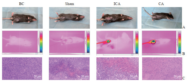 图 1 ICA小鼠模型的建立与验证Fig. 1 Establishment and validation of an ICA mouse modelA: Gross appearance of mice in each group; B: Infrared thermal imaging monitoring of tumor temperature profiles in each group; C: Hematoxylin-eosin staining of tumor tissue in each group. ICA: Incomplete cryoablation; BC: Black control; Sham: Sham-operation; CA: Cryoablation.
 下载:
全尺寸图片
下载:
全尺寸图片
2.2 ICA促进残余肿瘤发生肺部转移
大体观察小鼠肺组织发现,ICA组小鼠肺脏表面散布有明显的灰白色转移结节;BC组、Sham组及CA组肺组织外观形态正常,未见肉眼可见的转移结节(图 2A)。H-E染色结果显示,ICA组小鼠肺组织切片中可见明显的肿瘤细胞浸润灶,正常的肺泡结构破坏;其余各组的肺组织中均未见微转移灶(图 2B)。上述结果提示,ICA可促进残余肿瘤向肺部转移。
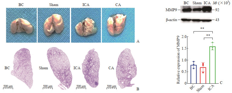 图 2 ICA促进LLC小鼠肺转移并上调MMP9表达Fig. 2 ICA promoted pulmonary metastasis and upregulated MMP9 expression in LLC miceA: Gross map of lung tissue of mice in each group; B: Hematoxylin-eosin staining of mouse lung tissue in each group; C: Western blotting detection of MMP9 content in residual tumor cells in each group and quantitative diagram of each group of Western blotting. **P < 0.01. n=3, x±s. ICA: Incomplete cryoablation; LLC: Lewis lung cancer; MMP9: Matrix metalloproteinase 9; BC: Black control; Sham: Sham-operation; CA: Cryoablation.下载:
全尺寸图片
图 2 ICA促进LLC小鼠肺转移并上调MMP9表达Fig. 2 ICA promoted pulmonary metastasis and upregulated MMP9 expression in LLC miceA: Gross map of lung tissue of mice in each group; B: Hematoxylin-eosin staining of mouse lung tissue in each group; C: Western blotting detection of MMP9 content in residual tumor cells in each group and quantitative diagram of each group of Western blotting. **P < 0.01. n=3, x±s. ICA: Incomplete cryoablation; LLC: Lewis lung cancer; MMP9: Matrix metalloproteinase 9; BC: Black control; Sham: Sham-operation; CA: Cryoablation.下载:
全尺寸图片
为探究ICA促进肿瘤细胞转移的分子机制,采用蛋白质印迹法检测各组肿瘤组织中MMP9的表达。结果显示,与BC组和Sham组相比,ICA组MMP9蛋白表达水平升高(均P<0.01,图 2C);BC组与Sham组间MMP9的表达水平差异无统计学意义(P>0.05)。该分子学结果与组织学观察结果一致,提示ICA可能通过上调MMP9表达促进残余肿瘤发生侵袭与转移。
2.3 ICA增加原代肿瘤细胞的迁移与侵袭能力
细胞划痕实验结果显示,在培养24、48 h后,ICA组的划痕愈合率均高于BC组和Sham组(均P<0.05),而BC组与Sham组在各时间点比较差异均无统计学意义(均P>0.05)。见图 3。
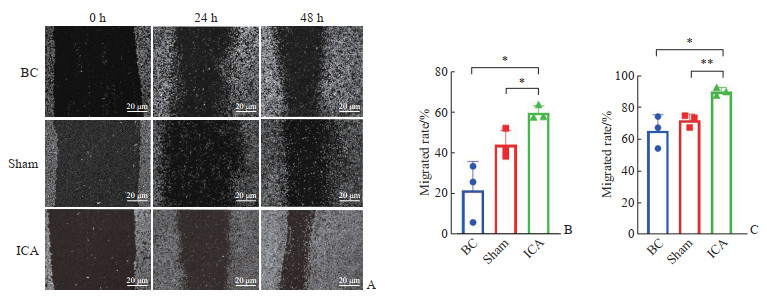 图 3 ICA增强了小鼠原代肿瘤细胞的划痕愈合能力Fig. 3 ICA increased wound healing abilities of primary tumor cells from miceA: Scratch experiment; B, C: Quantitative plots of 24 h (B) and 48 h (C). *P < 0.05, **P < 0.01. n=3, x±s. ICA: Incomplete cryoablation; BC: Black control; Sham: Sham-operation.下载:
全尺寸图片
图 3 ICA增强了小鼠原代肿瘤细胞的划痕愈合能力Fig. 3 ICA increased wound healing abilities of primary tumor cells from miceA: Scratch experiment; B, C: Quantitative plots of 24 h (B) and 48 h (C). *P < 0.05, **P < 0.01. n=3, x±s. ICA: Incomplete cryoablation; BC: Black control; Sham: Sham-operation.下载:
全尺寸图片
Transwell迁移与侵袭实验结果(图 4)显示,ICA组原代肿瘤细胞的穿膜能力增强:与BC组、Sham组相比,ICA组细胞在24 h和48 h的迁移细胞数及侵袭细胞数均增加(均P<0.05);而BC组与Sham组在各时间点的迁移、侵袭能力差异均无统计学意义(均P>0.05)。以上结果表明,ICA可提升残余肿瘤细胞的侵袭与转移潜能。
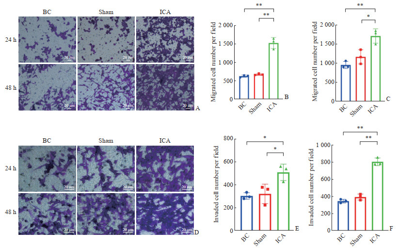 图 4 ICA增强了小鼠原代肿瘤细胞的迁移和侵袭能力Fig. 4 ICA enhanced migration and invasion capacities of primary mouse tumor cellsA: Representative microscopic images of migrating primary tumor cells stained with crystal violet at 24 h and 48 h; B, C: Quantification of migrated cells per field at 24 h (B) and 48 h (C), respectively; D: Representative microscopic images of invading primary tumor cells stained with crystal violet at 24 h and 48 h; E, F: Quantification of invaded cells per field at 24 h (E) and 48 h (F), respectively. *P < 0.05, **P < 0.01. n=3, x±s. ICA: Incomplete cryoablation; BC: Black control; Sham: Sham-operation.下载:
全尺寸图片
图 4 ICA增强了小鼠原代肿瘤细胞的迁移和侵袭能力Fig. 4 ICA enhanced migration and invasion capacities of primary mouse tumor cellsA: Representative microscopic images of migrating primary tumor cells stained with crystal violet at 24 h and 48 h; B, C: Quantification of migrated cells per field at 24 h (B) and 48 h (C), respectively; D: Representative microscopic images of invading primary tumor cells stained with crystal violet at 24 h and 48 h; E, F: Quantification of invaded cells per field at 24 h (E) and 48 h (F), respectively. *P < 0.05, **P < 0.01. n=3, x±s. ICA: Incomplete cryoablation; BC: Black control; Sham: Sham-operation.下载:
全尺寸图片
3 讨论
肿瘤消融治疗(如射频消融、冷冻消融及微波消融等)凭借创伤小、安全性高、并发症少及患者术后恢复快等优势,已成为多种晚期实体瘤的重要治疗手段[13-15]。然而,术后复发仍是制约其远期疗效的核心难题。为实现根治性治疗,Giraud等[16]提出,原发性肺腺癌和鳞状细胞癌的消融边界应分别超出肿瘤实质至少8 mm和6 mm。但在临床实践中,受限于病灶邻近大血管产生的“热沉效应(heat sink effect)”及特殊的解剖位置,往往难以实现完全消融[17]。既往研究已证实,不完全射频消融会加速残余肝细胞癌的恶性演进[18-20]。Zhao等[21]通过超声与红外热像仪双重监测,建立了皮下肝癌不完全射频消融小鼠模型,进一步明确了热应激对残余肿瘤生长的促进作用。基于此,本研究探讨了ICA所致的冷应激是否同样会诱发残余癌细胞的恶性进展。
冷冻消融利用极低温度诱导冰晶形成,致使肿瘤组织缺血、坏死并引发局部炎症反应。然而,针对ICA的物理界限,目前学术界尚未达成共识。本研究参考射频消融的相关标准[12],将病灶消融体积<70%界定为消融不全。实验中,我们借助红外热像仪精准定位并实时监控消融范围,成功构建了皮下LLC荷瘤小鼠ICA模型。组织学H-E染色结果显示,ICA组肿瘤内部呈现出典型的凝固性坏死区,且与正常肿瘤组织紧密相邻,形成明确的过渡带。这一病理特征证实了消融盲区内确有活性肿瘤细胞残存,表明模型构建成功。
在肿瘤侵袭转移机制的动物研究中,以往多倾向于采用尾静脉注射或原位成瘤模型以加速血液播散。本研究则采用皮下移植瘤模型,严格评估了原发灶受亚致死冷刺激后的自发转移潜能。体内实验表明,与BC组和Sham组相比,ICA处理促进了原发肿瘤转移。体外细胞划痕与Transwell侵袭实验进一步验证,ICA增强了原代肿瘤细胞的迁移与侵袭能力。该现象与不完全热消融诱导残余细胞迁移潜能上调的表型[22-24]高度一致,提示不同物理属性的亚致死消融可能触发了相似的促转移级联反应。
目前,关于消融后肿瘤进展的分子机制尚无定论。Tan等[25]在肝癌不完全射频消融模型中发现,亚致死热应激可通过激活血管内皮生长因子-血管内皮生长因子受体1通路增强细胞迁移能力。本研究则聚焦于MMP9,MMP9作为细胞外基质与基底膜降解的关键蛋白酶,是驱动肿瘤侵袭与转移的核心调控因子。实验结果显示,ICA组残余肿瘤组织中MMP9的表达水平上调,且伴随肺转移。推测ICA可能通过激活MMP9介导的细胞外基质降解通路重塑肿瘤局部微环境,从而放大残余癌细胞的侵袭性表型,最终驱动转移的发生。
尽管本研究揭示了ICA的促转移效应,但仍存在一定的局限性。首先,机制探讨暂局限于MMP9单一效应分子,尚未全面阐明介导MMP9异常激活的上游调控网络;其次,皮下移植瘤模型在基质成分及免疫浸润状态上,与真实的肺原位肿瘤微环境仍存在差异。未来,将结合转录组测序技术及多重基因干预手段,在原位模型中深度解析ICA促转移的全景分子网络,以期为临床寻找潜在的干预靶点。
综上所述,本研究证实了ICA可上调LLC残余肿瘤细胞中MMP9的表达,增强其迁移与侵袭能力并促进肺转移。这一发现为临床肿瘤学提出了重要警示:在实施冷冻消融治疗时,必须结合精准的影像学引导,最大限度地确保消融安全边界以实现完全消融,规避ICA引发的肿瘤复发与转移风险。
-
图 1 ICA小鼠模型的建立与验证
Fig. 1 Establishment and validation of an ICA mouse model
A: Gross appearance of mice in each group; B: Infrared thermal imaging monitoring of tumor temperature profiles in each group; C: Hematoxylin-eosin staining of tumor tissue in each group. ICA: Incomplete cryoablation; BC: Black control; Sham: Sham-operation; CA: Cryoablation.
下载:
全尺寸图片
图 2 ICA促进LLC小鼠肺转移并上调MMP9表达
Fig. 2 ICA promoted pulmonary metastasis and upregulated MMP9 expression in LLC mice
A: Gross map of lung tissue of mice in each group; B: Hematoxylin-eosin staining of mouse lung tissue in each group; C: Western blotting detection of MMP9 content in residual tumor cells in each group and quantitative diagram of each group of Western blotting. **P < 0.01. n=3, x±s. ICA: Incomplete cryoablation; LLC: Lewis lung cancer; MMP9: Matrix metalloproteinase 9; BC: Black control; Sham: Sham-operation; CA: Cryoablation.
下载:
全尺寸图片
图 3 ICA增强了小鼠原代肿瘤细胞的划痕愈合能力
Fig. 3 ICA increased wound healing abilities of primary tumor cells from mice
A: Scratch experiment; B, C: Quantitative plots of 24 h (B) and 48 h (C). *P < 0.05, **P < 0.01. n=3, x±s. ICA: Incomplete cryoablation; BC: Black control; Sham: Sham-operation.
下载:
全尺寸图片
图 4 ICA增强了小鼠原代肿瘤细胞的迁移和侵袭能力
Fig. 4 ICA enhanced migration and invasion capacities of primary mouse tumor cells
A: Representative microscopic images of migrating primary tumor cells stained with crystal violet at 24 h and 48 h; B, C: Quantification of migrated cells per field at 24 h (B) and 48 h (C), respectively; D: Representative microscopic images of invading primary tumor cells stained with crystal violet at 24 h and 48 h; E, F: Quantification of invaded cells per field at 24 h (E) and 48 h (F), respectively. *P < 0.05, **P < 0.01. n=3, x±s. ICA: Incomplete cryoablation; BC: Black control; Sham: Sham-operation.
下载:
全尺寸图片
-
[1] CHEUNG T T, MA K W, SHE W H. A review on radiofrequency, microwave and high-intensity focused ultrasound ablations for hepatocellular carcinoma with cirrhosis[J]. Hepatobiliary Surg Nutr, 2021, 10(2): 193-209. DOI: 10.21037/hbsn.2020.03.11. [2] ADWAN H, VOGL T J, BALABAN Ü, et al. Percutaneous thermal ablation therapy of hepatocellular carcinoma (HCC): microwave ablation (MWA) versus laser-induced thermotherapy (LITT)[J]. Diagnostics, 2022, 12(3): 564. DOI: 10.3390/diagnostics12030564. [3] VOGL T J, FREICHEL J, GRUBER-ROUH T, et al. Interventional oncological treatment of hepatocellular carcinoma (HCC) - A single-center long-term evaluation of thermoablation techniques like LITT, MWA, and TACE in a multimodal application over 26 years[J]. Heliyon, 2023, 9(4): e14646. DOI: 10.1016/j.heliyon.2023.e14646. [4] FACCIORUSSO A, ABD EL AZIZ M A, TARTAGLIA N, et al. Microwave ablation versus radiofrequency ablation for treatment of hepatocellular carcinoma: a meta-analysis of randomized controlled trials[J]. Cancers, 2020, 12(12): 3796. DOI: 10.3390/cancers12123796. [5] KWAK K, YU B, LEWANDOWSKI R J, et al. Recent progress in cryoablation cancer therapy and nanoparticles mediated cryoablation[J]. Theranostics, 2022, 12(5): 2175-2204. DOI: 10.7150/thno.67530. [6] AHMED M, BRACE C L, LEE F T Jr, et al. Principles of and advances in percutaneous ablation[J]. Radiology, 2011, 258(2): 351-369. DOI: 10.1148/radiol.10081634. [7] HYEON Y, CHARLES B. Comparison of percutaneous ablation technologies in the treatment of malignant liver tumors[J]. Semin Intervent Radiol, 2014, 31(2): 129-137. DOI: 10.1055/s-0034-1373788. [8] KONG J, YAO C, DING X, et al. ATPase inhibitory factor 1 promotes hepatocellular carcinoma progression after insufficient radiofrequency ablation, and attenuates cell sensitivity to sorafenib therapy[J]. Front Oncol, 2020, 10: 1080. DOI: 10.3389/fonc.2020.01080. [9] LI Z, JIANG M, ZHANG T, et al. GAS6-AS2 promotes hepatocellular carcinoma via miR-3619-5p/ARL2 axis under insufficient radiofrequency ablation condition[J]. Cancer Biother Radiopharm, 2021, 36(10): 879-887. DOI: 10.1089/cbr.2019.3541. [10] MA D, GAO X, LIU Z, et al. Exosome-transferred long non-coding RNA ASMTL-AS1 contributes to malignant phenotypes in residual hepatocellular carcinoma after insufficient radiofrequency ablation[J]. Cell Prolif, 2020, 53(9): e12795. DOI: 10.1111/cpr.12795. [11] MARKEZANA A, GOLDBERG S N, KUMAR G, et al. Incomplete thermal ablation of tumors promotes increased tumorigenesis[J]. Int J Hyperthermia, 2021, 38(1): 263-272. DOI: 10.1080/02656736.2021.1887942. [12] SUN T, SUN B, CAO Y, et al. Synergistic effect of OK-432 in combination with an anti-PD-1 antibody for residual tumors after radiofrequency ablation of hepatocellular carcinoma[J]. Biomed Pharmacother, 2023, 166: 115351. DOI: 10.1016/j.biopha.2023.115351. [13] CHEN Z, MENG L, ZHANG J, et al. Progress in the cryoablation and cryoimmunotherapy for tumor[J]. Front Immunol, 2023, 14: 1094009. DOI: 10.3389/fimmu.2023.1094009. [14] WU Y, CAO F, ZHOU D, et al. Cryoablation reshapes the immune microenvironment in the distal tumor and enhances the anti-tumor immunity[J]. Front Immunol, 2022, 13: 930461. DOI: 10.3389/fimmu.2022.930461. [15] HU K Q. Advances in clinical application of cryoablation therapy for hepatocellular carcinoma and metastatic liver tumor[J]. J Clin Gastroenterol, 2014, 48(10): 830-836. DOI: 10.1097/MCG.0000000000000201. [16] GIRAUD P, ANTOINE M, LARROUY A, et al. Evaluation of microscopic tumor extension in non-small-cell lung cancer for three-dimensional conformal radiotherapy planning[J]. Int J Radiat Oncol Biol Phys, 2000, 48(4): 1015-1024. DOI: 10.1016/s0360-3016(00)00750-1. [17] ABBAS G, SCHUCHERT M J, PENNATHUR A, et al. Ablative treatments for lung tumors: radiofrequency ablation, stereotactic radiosurgery, and microwave ablation[J]. Thorac Surg Clin, 2007, 17(2): 261-271. DOI: 10.1016/j.thorsurg.2007.03.007. [18] TENG W, LIU K W, LIN C C, et al. Insufficient ablative margin determined by early computed tomography may predict the recurrence of hepatocellular carcinoma after radiofrequency ablation[J]. Liver Cancer, 2015, 4(1): 26-38. DOI: 10.1159/000343877. [19] KODA M, MAEDA Y, MATSUNAGA Y, et al. Hepatocellular carcinoma with sarcomatous change arising after radiofrequency ablation for well-differentiated hepatocellular carcinoma[J]. Hepatol Res, 2003, 27(2): 163-167. DOI: 10.1016/s1386-6346(03)canlet.2018.02.000207-9. [20] KONG J, KONG J, PAN B, et al. Insufficient radiofrequency ablation promotes angiogenesis of residual hepatocellular carcinoma via HIF-1α/VEGFA[J]. PLoS One, 2012, 7(5): e37266. DOI: 10.1371/journal.pone.0037266. [21] ZHAO Z, WU J, LIU X, et al. Insufficient radiofrequency ablation promotes proliferation of residual hepatocellular carcinoma via autophagy[J]. Cancer Lett, 2018, 421: 73-81. DOI: 10.1016/j.canlet.2018.02.024. [22] ZHANG N, WANG L, CHAI Z T, et al. Incomplete radiofrequency ablation enhances invasiveness and metastasis of residual cancer of hepatocellular carcinoma cell HCCLM3 via activating β-catenin signaling[J]. PLoS One, 2014, 9(12): e115949. DOI: 10.1371/journal.pone.0115949. [23] ZHANG N, ZHU H, DONG Y H, et al. Establishment of an insufficient radiofrequency ablation orthotopic nude mouse model of hepatocellular carcinoma to study the invasiveness and metastatic potential of residual cancer[J]. Oncol Lett, 2019, 18(3): 2548-2553. DOI: 10.3892/ol.2019.10552. [24] CAO F, WAN C, XIE L, et al. Localized RNA interference therapy to eliminate residual lung cancer after incomplete microwave ablation[J]. Thorac Cancer, 2019, 10(6): 1369-1377. DOI: 10.1111/1759-7714.13079. [25] TAN L, CHEN S, WEI G, et al. Sublethal heat treatment of hepatocellular carcinoma promotes intrahepatic metastasis and stemness in a VEGFR1-dependent manner[J]. Cancer Lett, 2019, 460: 29-40. DOI: 10.1016/j.canlet.2019.05.041.
