

Value of artificial intelligence-based real-time ultrasound diagnostic system combined with strain elastography for differential diagnosis of breast nodules
-
摘要:目的
探讨超声人工智能(AI)实时诊断系统联合应变弹性成像(SE)对良恶性乳腺结节的鉴别诊断价值。
方法回顾性分析2024年1月至10月海军军医大学第一附属医院收治的280例女性患者的299个乳腺结节,以病理诊断结果为金标准,分析超声AI实时诊断系统、SE及两者联合的诊断效能。采用logistic回归、Z检验、Kappa检验进行统计学分析。
结果超声AI实时诊断系统与SE联合诊断的灵敏度(84.85%)、阳性预测值(98.82%)、阴性预测值(76.74%)、准确度(89.30%)均优于单一诊断方法,且特异度达98.02%。联合诊断的AUC值为0.941,高于超声AI实时诊断系统(0.863)与SE(0.890)(均P<0.05),与病理结果的一致性最佳(Kappa=0.776)。超声AI实时诊断系统与SE的AUC值呈中度正相关(r=0.471,P<0.05)。
结论超声AI实时诊断系统联合SE有良好的乳腺结节良恶性鉴别诊断效能,其高特异度与阳性预测值有助于减少过度诊疗。
Abstract:ObjectiveTo evaluate the value of an artificial intelligence (AI)-based real-time ultrasound diagnostic system combined with strain elastography (SE) for the differential diagnosis of benign and malignant breast nodules.
MethodsA retrospective analysis was conducted on 299 breast nodules from 280 female patients treated at The First Affiliated Hospital of Naval Medical University from Jan. to Oct. 2024. With pathological results as the gold standard, the diagnostic performance of the AI-based real-time ultrasound diagnostic system, SE, and their combination was compared. Statistical analyses were performed using logistic regression, Z-test, and Kappa test.
ResultsThe sensitivity (84.85%), positive predictive value (98.82%), negative predictive value (76.74%), and accuracy (89.30%) of the combined diagnosis were all superior to those of the single methods, with a specificity of 98.02%. The area under curve of the combined diagnosis was 0.941, which was significantly higher than those of the AI-based real-time ultrasound diagnostic system (0.863) and SE (0.890) (both P<0.05), and the combined diagnosis showed the best consistency with pathology (Kappa=0.776). There was a moderate positive correlation between the AI-based real-time ultrasound diagnostic system and SE (r=0.471, P<0.05).
ConclusionThe combination of the AI-based real-time ultrasound diagnostic system and SE can significantly improve the diagnostic efficacy for benign and malignant breast nodules. The high specificity and positive predictive value are conducive to reducing overdiagnosis and overtreatment.
-
乳腺癌作为全球女性发病率最高的恶性肿瘤,严重威胁着女性的生命健康[1]。乳腺结节是乳腺癌的重要前驱病变,其发病率近年来呈显著上升趋势[2]。据统计,在我国城市女性群体中,乳腺结节的总体检出率为27.9%,其中乳腺影像报告和数据系统(breast imaging-reporting and data system,BI-RADS)4~5级约占4.0%,且发病年龄逐渐年轻化[3-4]。早期准确鉴别乳腺结节的良恶性对于制定合理的治疗方案、改善预后至关重要。
超声检查凭借无创、便捷、实时、无辐射、经济等优势,成为乳腺结节筛查的首选检查方法[5]。然而,传统灰阶超声主要依据结节的形态、边界、回声、血流等特征进行判断,对于一些征象不典型的结节,其良恶性鉴别存在一定的局限性,且依赖于操作者的经验和主观判断。
超声人工智能(artificial intelligence,AI)实时诊断系统可以动态识别结节的特征,量化诊断指标,减少人为因素的干扰,提高诊断报告的一致性[6-8]。但是,目前市售超声AI实时诊断系统主要基于结节的灰阶特征所构建。研究显示,基于灰阶超声的深度学习模型在乳腺结节的鉴别诊断中具有稳定且接近高级职称医师水平的表现,外部验证下AUC值约为0.90[9-10];同时,应变弹性成像(strain elastography,SE)对病灶硬度的量化提供了互补信息[11],整体准确性良好。将超声AI实时诊断系统与SE相结合,综合灰阶和弹性应变信息,有望充分发挥两者的优势。本研究旨在探讨超声AI实时诊断系统联合SE在乳腺结节鉴别诊断中的应用价值。
1 资料和方法
1.1 病例资料
回顾性分析2024年1月至10月海军军医大学第一附属医院外科收治的378例女性乳腺结节患者的临床资料。纳入标准:(1)女性乳腺结节患者;(2)术前接受二维灰阶超声及SE检查;(3)病理结果明确(手术/穿刺活检证实)。排除标准:(1)既往有乳腺手术/放化疗史;(2)乳腺假体植入史;(3)图像质量差无法分析;(4)影像或病理资料不全。排除98例因影像/病理资料不全者,最终入组280例女性患者,年龄为17~92岁,平均年龄为(53.19±12.20)岁;19例含2个结节(均按独立样本纳入),共入组299个结节,结节最大径为0.17~9.90 cm,平均最大径为(2.12±1.37)cm。本研究通过海军军医大学第一附属医院伦理委员会审批。
1.2 仪器设备
采用Esatoe MyLab 8eHD超声诊断仪(Esaote ESAOTE S.p.A.),线阵探头5~9 MHz,内置SE模块。AI分析平台V3.2.1为北京医准智能科技有限公司产品,能与超声诊断仪实时共享图像,可以进行实时检测和分析。
1.3 检查方法
患者取仰卧位,双手举过头顶暴露双侧乳腺,以乳头为中心进行放射状扫查,发现病灶后按《超声医学质量控制管理规范》保存图像。将图像共享至超声AI实时诊断系统后,系统基于卷积神经网络(convolutional neural network,CNN)算法,通过深度学习海量乳腺结节灰阶图像并自动识别病灶,同时依据美国放射学会乳腺超声影像报告与数据系统(2013版)[American College of Radiology ultrasound-breast imaging reporting and data system (2013 edition),ACR US-BIRADS 2013]输出分类建议。随后启动弹性成像模式,调节感兴趣区域(region of interest,ROI),并激活双幅显示模式。操作时探头节律性轻压-释放皮肤,保证绿色质量控制指示弹簧≥4个(Esatoe操作指南)。所有图像均由具有10年以上超声诊断经验的医师完成采集,并于同一深度勾勒病灶与周围正常腺体的ROI,再由系统自动计算病灶/正常腺体比值,即应变比(strain ratio,SR)。每个结节的同一部位重复测3次,取均值作为最终SR值。
1.4 样本量估算
以AUC值作为主要评价指标,采用配对ROC曲线差异检验(Hanley-McNeil近似检验)进行样本量估算与主要效应检验。参考既往研究[10-12]中AUC_AI为0.89、AUC_SE为0.90、AUC_联合为0.95。设α=0.05(双侧)、效能1-β=0.90,恶性∶良性比例约2∶1,计算所需总样本量为294(恶性约196,良性约98)。本研究实际纳入299个结节(恶性198个、良性101个),满足样本量要求。
1.5 统计学处理
采用SPSS 29.0软件进行数据分析。计量资料以x±s表示;计数资料以例数(个数)和百分数表示,组间比较采用χ2检验或Fisher确切概率法。以病理诊断结果为金标准,将ACR US-BIRADS 2013分级和SR值(连续型变量)作为自变量,通过二元logistic回归分析构建联合诊断模型。绘制ROC曲线并计算AUC值,以评估SE、超声AI实时诊断系统及两者联合的诊断效能,并采用Z检验进行比较分析,率的比较采用χ2检验。采用Spearman秩相关分析评估ACR US-BIRADS 2013分级与SR值的关联性,采用Kappa检验评估各种方法诊断结果与病理诊断结果的一致性(Kappa<0.40表示一致性差,0.40~0.75表示一致性中等,>0.75表示一致性较好)。检验水准(α)为0.05。
2 结果
2.1 病理诊断结果与ACR US-BIRADS 2013
分级299个乳腺结节中恶性198个(66.22%)、良性101个(33.78%)。恶性结节包括浸润性导管癌145个,原位癌22个,乳头状癌14个,黏液癌8个,浸润性小叶癌5个,梭形细胞癌、佩吉特病、转移癌、恶性叶状肿瘤各1例;良性结节包括纤维腺瘤55个,乳腺腺病21个,乳头状瘤13个,炎性病变8个,良性叶状肿瘤与潴留囊肿各2个。超声AI实时诊断系统的ACR US-BIRADS 2013分级见表 1。
表 1 超声AI实时诊断系统对乳腺结节的ACR US-BIRADS 2013分级Table 1 ACR US-BIRADS 2013 classification of breast nodules using AI-based real-time ultrasound diagnostic systemn Classification Malignant
N=198Benign
N=101Total
N=2990 1 3 4 2 1 2 3 3 4 27 31 4a 34 55 89 4b 60 9 69 4c 94 5 99 5 4 0 4 AI: Artificial intelligence; ACR US-BIRADS 2013: American College of Radiology ultrasound-breast imaging reporting and data system (2013 edition). 2.2 3种方法的诊断结果及与病理结果的一致性分析
198个恶性结节中,超声AI实时诊断系统诊断恶性158个,SE诊断恶性165个,联合诊断恶性168个。101个良性结节中,AI实时诊断系统诊断良性87个,SE诊断良性99个,联合诊断良性99个。超声AI实时诊断系统的诊断与病理结果的一致性中等(Kappa值为0.620),SE诊断与病理结果的一致性较好(Kappa值为0.757),两者联合诊断与病理结果的一致性较好(Kappa值为0.776)。见表 2。典型结节的超声与病理图像见图 1和图 2。
表 2 3种方法对乳腺结节诊断结果与病理结果的一致性分析Table 2 Consistency analysis of diagnostic results from 3 methods with pathological diagnosis in breast nodulesn Pathological result N AI-based real-time ultrasound diagnostic system Strain elastography Combined diagnosis Malignant Benign Malignant Benign Malignant Benign Malignant 198 158 40 165 33 168 30 Benign 101 14 87 2 99 2 99 Total 299 172 127 167 132 170 129 Kappa value 0.620 0.757 0.776 P value <0.001 <0.001 <0.001 AI: Artificial intelligence. 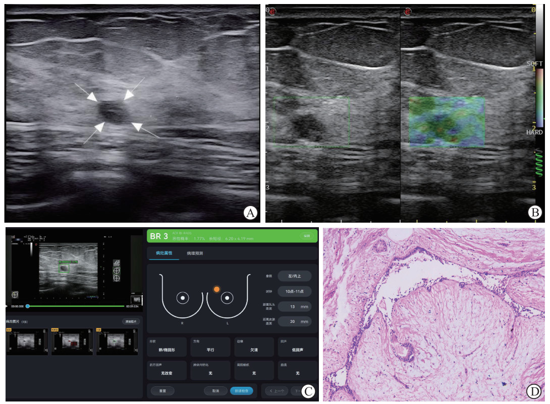 图 1 典型良性乳腺结节的超声与病理图像Fig. 1 Typical ultrasound and pathological images of a benign breast noduleA 46-year-old female presents with a hypoechoic nodule in the glandular layer of the upper inner quadrant of the left breast, with smooth margins and regular shape. A: 2-dimensional grayscale ultrasound image (the arrows indicate the nodule); B: Strain elastography image; C: AI-based real-time ultrasound diagnostic system; D: Pathological section image (hematoxylin-eosin staining, 100×). AI: Artificial intelligence.
 下载:
全尺寸图片
下载:
全尺寸图片
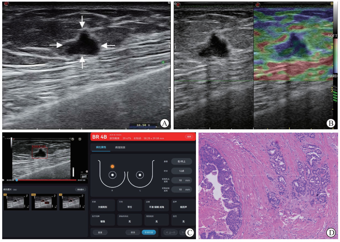 图 2 典型恶性乳腺结节的超声与病理图像Fig. 2 Typical ultrasound and pathological images of a malignant breast noduleA 66-year-old female has a markedly hypoechoic nodule in the upper quadrant of the right breast with ill-defined margins and irregular shape. A: 2-dimensional grayscale ultrasound image (the arrows indicate the nodule); B: Strain elastography image; C: AI-based real-time ultrasound diagnostic system; D: Pathological section image (hematoxylin-eosin staining, 100×). AI: Artificial intelligence.下载:
全尺寸图片
图 2 典型恶性乳腺结节的超声与病理图像Fig. 2 Typical ultrasound and pathological images of a malignant breast noduleA 66-year-old female has a markedly hypoechoic nodule in the upper quadrant of the right breast with ill-defined margins and irregular shape. A: 2-dimensional grayscale ultrasound image (the arrows indicate the nodule); B: Strain elastography image; C: AI-based real-time ultrasound diagnostic system; D: Pathological section image (hematoxylin-eosin staining, 100×). AI: Artificial intelligence.下载:
全尺寸图片
2.3 超声AI实时诊断系统、SE及两者联合的诊断效能分析
以病理诊断结果为金标准,分析ACR US-BIRADS 2013分级及SR的诊断效能,得出ACR US-BIRADS 2013分级>3级可作为恶性风险界值,SR最佳诊断良恶性的阈值为3.65。logistic回归分析显示,ACR US-BIRADS 2013分级和SR值均为结节恶性风险的独立预测因子(OR=3.654,95%CI 2.414~5.532,P<0.001;OR=4.753,95%CI 3.129~7.221,P<0.001),ACR US-BIRADS2013分级每增加1级恶性风险增加约3.65倍,SR值每增加1个单位恶性风险增加约4.75倍。采用logistic回归方法构建的联合模型方程为logitP=-10.473+1.296×(ACR US-BIRADS 2013分级)+1.559×SR。
Spearman秩相关分析显示,超声AI实时诊断系统和SE的AUC值呈中度正相关(r=0.471,P<0.05)。从整体诊断效能来看,联合诊断的AUC值(0.941)高于超声AI实时诊断系统(0.863)与SE(0.890)(均P<0.05)。两者联合诊断乳腺结节恶性的灵敏度(84.85%)最高,但与超声AI实时诊断系统(79.80%)和SE(83.33%)的差异无统计学意义(均P>0.05)。在特异度、阳性预测值和阳性预测值上,联合诊断与SE表现相当,均优于超声AI实时诊断系统(均P<0.05)。此外,联合诊断的准确度(89.30%)高于超声AI实时诊断系统(81.94%)(P<0.05)。见表 3和图 3。
表 3 3种方法对乳腺结节的诊断效能比较Table 3 Comparisons of diagnostic performance of 3 diagnostic methods for breast nodulesMethod AUC(95%CI) Pvalue Youden index Sensitivity/% Specificity/% PPV/% NPV/% Accuracy/% AI-based real-time ultrasound diagnostic system 0.863 (0.819, 0.907) <0.001 0.659 79.80 86.14 91.86 68.50 81.94 Strain elastography 0.890 (0.851, 0.930) <0.001 0.814 83.33 98.02* 98.80* 75.00* 88.29 Combined diagnosis 0.941 (0.914, 0.968)*△ <0.001 0.829 84.85 98.02* 98.82* 76.74* 89.30* *P<0.05 vs AI-based real-time ultrasound diagnostic system; △P<0.05 vs strain elastography. AI: Artificial intelligence; AUC: Area under curve; 95%CI: 95% confidence interval; PPV: Positive predictive value; NPV: Negative predictive value. 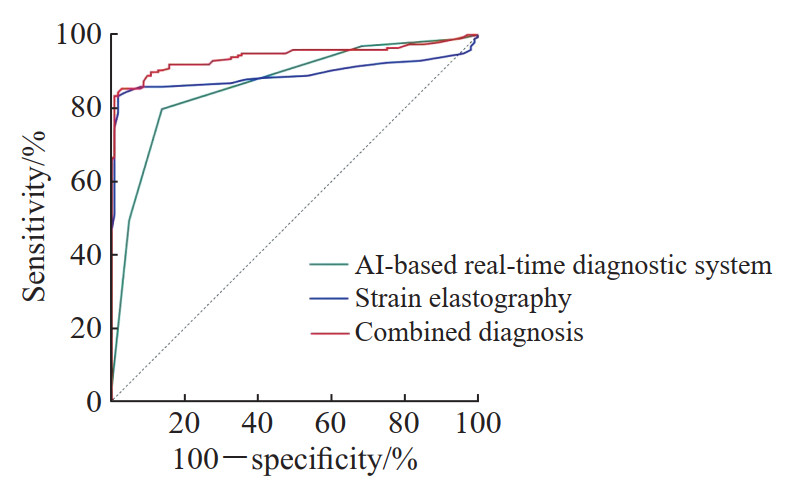 图 3 3种方法诊断恶性乳腺结节的受试者操作特征曲线Fig. 3 Receiver operating characteristic curves of 3 methods for diagnosing malignant breast nodulesAI: Artificial intelligence.下载:
全尺寸图片
图 3 3种方法诊断恶性乳腺结节的受试者操作特征曲线Fig. 3 Receiver operating characteristic curves of 3 methods for diagnosing malignant breast nodulesAI: Artificial intelligence.下载:
全尺寸图片
3 讨论
近年来,随着早癌筛查意识的提高和超声成像技术的进步,乳腺结节的检出率呈显著上升趋势[2]。有研究显示到2050年,乳腺癌新增病例数和死亡数将分别增加38%和68%[13],这对人类发展指数较低的国家影响尤为严重。因此,如何早期精准地鉴别乳腺结节的良恶性,已成为临床诊疗工作中亟待解决的问题。
超声检查因无创、实时和经济等优势,被国内外指南推荐为乳腺癌的首选筛查手段[14-15]。然而,传统灰阶超声依赖于操作者经验,观察者内和观察者间均可能存在差异,AI与超声的联合应用可以有效提升报告准确性。Ji等[16]研究显示,在AI辅助下,超声医师对乳腺结节的分类准确度显著提升[16]。虽然已有大量文献证实超声AI及弹性成像在乳腺结节鉴别诊断中的高效能[9, 17-18],但多为聚焦于单一技术的研究。本研究通过配对设计和一致性分析,为超声AI联合SE优于单独诊断提供了新的循证依据,具有重要的补充价值与实践意义。
本研究结果证实,将超声AI实时诊断系统与SE结合能够显著提升对乳腺结节的鉴别诊断效能,特别是在保持高特异度的前提下提高了灵敏度和准确度。超声AI实时诊断系统基于大量灰阶图像的深度学习,能有效识别结节的形态特征。本研究中,超声AI实时诊断系统鉴别乳腺结节良恶性的AUC值为0.863,表现出较好的诊断能力,但其灵敏度(79.80%)和特异度(86.14%)与Jin等[19]报道的4A类结节灵敏度92.1%、特异度79.6%略有区别,可能与样本选择和系统训练方法不同有关。部分良恶性结节在灰阶形态上存在影像重叠,本研究中1例浸润性导管癌和1例浸润性小叶癌由于灰阶特征均表现为边缘光整、形态规则、内部回声均匀,被误判为3类结节;1例导管内乳头状癌因结节位于导管内,无明显占位效应,且与周围导管内回声无明显差异,被误判为3类结节;70个良性结节由于形态不规则、内部回声不均匀、合并有钙化等高危征象,被误判为4类结节。
SE通过测量组织硬度反映生物力学信息。恶性结节通常质地较硬,良性结节较软。本研究中,SE表现出极高的特异度(98.02%),这意味着它能准确识别良性结节、避免假阳性。然而,其灵敏度(83.33%)较低,部分恶性结节(2例黏液癌、1例浸润性导管癌伴黏液分泌、8例原位癌)均因质地较软被误判为良性结节。
本研究中,Spearman秩相关分析显示超声AI实时诊断系统与SE的AUC值呈中度正相关(r=0.471,P<0.05),说明两者在一定程度上能识别相似的特征,但也存在差异性,这些差异性提示两者可相互补充,为联合诊断提供了理论基础。本研究通过logistic回归构建的联合诊断模型AUC值提升至0.941,显著优于单一诊断方法,证明了AI提供的形态学信息与SE提供的生物力学信息之间存在有效的互补性。联合诊断在维持高特异度(98.02%)的同时将灵敏度提高至84.85%、准确度提高至89.30%,意味着联合诊断不仅能更准确地识别恶性结节,还能非常可靠地排除良性结节。另外,联合诊断98.82%的阳性预测值和0.776的Kappa值也进一步支持了其临床可靠性。高特异度和高阳性预测值对于临床决策尤为重要,有助于减少对良性结节的不必要活检和过度治疗,降低患者的生理、心理及经济负担。
本研究存在以下局限性:(1)作为单中心回顾性研究,可能存在选择偏倚,研究结果的外推性有待多中心、前瞻性研究验证;(2)SE检查对操作者有一定依赖性,尽管有质量控制保证,但仍可能存在操作差异;(3)超声AI实时诊断系统的性能受限于其训练数据集和算法本身;(4)本研究未对不同病理类型的结节进行亚组分析,未来需要扩大样本量、针对不同病理分型进行分层研究。
-
图 1 典型良性乳腺结节的超声与病理图像
Fig. 1 Typical ultrasound and pathological images of a benign breast nodule
A 46-year-old female presents with a hypoechoic nodule in the glandular layer of the upper inner quadrant of the left breast, with smooth margins and regular shape. A: 2-dimensional grayscale ultrasound image (the arrows indicate the nodule); B: Strain elastography image; C: AI-based real-time ultrasound diagnostic system; D: Pathological section image (hematoxylin-eosin staining, 100×). AI: Artificial intelligence.
下载:
全尺寸图片
图 2 典型恶性乳腺结节的超声与病理图像
Fig. 2 Typical ultrasound and pathological images of a malignant breast nodule
A 66-year-old female has a markedly hypoechoic nodule in the upper quadrant of the right breast with ill-defined margins and irregular shape. A: 2-dimensional grayscale ultrasound image (the arrows indicate the nodule); B: Strain elastography image; C: AI-based real-time ultrasound diagnostic system; D: Pathological section image (hematoxylin-eosin staining, 100×). AI: Artificial intelligence.
下载:
全尺寸图片
图 3 3种方法诊断恶性乳腺结节的受试者操作特征曲线
Fig. 3 Receiver operating characteristic curves of 3 methods for diagnosing malignant breast nodules
AI: Artificial intelligence.
下载:
全尺寸图片
表 1 超声AI实时诊断系统对乳腺结节的ACR US-BIRADS 2013分级
Table 1 ACR US-BIRADS 2013 classification of breast nodules using AI-based real-time ultrasound diagnostic system
n Classification Malignant
N=198Benign
N=101Total
N=2990 1 3 4 2 1 2 3 3 4 27 31 4a 34 55 89 4b 60 9 69 4c 94 5 99 5 4 0 4 AI: Artificial intelligence; ACR US-BIRADS 2013: American College of Radiology ultrasound-breast imaging reporting and data system (2013 edition). 表 2 3种方法对乳腺结节诊断结果与病理结果的一致性分析
Table 2 Consistency analysis of diagnostic results from 3 methods with pathological diagnosis in breast nodules
n Pathological result N AI-based real-time ultrasound diagnostic system Strain elastography Combined diagnosis Malignant Benign Malignant Benign Malignant Benign Malignant 198 158 40 165 33 168 30 Benign 101 14 87 2 99 2 99 Total 299 172 127 167 132 170 129 Kappa value 0.620 0.757 0.776 P value <0.001 <0.001 <0.001 AI: Artificial intelligence. 表 3 3种方法对乳腺结节的诊断效能比较
Table 3 Comparisons of diagnostic performance of 3 diagnostic methods for breast nodules
Method AUC(95%CI) Pvalue Youden index Sensitivity/% Specificity/% PPV/% NPV/% Accuracy/% AI-based real-time ultrasound diagnostic system 0.863 (0.819, 0.907) <0.001 0.659 79.80 86.14 91.86 68.50 81.94 Strain elastography 0.890 (0.851, 0.930) <0.001 0.814 83.33 98.02* 98.80* 75.00* 88.29 Combined diagnosis 0.941 (0.914, 0.968)*△ <0.001 0.829 84.85 98.02* 98.82* 76.74* 89.30* *P<0.05 vs AI-based real-time ultrasound diagnostic system; △P<0.05 vs strain elastography. AI: Artificial intelligence; AUC: Area under curve; 95%CI: 95% confidence interval; PPV: Positive predictive value; NPV: Negative predictive value. -
[1] DENG T, ZI H, GUO X P, et al. Global, regional, and national burden of breast cancer, 1990-2021, and projections to 2050:a systematic analysis of the Global Burden of Disease Study 2021[J]. Thorac Cancer, 2025, 16(9):e70052. DOI: 10.1111/1759-7714.70052. [2] BAO Z, ZHAO Y, CHEN S, et al. Evidence and assessment of parenchymal patterns of ultrasonography for breast cancer detection among Chinese women: a cross-sectional study[J]. BMC Med Imaging, 2021, 21(1): 152. DOI: 10.1186/s12880-021-00687-0. [3] LIU X, XING Y, ZU Y, et al. Detection and BI-RADS classification of breast nodules in urban women: China, 2021[J]. China CDC Wkly, 2025, 7(10): 347-352. DOI: 10.46234/ccdcw2025.056. [4] ZHANG W, ZHANG B L, HE J J, et al. Clinicopathological characteristics and treatment of young women with breast cancer in China: a nationwide multicenter 10-year retrospective study[J]. Gland Surg, 2021, 10(1): 175-185. DOI: 10.21037/gs-20-574. [5] SPRAGUE B L, ICHIKAWA L, EAVEY J, et al. Breast cancer risk characteristics of women undergoing whole-breast ultrasound screening versus mammography alone[J]. Cancer, 2023, 129(16): 2456-2468. DOI: 10.1002/cncr.34768. [6] LI P, YIN M, GUERRINI S, et al. Roles of artificial intelligence and high frame-rate contrast-enhanced ultrasound in the differential diagnosis of breast imaging reporting and data system 4 breast nodules[J]. Gland Surg, 2025, 14(3): 462-478. DOI: 10.21037/gs-24-187. [7] MA B, WU G, ZHU H, et al. The prediction model of high-frequency ultrasound combined with artificial intelligence-assisted scoring system improved the diagnosis of sclerosing adenosis and early breast cancer[J]. Breast Cancer (Dove Med Press), 2025, 17: 145-155. DOI: 10.2147/BCTT.S483496. [8] WANG J, TONG J, LI J, et al. Using the GoogLeNet deep-learning model to distinguish between benign and malignant breast masses based on conventional ultrasound: a systematic review and meta-analysis[J]. Quant Imaging Med Surg, 2024, 14(10): 7111-7127. DOI: 10.21037/qims-24-679. [9] LIU Q, QU M, SUN L, et al. Accuracy of ultrasonic artificial intelligence in diagnosing benign and malignant breast diseases: a protocol for systematic review and meta-analysis[J]. Medicine (Baltimore), 2021, 100(50): e28289. DOI: 10.1097/MD.0000000000028289. [10] SUN P, FENG Y, CHEN C, et al. An AI model of sonographer's evaluation+S-Detect+elastography+clinical information improves the preoperative identification of benign and malignant breast masses[J]. Front Oncol, 2022, 12: 1022441. DOI: 10.3389/fonc.2022.1022441. [11] WU H, WANG C, AN Q, et al. Comparing the accuracy of shear wave elastography and strain elastography in the diagnosis of breast tumors: a systematic review and meta-analysis[J]. Medicine, 2022, 101(44): e31526. DOI: 10.1097/MD.0000000000031526. [12] HU W, LI X, TANG Y, et al. Performance of artificial intelligence-assisted ultrasound elastography in classifying benign and malignant breast tumors: a systematic review and meta-analysis[J]. BMC Med Imaging, 2025, 25(1): 440. DOI: 10.1186/s12880-025-01982-w. [13] KIM J, HARPER A, MCCORMACK V, et al. Global patterns and trends in breast cancer incidence and mortality across 185 countries[J]. Nat Med, 2025, 31(4): 1154-1162. DOI: 10.1038/s41591-025-03502-3. [14] 中国抗癌协会乳腺癌整合防筛专业委员会. 中国致密型乳腺女性乳腺癌筛查专家共识(2025年版)[J]. 中国普外基础与临床杂志, 2025, 32(11): 1345-1360. DOI: 10.7507/1007-9424.202509027. [15] PARK K H, LOIBL S, SOHN J, et al. Pan-Asian adapted ESMO clinical practice guidelines for the diagnosis, treatment and follow-up of patients with early breast cancer[J]. ESMO Open, 2024, 9(5): 102974. DOI: 10.1016/j.esmoop.2024.102974. [16] JI H, ZHU Q, MA T, et al. Development and validation of a transformer-based CAD model for improving the consistency of BI-RADS category 3-5 nodule classification among radiologists: a multiple center study[J]. Quant Imaging Med Surg, 2023, 13(6): 3671-3687. DOI: 10.21037/qims-22-1091. [17] SHAHZAD R, FATIMA I, ANJUM T, et al. Diagnostic value of strain elastography and shear wave elastography in differentiating benign and malignant breast lesions[J]. Ann Saudi Med, 2022, 42(5): 319-326. DOI: 10.5144/0256-4947.2022.319. [18] SELIM Y A Y, SABIT H, ARNETH B, et al. Diagnostic accuracy of sonoelastography for breast lesions: a meta-analysis comparing strain and shear wave elastography[J]. J Imaging, 2025, 11(7): 221. DOI: 10.3390/jimaging11070221. [19] JIN Z Y, LI J K, NIU R L, et al. Application of an artificial intelligence-assisted diagnostic system for breast ultrasound: a prospective study[J]. Gland Surg, 2024, 13(12): 2221-2231. DOI: 10.21037/gs-24-213.
