

Construction of cartilage organoids based on extracellular vesicles derived from Lactobacillus rhamnosus GG
-
摘要:目的
评估鼠李糖乳杆菌来源细胞外囊泡(LGG-EV)的促骨髓间充质干细胞(BMSC)软骨分化作用,并利用其构建软骨类器官。
方法基于前期转录组测序数据,分析LGG-EV对BMSC软骨分化的调控作用。提取LGG-EV并与BMSC共培养,通过细胞摄取、细胞增殖及细胞迁移实验验证其功能。采用阿利新蓝染色和免疫荧光染色检测软骨特异性标志物,评估LGG-EV促BMSC软骨分化效果。将BMSC与甲基丙烯酰化明胶(GelMA)混合制备微球,通过活细胞/死细胞双染色、细胞骨架染色及免疫荧光染色,验证软骨类器官的构建效果与表型特征。
结果LGG-EV富集多条与软骨分化及细胞外基质形成相关的信号通路。共培养实验显示,LGG-EV可被BMSC摄取并促进BMSC增殖与迁移。在促软骨分化过程中,BMSC在LGG-EV作用下形成结构完整、光滑的细胞球,阿利新蓝染色和免疫荧光染色证实细胞表达软骨特异性标志物。借助GelMA微球载体,细胞在三维环境中保持良好活力与形态铺展特征,显著表达软骨特异性标志物,验证了软骨类器官的成功构建及其功能完整性。
结论本研究采用LGG-EV作为促软骨分化因子成功构建了软骨类器官,为软骨类器官和再生医学研究提供了重要的理论支持和技术创新思路。
Abstract:ObjectiveTo evaluate the potential of Lactobacillus rhamnosus GG-derived extracellular vesicles (LGG-EVs) in promoting chondrogenic differentiation of bone marrow mesenchymal stem cells (BMSCs) and to utilize LGG-EVs to construct cartilage organoids.
MethodsBased on previous transcriptomic sequencing data, the regulatory effects of LGG-EVs on BMSC chondrogenic differentiation were analyzed. LGG-EVs were extracted and co-cultured with BMSCs. BMSC functional validation was performed via cellular uptake, proliferation, and migration assays. Alcian blue staining and immunofluorescence detection of cartilage-specific markers were used to assess the chondrogenic induction effect of LGG-EVs on BMSCs. BMSCs were mixed with gelatin methacryloyl (GelMA) to prepare microspheres. Viability staining, cytoskeleton staining, and immunofluorescence were employed to systematically verify the construction of cartilage organoids.
ResultsLGG-EVs exhibited enrichment of multiple signaling pathways related to chondrogenic differentiation and extracellular matrix formation. Co-culture assays showed efficient uptake of LGG-EVs by BMSCs and LGG-EVs promoted cell proliferation and migration. During chondrogenic induction, BMSCs formed structurally intact and morphologically smooth spheroids when treated with LGG-EVs. Alcian blue and immunofluorescence staining confirmed high expression of cartilage-specific markers. Using GelMA microspheres as scaffolds, we found that cells maintained good viability and spreading in a 3-dimensional environment and expressed cartilage-related proteins, validating the successful construction and functional integrity of cartilage organoids.
ConclusionThis study successfully constructed cartilage organoids using LGG-EVs as chondrogenic inducers, providing theoretical support and technical innovation for cartilage organoid development and regenerative medicine.
-
骨关节炎(osteoarthritis,OA)是一种常见的慢性退行性关节疾病,主要表现为关节疼痛、僵硬及功能障碍,晚期可导致关节畸形和残疾,显著降低患者生活质量[1-2]。其病理特征主要包括关节软骨退变与磨损、软骨下骨增生与硬化及滑膜慢性炎症[3]。由于关节软骨缺乏血管和神经且软骨细胞代谢活性较低,自我修复能力有限,因此退变与磨损所导致的软骨缺损多不可逆[4]。现有软骨修复手段如自体软骨移植、微骨折术及生物材料填充等,虽能在一定程度上改善症状,但受限于供体组织匮乏及修复软骨质量欠佳,难以达到长期修复效果[5-7]。如何构建兼具生物学功能且可移植的软骨组织,实现软骨缺损的有效修复,是软骨缺损修复重建亟需突破的难题。
软骨类器官(cartilage organoid)是一种由干细胞或祖细胞衍生,在体外构建的三维软骨组织模型,能够较为真实地模拟体内软骨的微环境、结构及功能[8-9]。构建软骨类器官需要多种促分化因子协同作用,其中成软骨诱导因子通过调控细胞增殖、分化及细胞外基质合成促进软骨组织的形成与成熟[10]。TGF-β家族,尤其是TGF-β1和TGF-β3是诱导骨髓间充质干细胞(bone marrow mesenchymal stem cell,BMSC)成软骨的重要因子,广泛应用于软骨类器官构建[11-12]。然而,TGF-β的生产和纯化过程复杂,制备高纯度蛋白成本较高;此外,过量或持续暴露于TGF-β可能引发细胞毒性和局部炎症反应,一定程度上限制了其应用。细菌细胞外囊泡(bacterial extracellular vesicles,BEV)制备工艺简便、产量高,便于规模化生产,富含多种活性分子,具有良好的生物相容性且细胞毒性低,是一种具有多重优势的新兴促软骨分化因子[13]。基于课题组前期转录组测序数据,本研究评估了鼠李糖乳杆菌(Lactobacillus rhamnosus GG,LGG)来源细胞外囊泡(LGG-derived extracellular vesicle,LGG-EV)的促BMSC软骨分化潜力[14],进一步以LGG-EV作为促软骨分化因子构建软骨类器官。
1 材料和方法
1.1 细菌、细胞与试剂
LGG(ATCC 53103)购自美国Microbiologics公司,置于甘油中保存。按1∶10的比例在5 mL乳酸菌专用培养基(de Man, Rogosa, Sharpe;MRS培养基)中培养20 h,随后将1 mL培养物接种于50 mL MRS培养基中培养24 h,获得细菌发酵液。BMSC购自上海普诺赛生物工程有限公司,细胞在生长培养基(含1%青霉素/链霉素、10% FBS的α-MEM)中培养,置于37 ℃、5% CO2、湿润恒温的细胞培养箱中,每2 d进行换液,培养至细胞密度80%~90%时进行传代,使用第1~5代细胞进行促软骨分化实验。
MRS培养基(货号T1886)购自上海瑞楚生物科技有限公司;FBS(货号5669701)、1%青霉素/链霉素(货号15140-122)、胰蛋白酶-EDTA(0.25%,含酚红;货号25200072)、α-MEM(货号C12571500)、DMEM(货号C11995500)购自美国Gibco公司;抗坏血酸(货号1043003)、地塞米松(货号D4902)、L-脯氨酸(货号P0380)购自美国Sigma公司;TGF-β3(货号P10600)购自大连美仑生物技术有限公司;胰岛素-转铁蛋白-硒-氨基乙醇添加剂(ITS-X,货号ITSS-10201)购自赛业(广州)生物科技有限公司;性别决定区Y框蛋白9(sex-determining region Y box 9,SOX9)抗体(货号67439-1-Ig)、Ⅱ型胶原蛋白(collagen type Ⅱ,COL-Ⅱ)抗体(货号28459-1-AP)购自美国Proteintech公司;FITC标记鬼笔环肽(货号CA1620)、罗丹明标记鬼笔环肽(货号CA1610)购自北京索莱宝科技有限公司;4%多聚甲醛固定液(货号G1101)、PBS(货号G4202)、BCA蛋白定量检测试剂盒(货号G2026)、CCK-8(货号G4103)、阿利新蓝染液(货号G1027)、DiD细胞膜远红外荧光染色试剂盒(货号G1742)、Triton X-100(货号GC204003)、抗荧光淬灭封片剂(含DAPI,货号G1407)、Alexa Fluor® 488标记的山羊抗兔IgG(货号GB25303)、Alexa Fluor® 594标记的山羊抗小鼠IgG(货号GB28303)、calcein-AM/PI活细胞/死细胞双染试剂盒(货号G1707)购自武汉赛维尔生物科技有限公司;甲基丙烯酰化明胶(gelatin methacryloyl,GelMA;货号EFL-GM)购自苏州永沁泉智能设备有限公司;耗材均购自无锡耐思生物科技有限公司。
1.2 LGG-EV的提取、表征与内含物分析
首先将20 mL细菌发酵液进行低速离心(10 000×g)1 h去除LGG。离心结束后,将上清液转移至50 mL离心管中。随后将上清液通过孔径0.22 μm的滤膜进行过滤。对滤液进行超高速离心(150 000×g)3 h以沉淀LGG-EV,沉淀物用预冷的PBS重悬洗涤。将样品重复离心(150 000×g)3 h后,用1 mL预冷的PBS重悬,获得LGG-EV,置于-80 ℃保存备用。
1.2.1 形态表征
将LGG-EV滴到碳涂层的铜格栅上,室温下放置干燥。用2%乙酸铀酰溶液实现阴性染色,染色10 s后用滤纸轻轻吸除多余染液。完成染色后,将样品置于透射电子显微镜(JEM-1400,日本JEOL公司)下,在加速电压80 kV条件下进行观察和成像。
1.2.2 粒径表征
LGG-EV用PBS重悬后,使用纳米颗粒追踪分析仪(德国ZetaView公司)进行粒径分析。
1.2.3 浓度表征
用BCA试剂盒对LGG-EV进行蛋白浓度测定。依据试剂盒说明书绘制浓度标准曲线。将LGG-EV稀释,加入BCA工作液在60 ℃孵育30 min。使用酶标仪在562 nm波长处测定光密度值,通过标准曲线换算出样品中蛋白浓度。
1.2.4 miRNA测序及生物信息学分析
使用TRIzol试剂提取LGG-EV的总RNA。通过Nanodrop ND-100分光光度计(美国ThermoFisher公司)测定RNA浓度与纯度。然后将样品送到和元生物技术(上海)股份有限公司进行高通量测序。以各样本总RNA为模板,使用TruSeq Small RNA Sample Library Preparation Kit(美国Illumina公司)构建miRNA测序文库,筛选出长度为22 nt的miRNA片段。使用Illumina HiSeq测序平台完成测序。基于课题组前期对LGG-EV中miRNA的分析[14],进一步对所有miRNA的靶基因进行京都基因与基因组百科全书(Kyoto Encyclopedia of Genes and Genomes,KEGG)和基因本体(Gene Ontology,GO)富集分析。
1.3 LGG-EV与BMSC共培养实验
1.3.1 细胞摄取LGG-EV实验
为了验证LGG-EV能否被BMSC内化,利用DiD染料(1∶500稀释)标记LGG-EV 30 min,以150 000×g离心3 h去除多余染料,用α-MEM重悬。弃去原有生长培养基后将BMSC接种于玻底共聚焦小皿,加入DiD标记的LGG-EV共孵育6 h。之后用4%多聚甲醛固定15 min,0.1% Triton X-100通透15 min,FITC标记鬼笔环肽染料染色30 min,抗荧光淬灭封片剂(含DAPI)进行封片。在激光共聚焦显微镜(LSM-710,德国Zeiss公司)下进行成像。
1.3.2 CCK-8实验
通过CCK-8实验量化LGG-EV对BMSC增殖活性的影响,确定安全剂量范围。将BMSC接种于96孔板(5×10³个细胞/孔),添加0.01~10 μg/mL的LGG-EV进行共培养,于第3天进行CCK-8检测。加入CCK-8检测液后在37 ℃孵育1 h,使用酶标仪检测450 nm波长处的光密度值,以未添加LGG-EV共培养的BMSC为对照计算相对细胞活力。
1.3.3 细胞划痕实验
通过划痕愈合模型评估LGG-EV对BMSC迁移能力的调控作用。将BMSC细胞接种在12孔板内(5×10⁴个细胞/孔),使用生长培养基培养至细胞密度达95%以上。使用200 μL移液器吸头在培养板正中间画一条笔直的横线,并用PBS冲洗。更换为含有10 ng/mL LGG-EV的α-MEM培养24 h,于不同时间点在显微镜下观察。
1.3.4 BMSC的促软骨分化
将3×10⁶个BMSC接种于15 mL离心管底部并离心成团,添加促软骨分化培养基培养14 d,每3 d更换一次培养基,并在换液时弹动离心管底部使形成的软骨球充分悬浮。含TGF-β3促软骨分化培养基的配制:1%青霉素/链霉素、50 μg/mL抗坏血酸、0.1 μmol/L地塞米松、40 μg/mL L-脯氨酸、1% ITS-X、10 ng/mL TGF-β3,补充DMEM至50 mL。含LGG-EV促软骨分化培养基的配制:1%青霉素/链霉素、50 μg/mL抗坏血酸、0.1 μmol/L地塞米松、40 μg/mL L-脯氨酸、1% ITS-X、10 ng/mL LGG-EV,补充DMEM至50 mL。将软骨球冷冻切片,进行后续染色分析。
1.3.5 阿利新蓝染色
阿利新蓝染色可特异性标记硫酸化糖胺聚糖,反映其富集程度,用于验证BMSC的软骨分化情况。将冷冻切片的软骨球样本在室温下复温10 min,用PBS洗去包埋剂。滴加50 μL阿利新蓝染液覆盖组织,室温染色30 min后洗去染色液,利用中性树脂封片,在光学显微镜下观察和拍照。
1.3.6 免疫荧光染色
免疫荧光染色通过空间定位软骨特异性蛋白(SOX9在细胞核内的高表达提示BMSC已启动软骨谱系定向分化程序,COL-Ⅱ在胞外间隙的纤维网络组装提示功能性软骨基质的合成与沉积)为分化细胞的软骨表型提供证据。将冷冻切片的软骨球样本在室温下复温10 min,用PBS洗去包埋剂。滴加0.1% Triton X-100溶液覆盖组织通透15 min,用免疫组化笔将样本圈出,滴加10%山羊血清封闭2 h。封闭结束后,将一抗溶液(1∶200稀释)滴加至样品表面,4 ℃孵育过夜。第2天,取出样品恢复至室温,去除一抗,避光条件下滴加荧光二抗溶液(1∶500稀释),于37 ℃孵育1 h,用抗荧光淬灭封片剂(含DAPI)封片,并用指甲油加以固定,在激光共聚焦显微镜下观察染色效果。
1.4 软骨类器官的构建
将5×10⁶个BMSC与1 mL GelMA溶液混合(含0.25%光引发剂)装入1 mL注射器,另取一个注射器吸入10 mL石蜡油备用。将2个注射器装在单分散微球制造仪上,设定凝胶相与油相的流速分别为5 μL/min和200 μL/min,以对应流速挤出制备微凝胶;用PBS洗去石蜡油,随后将其置于含LGG-EV的促软骨分化培养基中进行三维培养。
1.4.1 活细胞/死细胞染色
通过calcein-AM/PI双染色液(活细胞显示绿色,死细胞显示红色)评估三维培养体系的生物相容性。移去培养板中的培养基,采用PBS清洗3次,加入calcein-AM/PI双染色液(1∶1 000稀释)浸没样本,将细胞在37 ℃、5% CO2条件下孵育30 min,然后用PBS终止染色。在激光共聚焦显微镜下观察染色结果。
1.4.2 细胞骨架染色
通过细胞骨架染色观察BMSC在三维微环境中的形态变化,判断细胞适应状态。移去培养板中的培养基,用PBS清洗3次,4%多聚甲醛固定30 min,0.1% Triton X-100通透30 min,罗丹明标记鬼笔环肽染料染色30 min,DAPI染色10 min。在激光共聚焦显微镜下进行成像。
1.4.3 免疫荧光染色
移去培养板中的培养基,用PBS清洗3次,4%多聚甲醛固定30 min,0.1% Triton X-100通透30 min,山羊血清封闭2 h。封闭结束后,将一抗溶液(1∶200稀释)滴加至样品表面,4 ℃孵育过夜。第2天,取出样品恢复至室温,去除一抗,避光条件下滴加荧光二抗溶液(1∶500稀释),于37 ℃孵育1 h,DAPI染色10 min,使用激光共聚焦显微镜观察染色效果。
1.5 统计学处理
使用GraphPad Prism 10软件进行数据处理。计量资料以x±s表示,组间比较采用单因素方差分析,两两比较采用最小显著性差异法。检验水准(α)为0.05。
2 结果
2.1 LGG-EV的形态与功能
通过梯度离心提取LGG-EV,透射电子显微镜观察可见其具有典型的杯状双层膜结构,符合外泌体的形态特征(图 1A)。纳米颗粒追踪分析显示,LGG-EV的粒径主要分布在123 nm和176 nm附近(图 1B)。将DiD(红色荧光)标记的LGG-EV与BMSC共培养12 h,细胞内可观察到明显的红色点状荧光(图 1C)。功能实验显示,LGG-EV显著促进了BMSC的增殖和迁移能力(图 1D、1E)。这提示LGG-EV能被BMSC有效摄取,并可能调节其生物学行为。
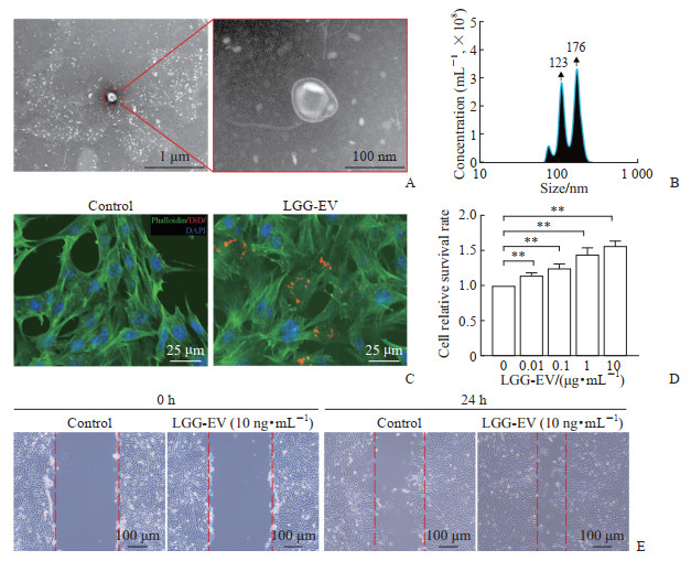 图 1 LGG-EV的形态与功能特征Fig. 1 Morphological and functional characteristics of LGG-EVsA: Morphology of LGG-EVs under transmission electron microscope; B: Nanoparticle tracking analysis of LGG-EVs; C: LGG-EVs can be taken up by BMSCs (immunofluorescence staining); D: LGG-EVs promote the proliferation of BMSCs (CCK-8 assay); E: LGG-EVs promote the migration of BMSCs (wound healing assay). **P<0.01. n=6, x±s. LGG-EV: Lactobacillus rhamnosus GG-derived extracellular vesicle; BMSC: Bone marrow mesenchymal stem cell; CCK-8: Cell counting kit 8; DiD: 1,1'-dioctadecyl-3,3,3',3'-tetramethylindodicarbocyanine perchlorate; DAPI: 4',6-diamidino-2-phenylindole.
 下载:
全尺寸图片
2.2 LGG-EV的内含物分析
基于课题组前期对LGG-EV中miRNA的分析,进一步通过KEGG富集分析筛选出前20条显著富集的信号通路,这些通路涵盖多条与细胞分化、增殖、迁移及基质合成密切相关的关键途径(图 2A)。结合促软骨分化相关研究[15-25],这些信号通路在调控BMSC软骨分化及软骨基质形成中发挥重要作用(表 1)。GO功能富集分析显示,LGG-EV富集于调控细胞增殖、迁移、信号转导及细胞外基质相关的生物过程(图 2B~2D)。上述结果表明,LGG-EV具备促BMSC软骨分化的潜力。
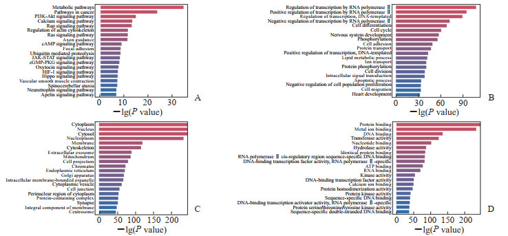 图 2 LGG-EV中miRNA靶基因的KEGG通路富集分析及GO功能注释Fig. 2 KEGG pathway enrichment analysis and GO functional annotation of miRNA target genes in LGG-EVsA: Top 20 of KEGG enrichment; B: Top 20 of GO biological process enrichment; C: Top 20 of GO cellular component enrichment; D: Top 20 of GO molecular function enrichment. LGG-EV: Lactobacillus rhamnosus GG-derived extracellular vesicle; miRNA: MicroRNA; KEGG: Kyoto Encyclopedia of Genes and Genomes; GO: Gene Ontology; PI3K: Phosphatidylinositol 3 kinase; Akt: Protein kinase B; cAMP: Cyclic adenosine monophosphate; JAK: Janus kinase; STAT: Signal transducer and activator of transcription; cGMP: Cyclic guanosine monophosphate; PKG: Protein kinase G; HIF-1: Hypoxia-inducible factor 1; ATP: Adenosine triphosphate.下载:
全尺寸图片
表 1 LGG-EV中miRNA靶基因富集的KEGG通路及其在软骨生成调控中的功能Table 1 KEGG pathways enriched by miRNA target genes in LGG-EVs and their functions in regulation of chondrogenesis
Pathway Inductive effect on BMSC differentiation into chondrocytes Reference PI3K-Akt pathway Promotes cell survival, proliferation, and differentiation [15-17] Calcium signaling pathway Regulates intracellular calcium ion concentration, influencing many downstream signals [18] Ras signaling pathway Regulates cell growth and differentiation [19] cAMP signaling pathway Regulates protein kinase A activity and transcription factors [20] Focal adhesion Connects cells to the extracellular matrix and regulates mechanical signal transduction [21-22] Ubiquitin mediated proteolysis Regulates protein degradation to maintain cellular protein homeostasis [23] HIF-1 signaling pathway Regulates gene expression to adapt to hypoxic (low oxygen) environments [24] Hippo signaling pathway Regulates cell proliferation and apoptosis [18] Apelin signaling pathway Regulates angiogenesis and cell metabolism [25] LGG-EV: Lactobacillus rhamnosus GG-derived extracellular vesicle; miRNA: MicroRNA; KEGG: Kyoto Encyclopedia of Genes and Genomes; PI3K: Phosphatidylinositol 3 kinase; Akt: Protein kinase B; cAMP: Cyclic adenosine monophosphate; HIF-1: Hypoxia inducible factor 1. 2.3 LGG-EV促BMSC软骨分化
采用LGG-EV替代TGF-β3添加至促软骨分化培养基中(图 3A)。生长培养基组(未添加诱导因子)未能形成细胞球,而添加TGF-β3和LGG-EV的促软骨分化培养基组均在离心管底部形成了形态光滑、结构完整的细胞球(图 3B)。阿利新蓝染色结果显示,形成的细胞球内富含糖胺聚糖成分(图 3C)。免疫荧光检测进一步证明,细胞球内表达软骨转录因子SOX9及软骨特异性基质蛋白COL-Ⅱ(图 3D)。以上结果表明,LGG-EV作为促分化因子能够促进BMSC软骨分化。
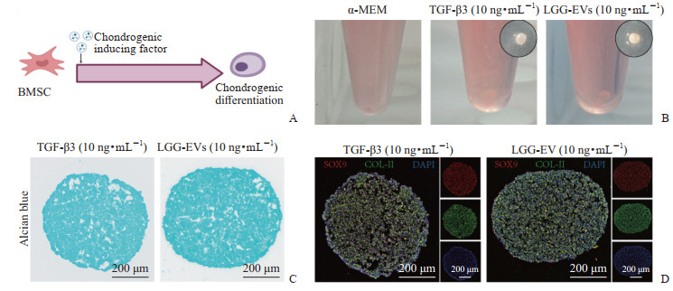 图 3 LGG-EV促进BMSC软骨分化Fig. 3 Chondrogenic differentiation of BMSCs promoted by LGG-EVsA: Schematic diagram of BMSC chondrogenic differentiation; B: Photographs of BMSC chondrogenic induction for 14 d; C: Alcian blue staining (14 d); D: Immunofluorescence staining of SOX9 and COL-Ⅱ (14 d). LGG-EV: Lactobacillus rhamnosus GG-derived extracellular vesicle; BMSC: Bone marrow mesenchymal stem cell; SOX9: Sex-determining region Y box 9 (a chondrogenic transcription factor); COL-Ⅱ: Collagen type Ⅱ (a cartilage extracellular matrix protein); α-MEM: α-minimum essential medium; TGF-β3: Transforming growth factor β3 (a chondrogenic induction factor).下载:
全尺寸图片
2.4 LGG-EV诱导软骨类器官构建
将BMSC与GelMA溶液均匀混合,采用单分散微球制造仪制备出形态规整、尺寸均一的微球(图 4A)。显微镜观察可见细胞在凝胶网络内呈均匀三维分布(图 4B)。利用含有LGG-EV的促软骨分化培养基持续培养14 d,活细胞/死细胞染色结果显示细胞存活率高,细胞形态健康、铺展良好(图 4C、4D)。免疫荧光染色结果显示,微球中的细胞表达软骨相关蛋白SOX9和COL-Ⅱ(图 4E)。上述结果提示,基于BMSC和GelMA制备的微球为构建软骨类器官提供了细胞载体和三维培养平台。
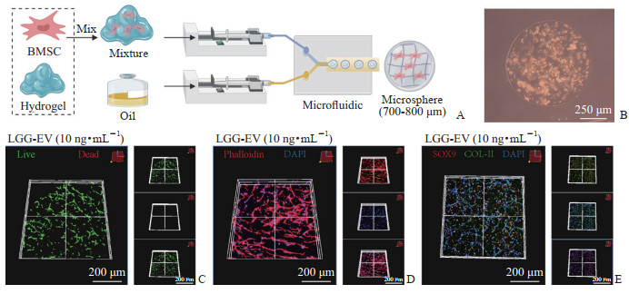 图 4 LGG-EV诱导BMSC构建软骨类器官Fig. 4 LGG-EVs induce the formation of chondrogenic organoids from BMSCsA: Schematic diagram of cartilage organoids fabricated using monodisperse microspheres; B: Actual image of the cartilage organoids; C: Live/dead staining (14 d); D: Cytoskeleton staining (14 d); E: Immunofluorescence staining of SOX9 and COL-Ⅱ (14 d). LGG-EV: Lactobacillus rhamnosus GG-derived extracellular vesicle; BMSC: Bone marrow mesenchymal stem cell; SOX9: Sex-determining region Y box 9 (a chondrogenic transcription factor); COL-Ⅱ: Collagen type Ⅱ (a cartilage extracellular matrix protein); DAPI: 4',6-diamidino-2-phenylindole.下载:
全尺寸图片
3 讨论
软骨组织自我修复能力有限,关节软骨损伤导致的软骨缺损通常不可逆[26]。当前临床主要采用自体软骨移植、微骨折术和生物材料填充进行修复:自体软骨移植可避免免疫排斥反应,修复组织接近天然软骨;微骨折术通过刺破骨面,刺激BMSC促进软骨再生,适用于小范围缺损;生物材料提供三维支架,支持细胞附着和组织生长,具有设计灵活、可调控降解速度和力学性能的优势[27-28]。这些方法在一定程度上缓解了患者症状,但由于供体资源有限、修复软骨质量难以保障、术后软骨退变以及结构和功能无法完全恢复等限制,目前难以实现理想的长期修复效果。此外,软骨具有复杂的层次结构和多样的生物学功能,如何构建既具稳定结构又具良好生物学功能的可移植软骨组织,同时结合患者的个体差异实现精准治疗,依然是软骨修复和骨关节炎治疗领域面临的重大挑战[5]。
软骨类器官通过三维立体培养技术模拟软骨组织的复杂结构和微环境,能够真实再现软骨生物学特性和功能状态,为软骨缺损的研究和治疗提供了新突破口[29-30]。其构建关键在于如何精准再现软骨细胞及相应的细胞外基质,并建立类似天然软骨的三维结构以促进细胞分化与功能表达。现有诱导因子普遍存在效率低、细胞毒性及免疫排斥等问题,限制了软骨类器官的发展。本研究利用LGG-EV作为诱导因子促进BMSC软骨分化,构建软骨类器官。miRNA测序结果显示LGG-EV富集多条与软骨生成相关的信号通路;透射电子显微镜和纳米颗粒追踪分析仪表征证实从菌液中提取了LGG-EV;共培养实验表明LGG-EV能被BMSC摄取,具有良好生物相容性和促增殖能力;阿利新蓝染色和免疫荧光染色检测SOX9和COL-Ⅱ证实了LGG-EV的促BMSC软骨分化能力;最后结合GelMA及单分散微球技术,成功构建了软骨类器官。
本研究提出的软骨类器官构建方法与目前已有的软骨类器官构建方法相比具有以下优势:(1)LGG-EV作为天然纳米囊泡,结构稳定,富含多样化成软骨分化信号分子,可激活BMSC关键信号通路,促进精准定向分化及软骨组织再生;(2)LGG-EV提取工艺简便、来源广泛且成本低,具备良好规模化生产潜力,适合临床推广;(3)LGG-EV源自益生菌,免疫原性低,显著降低了免疫排斥风险,提升了组织工程的安全性和耐受性;(4)LGG-EV具备优异的生物相容性与抗炎作用,可调控炎症微环境,缓解慢性炎症对软骨修复的抑制[14]。基于上述优势,本研究采用LGG-EV作为促软骨分化因子成功构建软骨类器官,为软骨组织工程和再生医学研究提供了重要的理论支持和技术创新思路。
本研究还存在一定的局限性,需在后续实验中进一步完善:(1)分化因子使用浓度。过度使用促软骨分化因子会引发异常软骨内骨化现象,进而影响软骨组织稳定性和功能。因此,后续实验需对分化因子用量和作用时间进行调控,以进一步提升软骨类器官构建质量。(2)分化时间。虽然本研究在体外成功构建并验证了软骨类器官的部分功能,但其体内修复效果及长期生物学行为尚未充分评估,最佳诱导时间及移植效果需通过动物实验和临床前研究进一步明确。(3)尺寸限制。目前构建的软骨类器官尺寸较小,在小尺寸条件下可以维持较好的营养供应和废物排出,但大尺寸类器官因缺乏血管系统和养分输送机制,长期培养与功能维持面临较大挑战。如何突破大尺寸软骨类器官的培养瓶颈,实现稳定、长效的组织生长和功能保持,是未来研究的重要方向。(4)软骨微环境复杂性。自然软骨组织的微环境不仅由软骨细胞和细胞外基质组成,还包含多种细胞(如成骨细胞、免疫细胞、血管内皮细胞)以及细胞因子、机械刺激等多重因素的共同作用。目前构建的类器官难以充分模拟这一高度复杂和动态变化的软骨微环境,未来需结合更多生物材料和微环境调控技术进一步完善类器官的仿生度和功能表现。
软骨类器官的研究仍处于起步阶段,未来研究应聚焦于精细复制软骨组织结构,并整合软骨微环境中多种细胞成分及物理化学因子的协同作用,如免疫细胞、成骨细胞以及力学刺激等,以提升软骨类器官的结构复杂性和功能完整性[31]。随着生物三维打印、生物材料科学、人工智能等多项前沿技术的不断进步,对软骨组织进行高精度扫描和数字化建模成为可能,实现了体外精准构建与天然软骨相似的三维微结构,显著提升了软骨类器官的空间分辨率和仿生性能[32]。此外,结合微流控芯片技术与生物学元件,类器官芯片作为模拟软骨微环境的创新平台得以快速发展[33]。软骨类器官芯片能够在微流控芯片上集成多种软骨相关细胞、细胞外基质及生理性机械刺激,成功复制生物体内复杂多变的软骨微环境。随着多学科技术的深度融合与跨界创新,软骨类器官及芯片技术正在不断完善,并将极大拓展其在骨关节炎及其他软骨相关疾病机制研究与临床转化中的应用潜力。相信在不久的将来,软骨类器官的发展与应用将为骨关节炎的发病机制解析、药物筛选及精准治疗提供强有力的技术支撑与理论基础。
-
图 1 LGG-EV的形态与功能特征
Fig. 1 Morphological and functional characteristics of LGG-EVs
A: Morphology of LGG-EVs under transmission electron microscope; B: Nanoparticle tracking analysis of LGG-EVs; C: LGG-EVs can be taken up by BMSCs (immunofluorescence staining); D: LGG-EVs promote the proliferation of BMSCs (CCK-8 assay); E: LGG-EVs promote the migration of BMSCs (wound healing assay). **P<0.01. n=6, x±s. LGG-EV: Lactobacillus rhamnosus GG-derived extracellular vesicle; BMSC: Bone marrow mesenchymal stem cell; CCK-8: Cell counting kit 8; DiD: 1,1'-dioctadecyl-3,3,3',3'-tetramethylindodicarbocyanine perchlorate; DAPI: 4',6-diamidino-2-phenylindole.
下载:
全尺寸图片
图 2 LGG-EV中miRNA靶基因的KEGG通路富集分析及GO功能注释
Fig. 2 KEGG pathway enrichment analysis and GO functional annotation of miRNA target genes in LGG-EVs
A: Top 20 of KEGG enrichment; B: Top 20 of GO biological process enrichment; C: Top 20 of GO cellular component enrichment; D: Top 20 of GO molecular function enrichment. LGG-EV: Lactobacillus rhamnosus GG-derived extracellular vesicle; miRNA: MicroRNA; KEGG: Kyoto Encyclopedia of Genes and Genomes; GO: Gene Ontology; PI3K: Phosphatidylinositol 3 kinase; Akt: Protein kinase B; cAMP: Cyclic adenosine monophosphate; JAK: Janus kinase; STAT: Signal transducer and activator of transcription; cGMP: Cyclic guanosine monophosphate; PKG: Protein kinase G; HIF-1: Hypoxia-inducible factor 1; ATP: Adenosine triphosphate.
下载:
全尺寸图片
图 3 LGG-EV促进BMSC软骨分化
Fig. 3 Chondrogenic differentiation of BMSCs promoted by LGG-EVs
A: Schematic diagram of BMSC chondrogenic differentiation; B: Photographs of BMSC chondrogenic induction for 14 d; C: Alcian blue staining (14 d); D: Immunofluorescence staining of SOX9 and COL-Ⅱ (14 d). LGG-EV: Lactobacillus rhamnosus GG-derived extracellular vesicle; BMSC: Bone marrow mesenchymal stem cell; SOX9: Sex-determining region Y box 9 (a chondrogenic transcription factor); COL-Ⅱ: Collagen type Ⅱ (a cartilage extracellular matrix protein); α-MEM: α-minimum essential medium; TGF-β3: Transforming growth factor β3 (a chondrogenic induction factor).
下载:
全尺寸图片
图 4 LGG-EV诱导BMSC构建软骨类器官
Fig. 4 LGG-EVs induce the formation of chondrogenic organoids from BMSCs
A: Schematic diagram of cartilage organoids fabricated using monodisperse microspheres; B: Actual image of the cartilage organoids; C: Live/dead staining (14 d); D: Cytoskeleton staining (14 d); E: Immunofluorescence staining of SOX9 and COL-Ⅱ (14 d). LGG-EV: Lactobacillus rhamnosus GG-derived extracellular vesicle; BMSC: Bone marrow mesenchymal stem cell; SOX9: Sex-determining region Y box 9 (a chondrogenic transcription factor); COL-Ⅱ: Collagen type Ⅱ (a cartilage extracellular matrix protein); DAPI: 4',6-diamidino-2-phenylindole.
下载:
全尺寸图片
表 1 LGG-EV中miRNA靶基因富集的KEGG通路及其在软骨生成调控中的功能
Table 1 KEGG pathways enriched by miRNA target genes in LGG-EVs and their functions in regulation of chondrogenesis
Pathway Inductive effect on BMSC differentiation into chondrocytes Reference PI3K-Akt pathway Promotes cell survival, proliferation, and differentiation [15-17] Calcium signaling pathway Regulates intracellular calcium ion concentration, influencing many downstream signals [18] Ras signaling pathway Regulates cell growth and differentiation [19] cAMP signaling pathway Regulates protein kinase A activity and transcription factors [20] Focal adhesion Connects cells to the extracellular matrix and regulates mechanical signal transduction [21-22] Ubiquitin mediated proteolysis Regulates protein degradation to maintain cellular protein homeostasis [23] HIF-1 signaling pathway Regulates gene expression to adapt to hypoxic (low oxygen) environments [24] Hippo signaling pathway Regulates cell proliferation and apoptosis [18] Apelin signaling pathway Regulates angiogenesis and cell metabolism [25] LGG-EV: Lactobacillus rhamnosus GG-derived extracellular vesicle; miRNA: MicroRNA; KEGG: Kyoto Encyclopedia of Genes and Genomes; PI3K: Phosphatidylinositol 3 kinase; Akt: Protein kinase B; cAMP: Cyclic adenosine monophosphate; HIF-1: Hypoxia inducible factor 1. -
[1] Tang S, Zhang C, Oo W M, et al. Osteoarthritis[J]. Nat Rev Dis Primers, 2025, 11(1):10. DOI: 10.1038/s41572-025-00594-6. [2] Halabitska I, Petakh P, Kamyshnyi O. Metformin as a disease-modifying therapy in osteoarthritis: bridging metabolism and joint health[J]. Front Pharmacol, 2025, 16: 1567544. DOI: 10.3389/fphar.2025.1567544. [3] Fuggle N, Laslop A, Rizzoli R, et al. Treatment of osteoporosis and osteoarthritis in the oldest old[J]. Drugs, 2025, 85(3): 343-360. DOI: 10.1007/s40265-024-02138-w. [4] Chen Y, Sun W, Wen Y, et al. A cationic polymer drives glycosaminoglycan assembly and secretion for preclinical osteoarthritis therapy[J]. Sci Transl Med, 2025, 17(804): eadl5623. DOI: 10.1126/scitranslmed.adl5623. [5] Lu J, Gao Y, Cao C, et al. 3D bioprinted scaffolds for osteochondral regeneration: advancements and applications[J]. Mater Today Bio, 2025, 32: 101834. DOI: 10.1016/j.mtbio.2025.101834. [6] Kaibara T, Kondo E, Sotome A, et al. A 10-year follow-up of autogenous osteochondral transplantation combined with medial opening-wedge high tibial osteotomy for large medial femoral condyle chondral delamination: a case report[J]. Jt Dis Relat Surg, 2025, 36(2): 444-455. DOI: 10.52312/jdrs.2025.2215. [7] Li D, Liu F, Hou Y, et al. Changes in periprosthetic bone mineral density following arthroplasty: an in-depth review and current perspectives[J]. Curr Osteoporos Rep, 2025, 23(1): 30. DOI: 10.1007/s11914-025-00921-6. [8] Wang Q, Mu M, Yu W, et al. Engineering polyphenol-based osteogenic system for bone and cartilage repair: transplantation, tissue engineering, and organoid[J]. J Adv Res, 2026, 82: 1027-1048. DOI: 10.1016/j.jare.2025.06.067. [9] Chen Z, Zhang H, Huang J, et al. DNA-encoded dynamic hydrogels for 3D bioprinted cartilage organoids[J]. Mater Today Bio, 2025, 31: 101509. DOI: 10.1016/j.mtbio.2025.101509. [10] Ripamonti U. Global morphogenesis regulating tissue architecture and organogenesis[J]. Biomater Adv, 2025, 172: 214262. DOI: 10.1016/j.bioadv.2025.214262. [11] Soni N, Niranjane P, Purohit A. Role of growth factors in nasal cartilage development and molding: a comprehensive review[J]. Cureus, 2024, 16(8): e67202. DOI: 10.7759/cureus.67202. [12] Wu M, Wu S, Chen W, et al. The roles and regulatory mechanisms of TGF-β and BMP signaling in bone and cartilage development, homeostasis and disease[J]. Cell Res, 2024, 34(2): 101-123. DOI: 10.1038/s41422-023-00918-9. [13] Niu L, Chen W, Yin Z, et al. Bacterial extracellular vesicles in osteoarthritis: a new bridge of the gut-joint axis[J]. Gut Microbes, 2025, 17(1): 2489069. DOI: 10.1080/19490976.2025.2489069. [14] Liu H, Wu Y, Wang F, et al. Bone-targeted engineered bacterial extracellular vesicles delivering miRNA to treat osteoporosis[J]. Compos Part B Eng, 2023, 267: 111047. DOI: 10.1016/j.compositesb.2023.111047. [15] Gao C, Dai W, Liu D, et al. Adaptive hydrogel loaded with pre-coordinated stem cells for enhanced osteoarthritis therapy[J]. Bioact Mater, 2025, 51: 613-633. DOI: 10.1016/j.bioactmat.2025.05.018. [16] Huang C, Zhong G, Xiao J, et al. An injectable kartogenin-incorporated hydrogel supports mesenchymal stem cells for cartilage tissue engineering[J]. Bioengineering (Basel), 2025, 12(5): 434. DOI: 10.3390/bioengineering12050434. [17] Wang M, Lian J, Ye M, et al. Pain mediator NGF improves chondrocyte extracellular matrix synthesis via PI3K/AKT pathway[J]. J Orthop Surg Res, 2025, 20(1): 207. DOI: 10.1186/s13018-025-05503-x. [18] Ying J, Wang P, Zhang S, et al. Transforming growth factor-beta1 promotes articular cartilage repair through canonical Smad and Hippo pathways in bone mesenchymal stem cells[J]. Life Sci, 2018, 192: 84-90. DOI: 10.1016/j.lfs.2017.11.028. [19] He Y, Zhu W, Shin M H, et al. cFOS-SOX9 axis reprograms bone marrow-derived mesenchymal stem cells into chondroblastic osteosarcoma[J]. Stem Cell Reports, 2017, 8(6): 1630-1644. DOI: 10.1016/j.stemcr.2017.04.029. [20] Chuang S C, Chen C H, Chou Y S, et al. G protein-coupled estrogen receptor mediates cell proliferation through the cAMP/PKA/CREB pathway in murine bone marrow mesenchymal stem cells[J]. Int J Mol Sci, 2020, 21(18): 6490. DOI: 10.3390/ijms21186490. [21] Zhou C, Wang Q, Zhang D, et al. Compliant substratum modulates vinculin expression in focal adhesion plaques in skeletal cells[J]. Int J Oral Sci, 2019, 11(2): 18. DOI: 10.1038/s41368-019-0052-3. [22] Reesink H L, Sutton R M, Shurer C R, et al. Galectin-1 and galectin-3 expression in equine mesenchymal stromal cells (MSCs), synovial fibroblasts and chondrocytes, and the effect of inflammation on MSC motility[J]. Stem Cell Res Ther, 2017, 8(1): 243. DOI: 10.1186/s13287-017-0691-2. [23] Liu Y, Molchanov V, Yang T. Enzymatic machinery of ubiquitin and ubiquitin-like modification systems in chondrocyte homeostasis and osteoarthritis[J]. Curr Rheumatol Rep, 2021, 23(8): 62. DOI: 10.1007/s11926-021-01022-w. [24] Jiang T, Heng S, Huang X, et al. Biomimetic poly(poly(ε-caprolactone)-polytetrahydrofuran urethane) based nanofibers enhanced chondrogenic differentiation and cartilage regeneration[J]. J Biomed Nanotechnol, 2019, 15(5): 1005-1017. DOI: 10.1166/jbn.2019.2748. [25] Şişli H B, Şenkal Turhan S, Bulut E, et al. The role of APLNR signaling in the developmental regulation of mesenchymal stem cell differentiation from human pluripotent stem cells[J]. Adv Biol, 2024, 8(1): e2300217. DOI: 10.1002/adbi.202300217. [26] Lou W, Qiu X, Qin Y, et al. 3D-printed advanced scaffold armed with exosomes derived from human skeletal stem cell identified by single-cell RNA sequencing enhances osteochondral regeneration[J]. Bioact Mater, 2025, 51: 231-256. DOI: 10.1016/j.bioactmat.2025.04.028. [27] Naranđa J, Bračič M, MAVER U, et al. Recent advancements in smart hydrogel-based materials in cartilage tissue engineering[J]. Materials (Basel), 2025, 18(11): 2576. DOI: 10.3390/ma18112576. [28] Wang Z, Xu J, Zhu J, et al. Osteochondral tissue engineering: scaffold materials, fabrication techniques and applications[J]. Biotechnol J, 2025, 20(1): e202400699. DOI: 10.1002/biot.202400699. [29] Shen C, Wang J, Li G, et al. Boosting cartilage repair with silk fibroin-DNA hydrogel-based cartilage organoid precursor[J]. Bioact Mater, 2024, 35: 429-444. DOI: 10.1016/j.bioactmat.2024.02.016. [30] Zhou Z, Cui J, Wu S, et al. Silk fibroin-based biomaterials for cartilage/osteochondral repair[J]. Theranostics, 2022, 12(11): 5103-5124. DOI: 10.7150/thno.74548. [31] Wang T, Chen Y, Zhu X, et al. IFT80 and TRPA1 cooperatively regulate bone formation by calcium signaling in response to mechanical stimuli[J]. Metabolism, 2025, 166: 156159. DOI: 10.1016/j.metabol.2025.156159. [32] Halwes M, Vidler C, Aguilar L C, et al. Tuneable hydrogel porosity via dynamic tailoring of spinodal decomposition[J]. Adv Sci (Weinh), 2025, 12(35): e04265. DOI: 10.1002/advs.202504265. [33] Ashok D, Singh J, Howard H R, et al. Interfacial engineering for biomolecule immobilisation in microfluidic devices[J]. Biomaterials, 2025, 316: 123014. DOI: 10.1016/j.biomaterials.2024.123014.
