 2016, Vol. 43
文章信息
- 胃腺癌组织中LRIG1蛋白的表达及意义
- Expression and Significance of LRIG1 Protein in Gastric Adenocarcinoma Tissues
- 肿瘤防治研究, 2016, 43(6): 489-491
- Cancer Research on Prevention and Treatment, 2016, 43(6): 489-491
- http://www.zlfzyj.com/CN/10.3971/j.issn.1000-8578.2016.06.011
- 收稿日期: 2015-09-14
- 修回日期: 2015-11-20
引用本文 |


胃癌是常见的消化道恶性肿瘤,其发病率在我国居于各种肿瘤首位,严重威胁人类健康[1]。胃癌的发生发展涉及多种基因和信号通路的改变,但其发病机制尚不明确。富亮氨酸重复序列免疫球蛋白样结构域1(tandem leucine-rich repeats andimmunoglobulin-like domains-1,LRIG1)是由Nilsson等[2]在研究EGFR负反馈环路中发现的。相关研究证实,LRIG1基因在人类的多种组织中都有表达,但在各种肿瘤组织中的表达却不尽相同,在大多数肿瘤组织中表达下调,也有的比正常组织表达高[3]。但是,关于LRIG1基因在胃癌中表达的相关文献却很少。本研究采用免疫组织化学EnVision二步法检测116例胃腺癌和相应癌旁正常胃组织中LRIG1的表达情况,并结合临床病理参数进行分析,为探讨LRIG1基因在胃癌发生发展中的可能作用提供依据。
1 资料与方法 1.1 组织标本116例胃腺癌及相应癌旁正常胃组织标本(距肿瘤边缘5 cm以外)全部取自辽宁医学院附属第一医院2013年7月—2015年1月接受胃大部分切除术的住院患者,其中高分化16例,中分化42例,低分化58例;男78例,女38例;年龄45~83岁,中位年龄61岁。所有患者在术前均未接受放疗、化疗及免疫治疗。
1.2 试剂兔抗人LRIG1多克隆抗体(英国Abcam公司);PV-9000试剂盒(北京中杉金桥生物技术有限公司);DAB显色剂(北京中杉金桥生物技术有限公司)。
1.3 方法 1.3.1 免疫组织化学病理切片于60℃恒温干燥箱中烤片2 h,常规脱蜡、水化,蒸馏水漂洗5 min,PBS漂洗4 min,3次;切片置于煮沸的pH6.0枸橼酸盐缓冲液中进行高压抗原修复2 min,室温冷却后PBS漂洗4 min,3次;3%H2O2室温封闭10 min,PBS漂洗4 min,3次;滴加兔抗人LRIG1多克隆抗体(稀释浓度1:400),4℃过夜;次日室温放置1 h,PBS漂洗4 min,3次;滴加二抗增强剂,37℃孵育30 min,PBS漂洗4 min,3次;滴加二抗PV-9000,37℃孵育30 min,PBS漂洗4 min,3次;DAB显色,苏木精对比染色5 min,分化3~5 s,脱水、透明、中性树胶封片。显微镜下拍照观察结果。
1.3.2 结果判定标准LRIG1蛋白主要定位于细胞质。参考相关文献[4]采用染色范围与染色强度双重判定法。染色范围:每张切片随机选取5个高倍视野,计数阳性细胞数占同类细胞数的百分比,<5%计0分,5%~25%计1分,>25%~50%计2分,>50%计3分。染色强度:无染色计0分,淡黄色计1分,棕黄色计2分,棕褐色计3分。总积分=染色范围×染色强度。结果判断:总积分0~1分为阴性,>1分为阳性。
1.4 统计学方法采用SPSS17.0统计学软件对实验数据进行统计分析,免疫组织化学结果采用四格表资料的χ2检验,计算结果以P<0.05为差异有统计学意义。
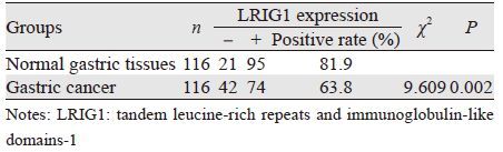
2 结果 2.1 LRIG1在胃腺癌及癌旁正常胃组织中的表达LRIG1蛋白主要表达于细胞质,在癌旁正常胃组织中的阳性表达率为81.9%(95/116),在胃腺癌组织中的阳性表达率为63.8%(74/116),明显低于正常胃组织,两者差异具有统计学意义(P<0.05),见表 1、图 1。
|

|
| A: LRIG1 expression in adjacent normal gastric tissues; B: LRIG1 expression in well-differentiated gastric adenocarcinoma tissues; C: LRIG1 expression in moderately differentiated gastric adenocarcinoma tissues; D: LRIG1 expression in poorly differentiated gastric adenocarcinoma tissues 图 1 LRIG1在胃腺癌及癌旁正常胃组织中的表达(DAB染色 ×400) Figure 1 LRIG1 expression in gastric adenocarcinoma and normal gastric tissues(DAB staining ×400) |
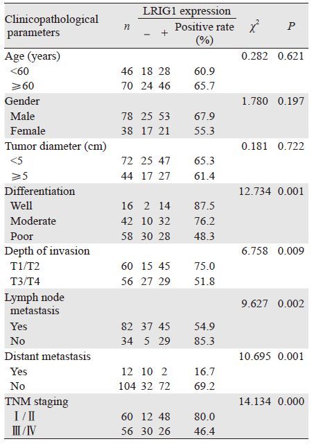
高分化组胃腺癌组织中LRIG1的阳性表达率高于中分化组,中分化组胃腺癌组织中LRIG1的阳性表达率高于低分化组;浸润深度方面,未侵及浆膜组胃腺癌组织中LRIG1的阳性表达率高于侵及浆膜组;淋巴结及远处转移方面,淋巴结和远处转移阳性组胃腺癌LRIG1的阳性表达率低于淋巴结及远处转移阴性组;TNM分期方面,Ⅰ/Ⅱ期组胃腺癌组织中LRIG1的阳性表达率高于Ⅲ/Ⅳ期组,差异均具有统计学意义(P<0.05)。LRIG1表达与患者年龄、性别及肿瘤大小无关(P>0.05),见表 2。
|
胃癌是我国发病率和死亡率最高的消化道恶性肿瘤,是我国重点防治癌症之一[5]。胃腺癌占所有胃恶性肿瘤的95%,是临床上发病率最高的胃恶性肿瘤,相对少见的是平滑肌肉瘤及胃淋巴癌[6]。胃腺癌是由胃腺体细胞恶化所致的,在胃黏膜癌变过程中常常涉及多种原癌基因的激活和抑癌基因的失活[7-8]。放化疗及外科手术等多元治疗手段对进展期胃癌仍不能很好的控制[9]。因此,从基因角度探讨胃癌发生发展的规律,明确胃癌发生发展过程中关键基因的激活或抑制状态并对其进行检测,有助于胃癌的早期诊断、治疗及预后判断[10-12]。LRIG1基因是新发现的一种候选抑癌基因,定位于人类染色体3p14.3,序列全长4 763bp,编码1 093个氨基酸。它编码的产物为一类跨膜糖蛋白,在人体各种组织中的表达不尽相同,在脑部表达最高,脾脏最低[13]。LRIG1在人类各种肿瘤组织中的表达亦不尽相同,在大多数肿瘤组织中表达下调,也有的比正常组织表达高[3]。Thomasson等[14]检测了LRIG1在肾癌中的表达,结果显示,与正常肾组织相比,LRIG1在肾癌中的表达显著降低。Hedman等[13]分析了LRIG1 mRNA在P31间皮瘤、PC-3前列腺癌和DLD-1结肠腺癌三种细胞系中的表达,发现在这三种细胞系中LRIG1的表达均明显下调,甚至在DLD-1中没有任何表达。而Bhattacharjee等[15]在白血病中检测到LRIG1表达上调,与大多数肿瘤组织中LRIG1表达下调或缺失不同。目前LRIG1在胃癌中表达的研究较少,其具体作用目前尚无定论。
本研究结果显示,LRIG1在胃腺癌组织中的表达显著低于相应癌旁正常胃组织,而且LRIG1在胃腺癌组织中的表达与肿瘤的分化程度、浸润深度、淋巴结转移、远处转移及临床分期有关,而与患者年龄、性别及肿瘤大小无关。此结果与之前国内外报道的LRIG1在大多数肿瘤中表达下调或缺失的结果相符。本研究的结果提示,LRIG1表达可能抑制胃腺癌的发生。本课题组前期的工作已经证实,LRIG1 mRNA在胃癌组织中的表达明显低于癌旁正常胃组织,LRIG1在胃癌发生发展中可能起着抑癌基因的作用[16]。我们在后续工作中将继续深入研究LRIG1基因在胃腺癌中表达下调的具体机制,为了解胃腺癌的发生发展机制及基因靶向治疗提供更多有力的证据。
| [1] | Liu JY, Chang JC, Liu Y. Expression of Cdc42 and β1 integrin and their clinical significance in gastric cancer[J]. Lin Chuang Yu Shi Yan Bing Li Xue Za Zhi, 2014, 30 (6) : 618–21. [刘家园, 常家聪, 刘弋. 胃癌组织中Cdc42、β1整合素的表达及临床意义[J]. 临床与实验病理学杂志,2014, 30 (6) : 618–21. ] |
| [2] | Nilsson J, Vallbo C, Guo D, et al. Cloning,characterization,and expression of human LIG1[J]. Biochem Biophys Res Commun, 2001, 284 (5) : 1155–61. |
| [3] | Xing XH, Wang XW, Guo DS. The relationship between LRIG1 gene and tumor[J]. Guangdong Yi Xue, 2010, 31 (23) : 3141–3. [邢细红, 王雄伟, 郭东升. LRIG1基因与肿瘤的关系[J]. 广东医学,2010, 31 (23) : 3141–3. ] |
| [4] | Skomedal H, Kristensen GB, Lie AK, et al. Aberrant expression of the cell cycle associated proteins TP53, MDM2, P21, P27, cdk4, cyclinD1, RB, and EGFR in cervical carcinomas[J]. Gynecol Oncol, 1999, 73 (2) : 223–8. |
| [5] | Bang YJ, Kim YW, Yang HK, et al. Adjuvant capecitabine and oxaliplatin for gastric cancer after D2gas-trectomy(CLASSIC): a phase 3 open-label randomized controlled trial[J]. Lancet, 2012, 379 (9813) : 315–21. |
| [6] | Sun M. The Expression and Its Significance of AGR2, p53, GST-π in Chronic Atrophic Gastritis and Gastric Adenocarcinoma[J]. Yi Xue Li Lun Yu Shi Jian, 2015, 28 (5) : 566–8. [孙敏. AGR2、 p53、GST-π在慢性萎缩性胃炎和胃腺癌中的表达及其意义[J]. 医学理论与实践,2015, 28 (5) : 566–8. ] |
| [7] | Shi YX, Su XL. p53, PUMA and MCL-1 Regulation of Apoptotic Pathway[J]. Yi Xue Zong Shu, 2014, 20 (11) : 1923–6. [师迎旭, 苏秀兰. p53、PUMA、MCL-1对于凋亡途径的调控[J]. 医学综述,2014, 20 (11) : 1923–6. ] |
| [8] | Robles AI, Harris CC. Clinical outcomes and correlates of TP53 mutations and cancer[J]. Cold Spring Harb Perspect Biol, 2010, 2 (3) : a001016. |
| [9] | Ren XF, Wang J, Li PF, et al. Relationship between VDR genetic polymorphisms,environmental risk factors and gastric cancer susceptibility:a case-control study[J]. Zhongguo Gong Gong Wei Sheng, 2014, 30 (7) : 948–51. [任晓峰, 王佳, 李鹏飞, 等. 维生素D受体基因VDR与胃癌易感性关系[J]. 中国公共卫生,2014, 30 (7) : 948–51. ] |
| [10] | Sempere LF. Integrating contextual miRNA and protein signatures for diagnostic and treatment decisions in cancer[J]. Expert Rev Mol Diagn, 2011, 11 (8) : 813–27. |
| [11] | Chen G, Lu XF. The Expression of PCNA, COX-2, p53 and EGFR in gastric cancer and Its relationship with prognosis[J]. Shandong Yi Yao, 2012, 52 (41) : 39–41. [陈刚, 陆晓峰. 胃癌组织中PCNA、COX-2、p53及EGFR的表达及其与预后的关系[J]. 山东医药,2012, 52 (41) : 39–41. ] |
| [12] | Zhang GY, Tian XL, Zhong Li, et al. Clinical Significance of microRNA-93 Expression in Gastric Tumor[J]. Zhong Liu Fang Zhi Yan Jiu, 2013, 40 (5) : 447–50. [张广钰, 田小林, 钟漓, 等. microRNA-93在胃癌中的表达及其临床意义[J]. 肿瘤防治研究,2013, 40 (5) : 447–50. ] |
| [13] | Hedman H, Nilsson J, Guo D, et al. Is LRIG1 a tumour suppressor gene at chromosome 3p14.3?[J]. Acta Oncol, 2002, 41 (4) : 352–4. |
| [14] | Thomasson M, Hedman H, Guo D, et al. LRIG1 and epidermal growth factor receptor in renal cell carcinoma:a quantitative RTPCR and immunohistochemical analysis[J]. Br J Cancer, 2003, 89 (7) : 1285–9. |
| [15] | Bhattacharjee A, Richards WG, Staunton J, et al. Classification of human lung carcinomas by mRNA expression profiling reveals distinct adenocarcinoma subclasses[J]. Proc Natl Acad Sci U S A, 2001, 98 (24) : 13790–5. |
| [16] | Dong C, Li H. Expression of Lrig1 and EGFR in gastric cancer tissue[J]. Zhongguo Gong Gong Wei Sheng, 2015, 30 (7) : 1–4. [董晨, 李红. 胃癌组织中Lrig1与EGFR基因表达[J]. 中国公共卫生,2015, 30 (7) : 1–4. ] |