文章信息
- 李晓莹, 赵玉虹, 郝一文
- LI Xiaoying, ZHAO Yuhong, HAO Yiwen
- 不同凝血指标在胃癌患者凝血功能中的应用评价
- Application assessment of different coagulation indexes in the coagulation function of gastric cancer patients
- 中国医科大学学报, 2021, 50(7): 602-607
- Journal of China Medical University, 2021, 50(7): 602-607
-
文章历史
- 收稿日期:2020-12-07
- 网络出版时间:2021-06-22 16:30
2. 中国医科大学附属第一医院输血科, 沈阳 110001
2. Department of Blood Transfusion, The First Hospital of China Medical University, Shenyang 110001, China
多数恶性肿瘤患者有明显的凝血功能异常,凝血功能异常与静脉血栓栓塞事件(venous thromboembolism,VTE)密切相关[1-2]。恶性肿瘤患者的凝血功能异常一般表现为血液高凝状态,其原因包括肿瘤自身的侵袭、转移等[3]。胃癌是最常见的恶性肿瘤之一,研究[4]表明,VTE的发生与胃癌的死亡率相关。目前,临床评价凝血功能最常用的指标是包含凝血酶时间的一系列指标。
血小板(platelet,PLT)计数可以在一定程度上反应凝血功能状态。然而,凝血是一个复杂的动态过程,是体内各种凝血成分和抗凝成分互相平衡的结果,常规凝血功能指标只能描述凝血过程的某个片段,无法全面描述。血栓弹力图(thromboela stogram,TEG)作为一种新的动态测量方法,能够全面地监测凝血过程[5]。因此,本研究采用常规凝血功能指标、PLT计数和TEG,探讨不同时期胃癌患者凝血功能状态的差异,并比较3种指标对胃癌患者高凝状态的检测价值。
1 材料与方法 1.1 研究对象查阅中国医科大学附属第一医院2018年7月1日至2019年10月31日胃癌患者的电子病历。所有纳入对象均为病历资料完整的新诊断胃癌患者,在常规凝血试验、PLT计数和TEG检测前,从未服用过抗凝药物,也从未进行过放化疗和手术治疗。这项研究排除了先前存在血液疾病或凝血障碍的患者。最终,165例患者纳入本研究,根据病理结果将其分为早期胃癌组(41例)和进展期胃癌组(124例)。
1.2 样本采集采集清晨空腹静脉血,采用3.2%枸橼酸钠1:9抗凝。
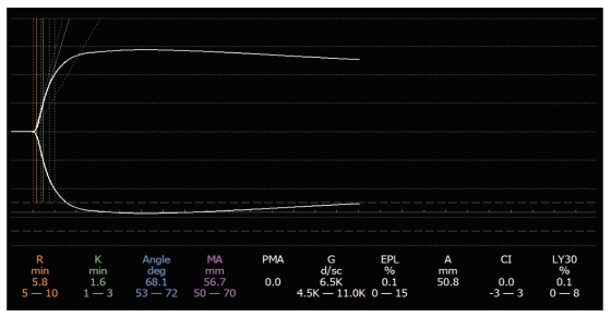
1.3 实验室检查采用全自动血凝分析仪检测常规凝血功能指标,包括凝血酶原时间(prothrombin time,PT,参考值范围为11~13.7 s)、活化部分凝血活酶时间(activated partial thromboplastin time,APTT,参考值范围为31.5~43.5 s)、凝血酶时间(thrombin time,TT,参考值范围为14~21 s)、国际标准化率(international standardized rate,INR,参考值范围为0.9~1.1)、纤维蛋白原(fibrinogen,Fib,参考值范围为2~4 g/L)和d -二聚体(参考值范围为0~0.5 μg/ml)。采用自动血液分析仪检测PLT计数(参考值范围为125×109~350×109/L)。采用TEG 5000型血栓弹力图仪(美国Heamoscope Inc公司)进行TEG分析,观察以下6个参数:凝血反应时间(reaction time,R,参考值范围为5~10 min)、凝血形成时间(K time,K,参考值范围为1~3 min)、凝固角(alpha角,参考值范围为53~72°)、最大振幅(maximum amplitude,MA,参考值范围为50~70 mm)、凝血指数(coagulation index,CI,参考值范围为-3~3)和MA确定后30 min纤维蛋白溶解率(lysis at 30 minutes,LY30,参考值范围为0%~8%)。这些指标的参考值范围参照仪器和试剂说明书。图 1展示了TEG的正常轨迹。根据先前的研究,在常规检测指标中,PT、APTT、TT、INR低于参考值范围,或PLT计数、Fib、d-二聚体高于参考值范围则提示存在高凝状态;在TEG检测中,R值、K值低于参考值范围,alpha角、MA值、CI值高于参考值范围,或LY30 < 0则提示存在高凝状态(以上任何一项指标出现如上异常则提示高凝状态) [6-8]。

|
| 图 1 TEG的正常轨迹 Fig.1 The normal trace of a TEG |
1.4 统计学分析
所有统计分析均采用SPSS 25.0软件进行,均为双侧检验。连续型变量根据分布情况,以x±s或M (P25~P75)来描述,采用t检验或Wilcoxon秩和检验进行组间比较。分类变量进行χ2检验。TEG参数与常规凝血功能指标、PLT计数根据分布情况进行Pearson’s或Spearman相关分析。P < 0.05为差异有统计学意义。
2 结果 2.1 研究对象基线特征本研究共纳入165例胃癌患者。根据病理结果将所有研究对象分为2组(早期胃癌组和进展期胃癌组)。早期胃癌患者41例,进展期胃癌患者124例。2组性别(P = 0.899)、年龄(P = 0.709)差异无统计学意义,肿瘤部位差异有统计学意义(P = 0.039)。肿瘤部位包括胃窦、胃体、胃角、胃底、贲门、幽门等,以胃窦、胃体为主。见表 1。
| Characteristics | Early gastric cancer group (n = 41) | Progressive gastric cancer group (n = 124) | P |
| Gender [n (%)] | 0.899 | ||
| Male | 27 (65.85) | 83 (66.94) | |
| Female | 14 (34.15) | 41 (33.06) | |
| Age [year,M (P25-P75)] | 65.00 (59.50-68.00) | 64.00 (57.25-69.00) | 0.709 |
| Tumor site [n (%)] | 0.039 | ||
| Gastric antrum | 17 (41.46) | 40 (32.26) | |
| Gastric body | 9 (21.95) | 25 (20.16) | |
| Gastric angle | 7 (17.07) | 8 (6.45) | |
| Cardia | 0 (0.00) | 6 (4.84) | |
| Gastric fundus | 0 (0.00) | 3 (2.42) | |
| Pylorus | 1 (2.44) | 0 (0.00) | |
| Other | 7 (17.07) | 42 (33.87) |
2.2 2组患者常规凝血功能指标、PLT计数和TEG结果的比较
进展期胃癌患者PLT计数和Fib显著高于早期胃癌患者(P < 0.001),TT显著低于早期胃癌患者(P = 0.005)。2组比较,PT、APTT、INR和d-二聚体差异无统计学意义(P > 0.05)。见表 2。
| Parameters | Early gastric cancer | Advanced gastric cancer | P |
| PLT (109/L) | 219.00 (174.50-251.50) | 270.00 (226.25-320.50) | < 0.001 |
| PT (x±s,s) | 13.10±0.72 | 13.04±0.73 | 0.653 |
| APTT (s) | 36.30 (34.30-39.55) | 36.30 (33.93-39.30) | 0.759 |
| TT (s) | 16.20 (15.90-17.20) | 15.90 (15.33-16.60) | 0.005 |
| INR | 1.00 (1.00-1.04) | 1.00 (1.00-1.03) | 0.938 |
| Fib (g/L) | 3.30 (2.86-3.77) | 3.92 (3.49-4.62) | < 0.001 |
| D-Dimer (μg /mL) | 0.32 (0.26-0.56) | 0.39 (0.28-0.73) | 0.102 |
与早期胃癌患者相比,进展期胃癌患者K值显著降低(P < 0.001),Alpha角、MA值和CI值显著增加(P < 0.001)。2组R值和LY30值差异无统计学意义(P > 0.05)。见表 3。
| Parameters | Early gastric cancer | Advanced gastric cancer | P |
| R time (min) | 6.00 (5.15-6.90) | 5.75 (4.90-6.48) | 0.160 |
| K time (min) | 1.80 (1.40-2.30) | 1.45 (1.10-1.80) | < 0.001 |
| Alpha angle (degree) | 65.70 (59.85-68.75) | 68.70 (64.85-73.75) | < 0.001 |
| MA (mm) | 60.20 (56.40-63.25) | 65.00 (60.90-68.78) | < 0.001 |
| CI | -0.20 (-1.55-1.25) | 1.15 (0.23-2.18) | < 0.001 |
| LY30 (%) | 0.00(0.00-0.30) | 0.00(0.00-1.00) | 0.832 |
2.3 TEG参数与常规凝血功能指标、PLT计数的相关性
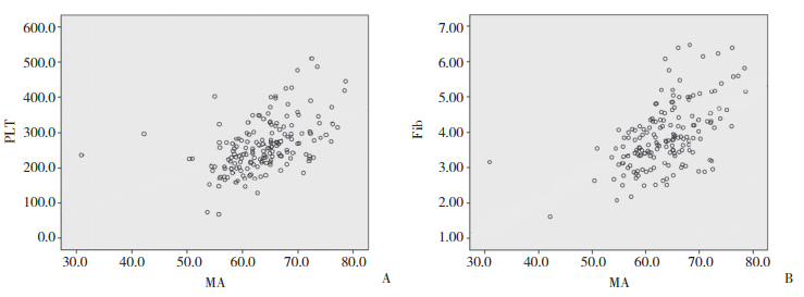
R值与APTT、TT呈正相关;K值与TT呈正相关,与PLT计数、Fib、d-二聚体呈负相关;Alpha角与PLT计数、Fib、d-二聚体呈正相关,与TT呈负相关;如图 2所示,MA值与PLT计数(r = 0.534,P < 0.001)、Fib (r = 0.525,P < 0.001)存在较强的线性正相关,而与TT、d-二聚体的线性相关关系相对较弱;CI与PLT计数、Fib、d-二聚体存在正相关关系,与APTT、TT存在负相关关系;LY30与PLT计数呈正相关。见表 4。

|
| A, PLT; B, Fib. 图 2 MA与PLT、Fib相关性分析的散点图 Fig.2 The scatter diagram of the correlation between MA and PLT, Fib |
| Parameters | Statistic | R time | K time | Alpha angle | MA | CI | LY30 |
| PLT | r | -0.111 | -0.426 | 0.444 | 0.534 | 0.437 | 0.195 |
| P | 0.156 | < 0.001 | < 0.001 | < 0.001 | < 0.001 | 0.012 | |
| PT | r | -0.048 | -0.038 | 0.020 | -0.044 | 0.006 | -0.008 |
| P | 0.542 | 0.625 | 0.803 | 0.574 | 0.940 | 0.923 | |
| APTT | r | 0.232 | 0.112 | -0.134 | -0.083 | -0.173 | -0.022 |
| P | 0.003 | 0.154 | 0.087 | 0.291 | 0.026 | 0.780 | |
| TT | r | 0.214 | 0.337 | -0.356 | -0.296 | -0.336 | 0.010 |
| P | 0.006 | < 0.001 | < 0.001 | < 0.001 | < 0.001 | 0.899 | |
| INR | r | -0.092 | -0.017 | -0.004 | -0.118 | -0.019 | 0.005 |
| P | 0.239 | 0.829 | 0.959 | 0.130 | 0.808 | 0.953 | |
| Fib | r | -0.133 | -0.414 | 0.418 | 0.525 | 0.414 | 0.116 |
| P | 0.088 | < 0.001 | < 0.001 | < 0.001 | < 0.001 | 0.138 | |
| D-dimer | r | -0.055 | -0.243 | 0.263 | 0.180 | 0.193 | 0.089 |
| P | 0.488 | 0.002 | 0.001 | 0.023 | 0.015 | 0.262 |
2.4 常规凝血功能指标、PLT计数与TEG对胃癌患者高凝状态识别价值的比较
常规凝血功能指标和PLT检测发现96例胃癌患者为高凝状态,69例为非高凝状态;TEG检测发现162例胃癌患者为高凝状态,3例为非高凝状态。常规凝血功能指标和PLT计数高凝检出率为58.18%,TEG高凝检出率为98.18%。3种方法对高凝状态的识别差异有统计学意义(P < 0.001)。见表 5。
| Test | Hypercoagulation | Nonhypercoagulation | P |
| TEG | 162 | 3 | < 0.001 |
| Conventional coagulation tests,PLT | 96 | 69 |
3 讨论
胃癌是发病率和病死率最高的恶性肿瘤之一[9]。肿瘤细胞可以释放促凝血因子并且可以诱导正常细胞的促凝特性,从而使机体出现高凝状态,而高凝状态是导致肿瘤患者发生VTE的主要危险因素之一[10-11]。研究[12]表明,胃癌患者的高凝状态与肿瘤分期和浸润程度等密切相关。TEG作为一种分析血凝块形成动力学的敏感方法,可以动态全面地监测凝血过程,已经被广泛应用于肿瘤患者高凝状态的评估[13-14],以及多种疾病的凝血功能监测[15-16]。
本研究中,首先应用常规凝血功能指标、PLT计数和TEG共同比较不同时期胃癌患者的凝血功能状态,结果显示,进展期胃癌患者较早期胃癌患者血液高凝状态更明显。这与在肾细胞癌中的研究结果相似[13]。然后把胃癌患者的TEG参数与常规凝血功能指标、PLT计数进行相关性分析。结果显示,在胃癌患者中,TEG的某些参数与常规凝血功能指标、PLT计数存在线性相关关系。这与研究人员在其他疾病中的研究结果相似[17]。在所有相关关系中,MA值与PLT计数、Fib的相关关系最强,这与BOWBRICK等[18]在健康对照组中的发现一致。MA值是反映血凝块形成最大强度的指标,主要与PLT计数数量和功能相关,也与Fib相关[8],本研究的结果表明,在胃癌患者中,该相关关系依然存在。此外,BAO等[19]发现MA值可以预测白血病患者的出血风险。因此,MA值可能是TEG鉴别和评估胃癌患者异常血流动力学变化的标志性参数。最后比较了常规凝血功能指标、PLT计数与TEG对胃癌患者高凝状态的识别能力。结果表明,TEG对胃癌患者高凝状态的识别能力优于常规凝血功能指标和PLT计数。
综上所述,本研究发现进展期胃癌患者较早期胃癌患者血液高凝状态更明显,对于进展期胃癌患者应该更加注重血液高凝状态的监测和VTE的预防控制;TEG的某些参数与常规凝血功能指标、PLT计数存在线性相关关系,提示TEG和常规凝血功能指标、PLT计数均可以应用于凝血功能评估,但是相关系数大体不高,因此,TEG和常规凝血功能指标、PLT计数不能相互替代;TEG对胃癌患者高凝状态的识别能力优于常规凝血功能指标和PLT计数,尚不能进行TEG检测的实验室和医院应予配备,以方便医生根据TEG和常规凝血功能指标、PLT计数结果综合评估胃癌患者的凝血功能状态,更好地预防VTE的发生,提高胃癌患者的生存质量。
| [1] |
AY C, PABINGER I, COHEN AT. Cancer-associated venous thromboembolism:burden, mechanisms, and management[J]. Thromb Haemost, 2017, 117(2): 219-230. DOI:10.1160/th16-08-0615 |
| [2] |
DONNELLAN E, KHORANA AA. Cancer and venous thromboembolic disease:a review[J]. Oncologist, 2017, 22(2): 199-207. DOI:10.1634/theoncologist.2016-0214 |
| [3] |
王静, 张侠. 恶性肿瘤患者高凝状态的危险因素、发生机制及实验室检测[J]. 检验医学与临床, 2015, 12(15): 2284-2287. DOI:10.3969/j.issn.1672-9455.2015.15.064 |
| [4] |
FUENTES HE, ORAMAS DM, PAZ LH, et al. Venous thromboembolism is an independent predictor of mortality among patients with gastric cancer[J]. J Gastrointest Cancer, 2018, 49(4): 415-421. DOI:10.1007/s12029-017-9981-2 |
| [5] |
GANTER MT, HOFER CK. Coagulation monitoring:current techniques and clinical use of viscoelastic point-of-care coagulation devices[J]. Anesth Analg, 2008, 106(5): 1366-1375. DOI:10.1213/ane.0b013e318168b367 |
| [6] |
杨震, 姜丹丹, 田庆, 等. 血栓弹力图在晚期肺癌初诊患者凝血功能状态评估中的应用[J]. 中国循证心血管医学杂志, 2012, 4(2): 155-157. DOI:10.3969/j.issn.1674-4055.2012.02.022 |
| [7] |
WANG Z, LI J, CAO Q, et al. Comparison between thromboelastography and conventional coagulation tests in surgical patients with localized prostate cancer[J]. Clin Appl Thromb Hemost, 2018, 24(5): 755-763. DOI:10.1177/1076029617724229 |
| [8] |
郝宝岚, 郝新建, 叶昱坪, 等. 血栓弹力图对胃癌患者凝血功能的检测及意义[J]. 实用医学杂志, 2017, 33(10): 1688-1690. DOI:10.3969/j.issn.1006-5725.2017.10.042 |
| [9] |
SIEGEL RL, MILLER KD, JEMAL A. Cancer statistics, 2019[J]. CA:a Cancer J Clin, 2019, 69(1): 7-34. DOI:10.3322/caac.21551 |
| [10] |
谭雍, 潘湘涛. 恶性血液肿瘤相关静脉血栓栓塞症的研究进展[J]. 临床医学研究与实践, 2020, 5(28): 195-198. DOI:10.19347/j.cnki.2096-1413.202028073 |
| [11] |
RUFF SM, WEBER KT, KHADER A, et al. Venous thromboembolism in patients with cancer undergoing surgical exploration[J]. J Thromb Thrombolysis, 2019, 47(2): 316-323. DOI:10.1007/s11239-018-1774-3 |
| [12] |
YANG C, MA R, JIANG T, et al. Contributions of phosphatidylserine-positive platelets and leukocytes and microparticles to hypercoagulable state in gastric cancer patients[J]. Tumour Biol, 2016, 37(6): 7881-7891. DOI:10.1007/s13277-015-4667-5 |
| [13] |
WANG X, SHI A, HUANG J, et al. Assessment of hypercoagulability using thromboelastography predicts advanced status in renal cell carcinoma[J]. J Clin Lab Anal, 2020, 34(1): e23017. DOI:10.1002/jcla.23017 |
| [14] |
OTHMAN M, KAUR H. Thromboelastography (TEG)[J]. Methods Mol Biol, 2017, 1646: 533-543. DOI:10.1007/978-1-4939-7196-1_39 |
| [15] |
HERNANDEZ CONTE A, PEROTTI D, FARAC L. Thromboelastrography (TEG) is still relevant in the 21st century as a point-of-care test for monitoring coagulation status in the cardiac surgical suite[J]. Semin Cardiothorac Vasc Anesth, 2017, 21(3): 212-216. DOI:10.1177/1089253217699282 |
| [16] |
王影, 阎妍, 王忠利. 血栓弹力图对急性弥散性血管内凝血诊断价值的研究[J]. 中国实验血液学杂志, 2020, 28(5): 1699-1703. DOI:10.19746/j.cnki.issn1009-2137.2020.05.045 |
| [17] |
钱雨. 血栓弹力图与凝血功能四项、血小板计数结果的相关性研究[J]. 名医, 2020, 4: 91-92. |
| [18] |
BOWBRICK VA, MIKHAILIDIS DP, STANSBY G. Influence of platelet count and activity on thromboelastography parameters[J]. Platelets, 2003, 14(4): 219-224. DOI:10.1080/0953710031000118849 |
| [19] |
BAO HX, DU J, CHEN BY, et al. The role of thromboelastography in predicting hemorrhage risk in patients with leukemia[J]. Medicine (Baltimore), 2018, 97(13): e0137. DOI:10.1097/md.0000000000010137 |
 2021, Vol. 50


