文章信息
- 刘倩, 赵芳坤, 孔珺
- LIU Qian, ZHAO Fangkun, KONG Jun
- 眼部干细胞研究热点的文献计量学分析
- Bibliometric analysis of ocular stem cells
- 中国医科大学学报, 2021, 50(3): 235-240
- Journal of China Medical University, 2021, 50(3): 235-240
-
文章历史
- 收稿日期:2020-08-31
- 网络出版时间:2021-03-18 14:35
干细胞具有无限分裂、增殖及分化成为体内多种细胞的能力,为多种疾病的治疗提供了新的思路[1]。视网膜色素变性、Stargardt黄斑营养不良及年龄相关性黄斑变性等眼部不可逆性致盲眼病目前在临床上没有有效的治疗方法,近年来干细胞分化光感受器细胞及视网膜色素上皮细胞进行视网膜下腔移植,成为此类疾病未来可能有效的治疗手段[2]。基于眼部干细胞相关研究的重要性与研究数量的众多性,对此研究领域的文献进行科学的分析十分必要。
文献计量学分析是一种利用统计方法对大量文献进行分析,进而衡量和评估研究影响力的重要工具[3]。VOSviewer和CiteSpace是用于构建可视化文献计量网络的软件工具[4]。为进一步认识眼部干细胞的研究热点和趋势,本研究应用文献计量学分析方法以及VOSviewer和CiteSpace工具,对国际上该领域近10年的相关文献进行梳理与可视化分析,获得该领域研究热点,以期对未来眼部干细胞的基础研究和临床应用提供参考和借鉴。
1 材料与方法 1.1 资料与数据收集以Web of Science核心数据库(www.webofscience.com)为数据源,按照以下检索策略:TS=(“stem cells” OR “stem cell” OR “progenitor cells” OR “progenitor cell”)AND TS=“eye”,时间跨度为“2010年至2019年”,文献类型为“article”,进行检索,将数据按照纯文本格式导出。
1.2 数据可视化分析将收集的数据导入VOSviewer软件(1.6.14版本)和CiteSpace软件,进行数据可视化分析。应用VOSviewer对关键词、研究者、研究机构、国家、杂志、论文发表时间和引文等条目进行关系网络构建以及可视化分析。此外,还重点关注了由关键词构建的共现分析,以明确眼部干细胞研究中的热点与趋势。
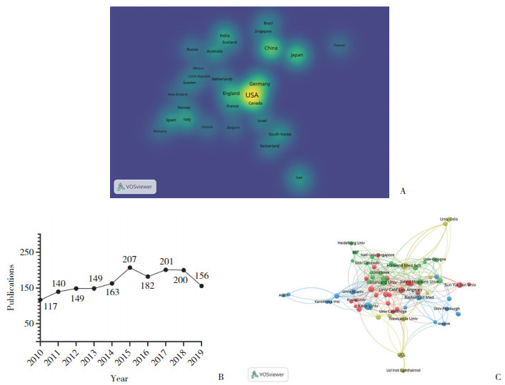
2 结果 2.1 文献分布特点2010年至2019年共有63个国家/地区参与了该领域的研究,共计发表 1 664篇文献。排名前10位国家的总发文数量占98.3%,其中,美国发文最多(689篇,41.4%),其次为中国(199篇,12.0%)、德国(159篇9.6%)。用VOSviewer 1.6.14软件绘制国家/地区密度知识图谱(图 1A),有31个国家/地区的发文量≥10篇,每个国家在图中都有对应的密度颜色,密度大小依赖于元素的数量以及重要性,密度越大,颜色越暖,颜色越暖代表其在眼部干细胞研究中贡献越多,权重越高,反之,颜色越冷权重越低。结果显示,美国居于本研究领域的主导地位。从年发文数量上看,2010年至2015年,每年发文数量逐年增长,2016年至2019年,每年发文数量稳定在150篇以上(图 1B)。文献分布于464种期刊,发文量前3位的杂志是《Investigative Ophthalmology & Visual Science》 《PLos One》 《Cornea》,分别占总发文量的7.3%、4.9%、3.8%,表 1所示为排名前10位的文献来源杂志。
| Rank | Journal | Count(%) | Citations |
| 1 | Investigative Ophthalmology & Visual Science | 122(7.3) | 2 382 |
| 2 | Plos One | 81(4.9) | 1 572 |
| 3 | Cornea | 63(3.8) | 797 |
| 4 | Experimental Eye Research | 57(3.4) | 800 |
| 5 | Development | 44(2.6) | 1 193 |
| 6 | Stem Cells | 37(2.2) | 1 422 |
| 7 | Developmental Biology | 37(2.2) | 651 |
| 8 | Scientific Reports | 35(2.1) | 391 |
| 9 | Molecular Vision | 34(2.0) | 484 |
| 10 | Ocular Surface | 27(1.6) | 363 |
2.2 研究机构合作网络
共1 813家机构参与了该领域的研究,其中发文量排名前3位的机构为日本Keio University,美国Harvard Medical School和Harvard University,发文量前10位的其他机构见表 2。通过VOSviewer绘制机构合作网络图谱(图 1C),选取发文量≥10篇的71个机构,其中有70个机构存在合作关系。图中每个节点表示1个机构,节点大小表示发文量。节点之间的联系表示协作,联系强度越大,合作越紧密。

|
| A, country density of publications in stem cell and eye research from 2010 to 2019; B, annual documents in stem cell and eye research from 2010 to 2019; C, cooperation network of organizations in stem cell and eye research from 2010 to 2019. 图 1 2010年至2019年眼部干细胞领域文献分布特点与机构合作网络图 Fig.1 The distribution of publications and cooperation network of organizations in stem cell and eye research from 2010 to 2019 |
| Rank | Organization | Country | Count(%) |
| 1 | Keio University | Japan | 39(2.3) |
| 2 | Harvard Medical School | USA | 30(1.8) |
| 3 | Harvard University | USA | 28(1.7) |
| 4 | Johns Hopkins University | USA | 26(1.6) |
| 5 | University College London | England | 26(1.6) |
| 6 | University of Toronto | Canada | 26(1.6) |
| 7 | L V Prasad Eye Institute | India | 25(1.5) |
| 8 | University of California Los Angeles | USA | 25(1.5) |
| 9 | The National Eye Institute | USA | 23(1.4) |
| 10 | Sun Yat-sen University | China | 23(1.4) |
2.3 作者及其研究领域
用VOSviewer软件绘制作者合作网络图谱,在8 592名作者中,发文量≥5篇的作者共有127名(图 2A),而其中有合作关系的作者数量为60名(图 2B),将最小聚类值设为10,共产生了以下3个聚类:以作者TSUBOTA K,OGAWA Y,SHIMMURA S等为主的绿色聚类;以作者SANGWAN VS,BASU S,HOLLAND EJ等为主的红色聚类,以及以作者CVEKL A,LIU Y等为主的蓝色聚类。

|
| A, main authors in stem cell and eye study from 2010 to 2019;B, co-authorship network of main authors in stem cell and eye study. 图 2 2010年至2019年眼部干细胞领域作者及作者合作网络图 Fig.2 Authors and co-authorship network of main authors in stem cell and eye study from 2010 to 2019 |
2.4 引用频次前10位的文章
表 3所示为2010年至2019年该领域发表的文章被引用次数最多的10篇文章标题、发表杂志、发表年度、被引用次数和主要观点。在引用次数排名前10位的文章里,有2篇发表于《Lancet》,其余分别发表于《Nature》 《Cell》等期刊上。
| Rank | Title | Journal | Year | Citations | Main conclusion |
| 1 | Self-organizing optic-cup morphogenesis in three- dimensional culture |
Nature | 2011 | 860 | A successful process in 3D culture of mouse embryonic stem cells to generate visual cup |
| 2 | Embryonic stem cell trials for macular degeneration: a preliminary report |
Lancet | 2012 | 794 | Subretinal space transplantation of human embryonic stem cell derived retinal pigment epithelial cells for the treatment of macular degeneration |
| 3 | Human embryonic stem cell-derived retinal pigment epithelium in patients with age-related macular degeneration and Stargardt’s macular dystrophy: follow-up of two open-label phase 1/2 studies |
Lancet | 2015 | 505 | A long-time safety and follow up of macular degeneration treatment with human embryonic stem cell derived retinal pigment epithelial transplantation |
| 4 | The Rd8 mutation of the Crb1 gene is present in vendor lines of C57BL/6N mice and embryonic stem cells,and confounds ocular induced mutant phenotypes |
Investigative Ophthalmology & Visual Science | 2012 | 339 | Crb1 mutation is usual in the substrain of C57BL/6N which is a common tool mouse to generate transgenic mice or embryonic stem (ES)cells |
| 5 | Generation of three-dimensional retinal tissue with functional photoreceptors from human iPSCs |
Nature Communications | 2014 | 245 | An eye disease model: a 3D retinal tissue generated by human induced pluripotent stem cells in vitro |
| 6 | Optic vesicle-like structures derived from human pluripotent stem cells facilitate a customized approach to retinal disease treatment |
Stem Cells | 2011 | 212 | Utilize human embryonic stem cell and human induced pluripotent stem cell to generate an Optic Vesicle-like structures |
| 7 | Sox2 roles in neural stem cells | International Journal of Biochemistry & Cell Biology | 2010 | 183 | The function of Sox2 in the development of eye and brain neuronal systems |
| 8 | Transplantation of adult mouse iPS cell-derived photoreceptor precursors restores retinal structure and function in degenerative mice |
PLos One | 2011 | 183 | A successful case in mouse induced pluripotent stem cell producing the functional photoreceptors to treat the retina degeneration |
| 9 | Neuroprotective effects of intravitreal mesenchymal stem cell transplantation in experimental glaucoma |
Investigative Ophthalmology & Visual Science | 2010 | 170 | Local transplantation of mesenchymal stem cell protects the optic nerve in an animal glaucoma model |
| 10 | Tet3 CXXC domain and dioxygenase activity cooperatively regulate key genes for xenopus eye and neural development |
Cell | 2012 | 149 | The function of Tet3 during the development of eye and neuro |
2.5 眼部干细胞研究热点及演变
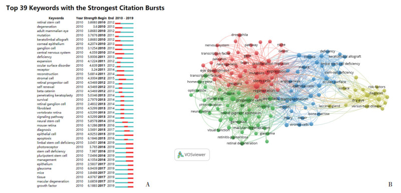
本研究用CiteSpace对关键词进行了突发分析,图 3A所示为从2010年开始的前39个突发的关键词开始和结束的时间、突发的强度,以及随着时间的改变突发关键词的改变。“photoreceptor”“pluripotent stem cell”“epithelial cell”“macular degeneration”“glaucoma”“limbal stem cell deficiency”“growth factor”等关键词突发一直持续至2019年。为了进一步发现眼部干细胞研究关键词之间的联系,本研究用VOSviewer在1 664篇文献中进行了关键词分析,在7 397个关键词中,设置最小出现频次20次,共有140个关键词达到要求,同一颜色的关键词属于同一聚类,共形成了红、绿、蓝、黄4个聚类(图 3B)。

|
| A, burst analysis of keywords in stem cell and eye study from 2010 to 2019;B, co-occurrence network of keywords in stem cell and eye study from 2010 to 2019. 图 3 2010年至2019年眼部干细胞领域关键词突发分析与关键词共现分析图 Fig.3 Burst analysis of keywords and co-occurrence network of keywords in stem cell and eye study from 2010 to 2019 |
3 讨论
本研究应用VOSviewer对Web of Science核心合集中近10年关于眼部干细胞研究的1 664篇文章进行了文献计量学分析,初步了解了该领域的研究发展状况。文章分布结果显示:(1)美国作为该领域发文量第一的国家,其文章累计引用频次16 885次,同样位居第一,中国和德国虽然排名第二和第三,但其发文量和引用频次远低于美国,国家密度图结果也凸显了美国在该领域中的地位;(2)前5年发文数量持续增长,后5年发文数量稳定维持在≥150篇/年,说明眼部干细胞的研究一直受到研究者的青睐;(3)杂志的分布分析中,《Investigative Ophthalmology & Visual Science》以最高的发文量和引用数位居第一。
从机构分布的地理位置来看,发文量前10位的机构中有5个机构位于美国,然后各有1个机构位于日本、英国、加拿大、印度和中国。从单一机构发文量来看,位于日本的Keio University以发文数39(2.3%)的优势位居第一。而机构合作网络图(图 1C)显示,不同机构间存在相互联系和合作,位于同一颜色聚类距离越近的机构联系越紧密。
从作者合作分析来看,众多作者中存在合作关系的作者共产生了3个聚类,同一颜色代表同一聚类,位于同一聚类的作者合作紧密,研究方向及主题具有一定的相似性。聚类1(红色):以作者SANGWAN VS,BASU S,HOLLAND EJ等为主,其主要研究内容为角膜缘干细胞缺乏的治疗[5]、角膜损伤与角膜再生[6]、眼表干细胞移植[7]。聚类2(绿色):以作者TSUBOTA K,OGAWA Y,SHIMMURA S等为主,其主要研究内容为造血干细胞移植后并发症的诊断、疾病机制和治疗[8]相关,造血干细胞移植后并发症干眼症的治疗与机制研究[9],泪腺和角膜内皮的再生[10],诱导多能干细胞的免疫学特性[11]。聚类3(蓝色):以作者CVEKL A,ASHERY-PADAN R等为主,其主要研究内容为眼部组织的正常发育过程,转录因子(如PAX6等)在眼部发育中的作用等,如晶状体[12]、视网膜[13]、视网膜色素上皮细胞分化与脉络膜血管系统[14]发育。
关键词凝聚了论文主要思想和要点,它出现频次的多少反映了研究者对该领域关注度的大小。出现频次越高的关键词,越有可能成为研究的热点。关键词的突发是指特定时间段内的数据量显著异常于其他时间段,即关键词出现的频次明显增多。图 3A展示了近10年眼部干细胞研究中的39个突发关键词,关键词的突发分析提示了特定时间段中研究者共同关注的热点以及随着时间突发关键词的转变,有助于纵向动态观察热点的变化。“photoreceptor”“pluripotent stem cell”“macular degeneration” “glaucoma” “limbal stem cell deficiency”等是一直延续至2019年的突发关键词,说明现在仍是该领域内研究的热点。而VOSviewer的关键词共现分析将高频关键词按照相关度进行聚类,便于分类分析眼部干细胞研究的热点。VOSviewer的关键词共现分析一共产生4个聚类:聚类1(红色)的关键词主要研究的是关于正常眼部发育中的基因表达和细胞分化的过程;聚类2(绿色)主要体现的是在体外模拟体内眼部发育环境,使用干细胞再生眼部组织,治疗眼部变性型疾病;聚类3(蓝色)是关于干细胞在眼表疾病治疗中的应用;聚类4(黄色)主要是关于造血干细胞移植后常见并发症慢性移植物抗宿主病和干眼症相关研究。
本研究将文献计量学与可视化制图分析相结合,更好地展示了眼部干细胞相关的研究进展以及影响力排名靠前的国家、机构和研究者,并展示了该领域中有影响力和高被引用的论文,为研究人员提供了有用的信息。本研究的不足之处在于,在作者聚类分析时,重点分析了联系比较紧密的3个聚类中的作者,而未纳入独立作者的研究方向。此外,本研究纳入的文献语言为英语,可能会在一定程度上影响分析的全面性。
综上所述,2010年至2019年在眼部干细胞研究中,影响力最大的国家、杂志和机构分别为美国、《Investigative Ophthalmology & Visual Science》和Keio University。眼部干细胞研究的热点和趋势主要聚集在眼部正常发育的调控、体外干细胞分化眼部组织、干细胞眼表疾病的治疗和干细胞移植后眼部并发症这4个方面。
| [1] |
JIN J. Stem cell treatments[J]. JAMA, 2017, 317(3): 330. DOI:10.1001/jama.2016.17822 |
| [2] |
GAGLIARDI G, BEN M'BAREK K, GOUREAU O. Photoreceptor cell replacement in macular degeneration and retinitis pigmentosa: a pluripotent stem cell-based approach[J]. Prog Retin Eye Res, 2019, 71: 1-25. DOI:10.1016/j.preteyeres.2019.03.001 |
| [3] |
COOPER ID. Bibliometrics basics[J]. J Med Libr Assoc, 2015, 103(4): 217-218. DOI:10.3163/1536-5050.103.4.013 |
| [4] |
ECK NJ, WALTMAN L. Software survey: vosviewer, a computer program for bibliometric mapping[J]. Scientometrics, 2010, 84(2): 523-538. DOI:10.1007/s11192-009-0146-3 |
| [5] |
BASU SY, FERNANDEZ MM, DAS S, et al. Clinical outcomes of xeno-free allogeneic cultivated limbal epithelial transplantation for bilateral limbal stem cell deficiency[J]. Br J Ophthalmol, 2012, 96(12): 1504-1509. DOI:10.1136/bjophthalmol-2012-301869 |
| [6] |
SUSAIMANICKAM PJ, MADDILETI S, PULIMAMIDI VK, et al. Generating minicorneal organoids from human induced pluripotent stem cells[J]. Development, 2017, 144(13): 2338-2351. DOI:10.1242/dev.143040 |
| [7] |
CHEUNG AY, PATEL S, KURJI KH, et al. Ocular surface stem cell transplantation for treatment of keratitis-ichthyosis-deafness syndrome[J]. Cornea, 2019, 38(1): 123-126. DOI:10.1097/ICO.0000000000001802 |
| [8] |
MUKAI S, OGAWA Y, SAYA H, et al. Therapeutic potential of tranilast for the treatment of chronic graft-versus-host disease in mice[J]. PLoS One, 2018, 13(10): e0203742. DOI:10.1371/journal.pone.0203742 |
| [9] |
OGAWA Y, HE H, MUKAI S, et al. Heavy chain-hyaluronan/pentraxin 3 from amniotic membrane suppresses inflammation and scarring in murine lacrimal gland and conjunctiva of chronic graft-versus-host disease[J]. Sci Rep, 2017, 7(1): 42195. DOI:10.1038/srep42195 |
| [10] |
HATOU S, YOSHIDA S, HIGA K, et al. Functional corneal endothelium derived from corneal stroma stem cells of neural crest origin by retinoic acid and wnt/β-catenin signaling[J]. Stem Cells Dev, 2013, 22(5): 828-839. DOI:10.1089/scd.2012.0286 |
| [11] |
FUJII S, YOSHIDA S, INAGAKI E, et al. Immunological properties of neural crest cells derived from human induced pluripotent stem cells[J]. Stem Cells Dev, 2019, 28(1): 28-43. DOI:10.1089/scd.2018.0058 |
| [12] |
ZHAO YL, ZHENG DY, CVEKL A. Profiling of chromatin accessibility and identification of general Cis-regulatory mechanisms that control two ocular lens differentiation pathways[J]. Epigenetics Chromatin, 2019, 12(1): 27. DOI:10.1186/s13072-019-0272-y |
| [13] |
MENUCHIN-LASOWSKI Y, OREN-GILADI P, XIE Q, et al. Sip1 regulates the generation of the inner nuclear layer retinal cell lineages in mammals[J]. Dev Camb Engl, 2016, 143(15): 2829-2841. DOI:10.1242/dev.136101 |
| [14] |
COHEN-TAYAR Y, COHEN H, MITIAGIN Y, et al. Pax6 regulation of Sox9 in the mouse retinal pigmented epithelium controls its timely differentiation and choroid vasculature development[J]. Development, 2018, 145(15): dev163691. DOI:10.1242/dev.163691 |
 2021, Vol. 50


