文章信息
- 段禹舟, 李龙, 高伟, 李连祥, 吴安华
- DUAN Yuzhou, LI Long, GAO Wei, LI Lianxiang, WU Anhua
- 蝶窦分隔确定海绵窦段颈内动脉位置在神经内镜手术中的应用
- Application of a sphenoid sinus septum to determine the position of the internal carotid artery in the cavernous segment in neuroendoscopic surgery
- 中国医科大学学报, 2021, 50(2): 102-107
- Journal of China Medical University, 2021, 50(2): 102-107
-
文章历史
- 收稿日期:2020-05-07
- 网络出版时间:2021-01-13 16:32
目前,经蝶入路手术已经成为超过90%的垂体瘤和其他鞍区病变的首选术式[1]。内镜经蝶入路手术具有术野明亮、肿瘤切除精确和术后并发症少的优点[2-3]。常见的并发症包括视神经损伤、脑脊液漏等,其中颈内动脉(internal carotid artery,ICA)损伤发生率很低(0.2%~2%),但最为凶险[4-9]。ICA C3段呈“S”形弯曲转弯,由于形态导致此处血流冲击力更大,并且此处ICA与骨质菲薄的蝶骨距离较近,更容易发生损伤[10-11]。所以在手术中明确ICA C3段位置,对内镜手术的顺利进行至关重要。
蝶窦变异程度较大[12-13],既往研究[14-17]表明,ICA的形状与蝶窦存在一定的相关性,因此,在经蝶手术前和手术中识别蝶窦的类型对于避免ICA的损伤尤为重要[16-18]。本研究为探究蝶窦分隔和ICA C3段的解剖关系,进行了1项回顾性分析。对患者的影像学资料进行融合处理,将不同形状的蝶窦分隔进行分类归纳,对可能影响蝶窦分隔和ICA位置关系的相关因素进行分析,并根据内镜手术中的实际图像进行验证。
1 材料与方法 1.1 研究设备术前的鼻咽部计算机断层扫描(computer tomography,CT)和头颅磁共振血管造影(magnetic resonance angiography,MRA)图像被导入到美敦力神经导航工作站S7,对图像进行融合和三维结构处理。
1.2 研究对象和分组收集2014年6月至2019年9月于中国医科大学附属第一医院神经外科进行手术的110例患者的临床资料。记录患者性别、年龄、病理、肿瘤位置、肿瘤大小、手术入路等信息,并对图像进行归类和测量。
1.2.1 蝶窦分隔ICA指向的判定基于融合图像的冠状位,找到ICA C3段前曲处的位置,当蝶窦分隔连接至鞍底的骨性分隔的位置在ICA外缘之间时,认为其指向ICA。
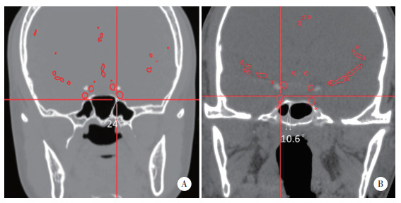
1.2.2 蝶窦分隔和中线之间夹角的测量将所有蝶窦分隔用Photoshop测量出蝶窦分隔与中线之间的夹角大小,在测量角度时选取蝶窦分隔与鞍底的连接处至其第1个拐点和中线的夹角,蝶窦分隔线起于颅底交界处,止于颅底交界处的第1个拐点,另一条线起于与中线平行的交界处。然后测量这两条线之间的夹角。见图 1。
|
| A, type Ⅱ; B, type Ⅰ. 图 1 蝶窦分隔和中线之间夹角的测量方式 Fig.1 Measurement of the angle between the sphenoid sinus separation and the midline |
1.2.3 蝶窦分隔的分类方法
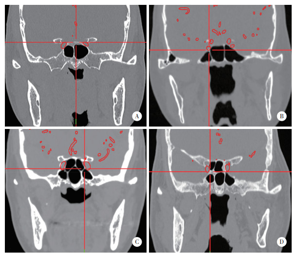
分类方法根据ICA C3段鼻咽部CT图像在冠状位中附着于鞍底的蝶窦骨性分隔的类型。并在神经内镜手术病例中进行验证。“O”型,仅有1个骨性蝶窦(鞍底无明显隔)或无蝶窦;“Ⅰ”型,有1个骨性分隔连接在鞍底上;“Ⅱ”型,有2个或2个以上平行的骨性分隔连接在鞍底上;“Y”型有分叉的骨性分隔连接在鞍底上,包括既有分叉的分隔,又有平行的分隔;“X”型有交叉的骨性分隔连接在鞍底上。对于既有交叉的分隔,又有平行分隔的病例,根据其分隔的位置,左侧或者右侧,分别记入分别标记为“XI”、“XX”等。同理,对于包括既有分叉的分隔,又有平行的分隔,根据其分隔的位置,左侧或者右侧标记为“IY”、“YY”。见图 2。
|
| A, type Ⅰ; B, type Ⅱ; C, type YY; D, type IX. 图 2 蝶窦分隔的分类 Fig.2 Classification of sinus septa |
1.3 统计学分析
采用SPSS 25.0进行统计分析。计算Pearson相关系数,用来验证各个因素与判断ICA位置可能存在的相关性关系。P < 0.05为差异有统计学意义。
2 结果 2.1 蝶窦分隔分类汇总与指向性汇总本研究共纳入110例患者,收集复发与否、性别、年龄、肿瘤位置、病理等详细原始病历信息。见表 1。其中,“Y”型4例,“YI”型21例,“YY”型4例,“Ⅰ”型21例,“Ⅱ”型22例,“O”型11例,“X”型5例,“XI”型2例。没有“XY”型的病例。14例因严重侵犯蝶窦间隔而被排除。也有6例由于肿瘤的侵袭而使蝶窦分隔不能进行分类,但仍能看到蝶骨隔的残余部分,可进行统计分析。
| Item | Clinical informations |
| Gender | |
| Male | 39 |
| Female | 57 |
| Age(year) | |
| Average | 47.7 |
| Pathology | |
| Pituitary | 38 |
| Chordoma | 25 |
| Craniopharyngioma | 9 |
| Others | 26 |
| Tumor position | |
| Sphenoid sinus | 50 |
| Clivus | 20 |
| Others | 15 |
| Maximum diameter of tumor(mm) | |
| Average | 42 |
| Surgical approach | |
| Neuroendoscopic transsephenoidal | 81 |
| Microscopic transsephenoidal | 4 |
| Others | 11 |
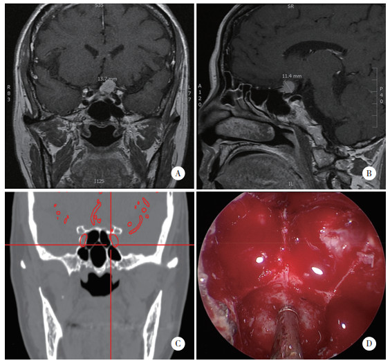
在内镜实际图像中进行验证。患者的增强磁共振冠状位和矢状位图像显示,肿瘤位于鞍结节,增强明显;融合图像(CT和MRA)显示蝶窦分隔及ICA C3段。双侧蝶窦分隔直接指向ICA C3段;在神经内窥镜影像术中,可见蝶窦分隔实际形态与冠状位图像十分相符。见图 3。
|
| A, MRI coronary images; B, MRI sagittal images; C, fusion images; D, uroendoscopy video. 图 3 某鞍结节脑膜瘤病例相关信息 Fig.3 A case of tuberculum sellae meningioma |
对蝶窦分隔是否指向ICA C3段进行了分类汇总。85例中,有51例双侧均存在分隔,其中双侧均不指向ICA 3例,一侧指向ICA 23例,双侧均指向ICA 25例;仅单侧存在分隔ICA 34例,不指向ICA 14例,指向ICA 20例,即有68例(80%)至少有1个蝶窦分隔指向ICA C3段。说明蝶窦分隔指向ICA的情况普遍存在,在术中具有实际应用的价值。
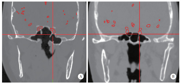
在本研究中,发现2种相对特殊的情况:第1种情况,在复发性肿瘤,术中可见原有的蝶窦分隔被既往手术破坏,在此时蝶窦分隔残余的部分仍可作为判断ICA位置的解剖标志点(图 4A);第2种情况,蝶窦分隔并不直接指向ICA C3段,而是ICA C3段位于Y型蝶窦分隔的2个分支之间,这种情况下蝶窦分隔也具有判断ICA位置的作用(图 4B)。
|
| A, in recurrent cases, the sphenoid septum still points to the ICA C3 segment; B, the ICA C3 segment is located between two sphenoid septum. 图 4 复发肿瘤蝶窦分隔残根和ICA位于蝶窦分隔分叉之间的情况 Fig.4 A case of recurrent tumor with sphenoid septum residual roots and a case of ICA between the sphenoid septum bifurcation |
2.2 蝶窦分隔是否指向ICA与相关因素的分析
“O”型病例通常具有ICA凸起,因此在手术中很容易确定ICA的位置。除上述病例外,共有85例被用于分析。因ICA为双侧存在,而蝶窦分隔出现情况不一定,有时可能出现双侧均存在蝶窦分隔,一侧没有蝶窦分隔,或者同侧具有多个蝶窦分隔。所以将数据根据蝶窦分隔的位置(左侧或右侧)和分隔指向性,对所有的分隔进行归类汇总。并且在同侧有多个分隔的病例中,只研究离ICA最近的一个。对于侵犯蝶窦分隔的类似脊索瘤等病例,除过度侵袭无法辨认清楚和复发的肿瘤病例外,在能分辨出保留有蝶窦分隔连接至鞍底残根的情况下,仍然纳入分析。汇总后显示,左侧共有69例蝶窦分隔,右侧共有67例蝶窦分隔。之后根据所获的结果计算各相关因素的列联系数。多因素分析结果显示,蝶窦分隔是否指向ICA与性别、年龄、肿瘤部位、病理类型、是否侵袭蝶骨和是否为复发肿瘤均不相关(P > 0.05)。见表 2。
| Item | Left(n = 69) | Right(n = 67) | |||
| C | P | C | P | ||
| Gender | 0.038 | 0.755 | 0.038 | 0.753 | |
| Age | 0.646 | 0.055 | 0.576 | 0.644 | |
| Tumor position | 0.321 | 0.093 | 0.227 | 0.457 | |
| Pathology | 0.348 | 0.090 | 0.291 | 0.286 | |
| Tumor invasion | 0.053 | 0.661 | 0.061 | 0.615 | |
| Tumor recurrence | 0.134 | 0.261 | 0.101 | 0.405 | |
| C,coefficient of contingency. | |||||
2.3 蝶窦分隔指向性与蝶窦分隔与中线之间的夹角角度相关性分析
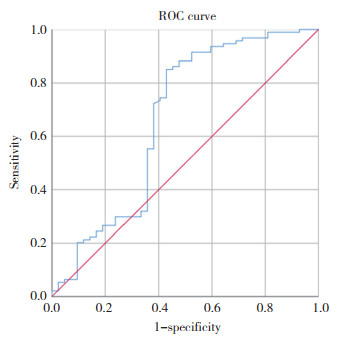
为了分析相关性,排除了其他相关因素的影响后,共分析了85例共136个蝶窦分隔。根据独立分量分析的角度和指向性绘制了受试者操作特征(receiver operating characteristic,ROC)曲线,并计算了评价值。ROC曲线显示95% CI:0.551~0.778,P = 0.002,Cut-off值为15.35°,蝶窦分隔指向性与蝶窦分隔和中线之间的夹角角度有关。当蝶窦分隔与中线之间的夹角角度 > 15.35°时,在ICA C3段的层面上,蝶窦分隔很大可能性会指向ICA C3段。见图 5。
|
| 图 5 根据ICA的角度和指向性的绘制ROC曲线 Fig.5 An ROC curve was drawn based on the angle and directivity to the ICA |
3 讨论
蝶窦是内镜经蝶手术的必经之路,其变异程度大,每个患者的蝶窦形状变化、气化程度均不同[12-13],需术者辨别其附近的解剖结构以减小创伤,既往的解剖学和放射学研究[14-17]已经表明ICA的形状与蝶窦存在一定的相关性,因此,在经蝶手术前和手术中识别蝶窦的类型对于避免ICA的损伤尤为重要。目前,在我国许多大型医疗中心都配备有神经导航系统。在神经导航的指导下,外科医生可以识别许多重要的结构,但是对于一些地市级别医疗中心没有配制神经导航设备,或者在神经导航设备无法使用、神经导航在术中出现误差时,外科医生需要利用骨窗的冠状位和矢状位CT图像来分析和判断蝶窦的形态和分隔的位置。
目前对蝶窦及分隔的分类多基于普通CT的轴位像,所得结论多为统计数量方面的分类结果。如不同类型的蝶窦分隔与ICA凸起或视神经凸起之间的存在关系[16-17]。本研究采用了一种更方便、更详细的方法对蝶窦分隔进行分类,专注于ICA最容易在经蝶内镜手术中发生损伤的C3段,ICA在这一段有一个前向转弯,动脉血对ICA的压力冲击大于其他段。ICA C3段是最接近颅底的部分,其周围的骨质结构薄弱。在这个区域颅底通常与某个蝶窦分隔相连,有时还有可能形成一个小而且坚固的框架,并且基于更符合术中实际图像的冠状位CT图像。相比之前的研究,本研究介绍的分类方式更适合于临床应用,有着更广泛的适用性。
本研究结果显示,当蝶窦分隔与中线之间的夹角角度 > 15.35°时,在ICA C3段的层面上,蝶窦分隔很大可能性会指向ICA,此时蝶窦分隔可以作为内镜经蝶手术中的重要解剖标志点。尤其适用于切除包绕ICA C3段的肿瘤和对骨质破坏严重的肿瘤,以及复发的肿瘤。结合医生的判断和实际内镜影像,以及辅助设备相互确认,可以最大程度增加手术的安全性。
综上,建议外科医生在制定手术计划和手术过程中密切注意蝶窦分隔。将蝶窦分隔作为判断ICA位置的标志结构而不仅仅是内镜手术中干扰视线的障碍,此外,在内镜手术中应以恰当的方式处理蝶窦分隔。在磨除蝶窦分隔,尤其是在去除指向ICA的蝶窦分隔时,应格外注意。
| [1] |
WANG AJ, ZAIDI HA, LAWS ED JR, et al. History of endonasal skull base surgery[J]. J Neurosurg Sci, 2016, 60(4): 441-453. |
| [2] |
AZAB WA, ABDELNABI EA, MOSTAFA KH, et al. Effect of sphenoid sinus pneumatization on the surgical windows for extended endoscopic endonasal transsphenoidal surgery[J]. World Neurosurg, 2020, 133: e695-e701. DOI:10.1016/j.wneu.2019.09.126 |
| [3] |
ASEMOTA AO, ISHⅡ M, BREM H, et al. Comparison of complications, trends, and costs in endoscopic vs microscopic pituitary surgery:analysis from a US health claims database[J]. Neurosurgery, 2017, 81(3): 458-472. DOI:10.1093/neuros/nyx350 |
| [4] |
DEHDASHTI AR, GANNA A, KARABATSOU K, et al. Pure endoscopic endonasal approach for pituitary adenomas:early surgical results in 200 patients and comparison with previous microsurgical series[J]. Neurosurgery, 2008, 62(5): 1006-1015. DOI:10.1227/01.Neu.0000297072.75304.89 |
| [5] |
VALENTINE R, WORMALD PJ. Controlling the surgical field during a large endoscopic vascular injury[J]. Laryngoscope, 2011, 121(3): 562-566. DOI:10.1002/lary.21361 |
| [6] |
SYLVESTER PT, MORAN CJ, DERDEYN CP, et al. Endovascular management of internal carotid artery injuries secondary to endonasal surgery:case series and review of the literature[J]. J Neurosurg, 2016, 125(5): 1256-1276. DOI:10.3171/2015.6.JNS142483 |
| [7] |
LI AJ, LIU WS, CAO PC, et al. Endoscopic versus microscopic transsphenoidal surgery in the treatment of pituitary adenoma:a systematic review and meta-analysis[J]. World Neurosurg, 2017, 101: 236-246. DOI:10.1016/j.wneu.2017.01.022 |
| [8] |
ALQAHTANI A, CASTELNUOVO P, NICOLAI P, et al. Injury of the internal carotid artery during endoscopic skull base surgery[J]. Otolaryngol Clin N Am, 2016, 49(1): 237-252. DOI:10.1016/j.otc.2015.09.009 |
| [9] |
GUVENC G, KIZMAZOGLU C, PINAR E, et al. Outcomes and complications of endoscopic versus microscopic transsphenoidal surgery in pituitary adenoma[J]. J Craniofacial Surg, 2016, 27(4): 1015-1020. DOI:10.1097/scs.0000000000002684 |
| [10] |
SHAKUR SF, ALETICH VA, AMIN-HANJANI S, et al. Quantitative assessment of parent vessel and distal intracranial hemodynamics following pipeline flow diversion[J]. Interv Neuroradiol, 2017, 23(1): 34-40. DOI:10.1177/1591019916668842 |
| [11] |
SHIKARY T, ANDALUZ N, MEINZEN-DERR J, et al. Operative learning curve after transition to endoscopic transsphenoidal pituitary surgery[J]. World Neurosurg, 2017, 102: 608-612. DOI:10.1016/j.wneu.2017.03.008 |
| [12] |
FERNANDEZ-MIRANDA JC, PREVEDELLO DM, MADHOK R, et al. Sphenoid septations and their relationship with internal carotid arteries:anatomical and radiological study[J]. Laryngoscope, 2009, 119(10): 1893-1896. DOI:10.1002/lary.20623 |
| [13] |
TWIGG V, CARR SD, BALAKUMAR R, et al. Radiological features for the approach in trans-sphenoidal pituitary surgery[J]. Pituitary, 2017, 20(4): 395-402. DOI:10.1007/s11102-017-0787-9 |
| [14] |
DAL SECCHI M, DOLCI R, TEIXEIRA R, et al. An analysis of anatomic variations of the sphenoid sinus and its relationship to the internal carotid artery[J]. Int Arch Otorhinolaryngol, 2018, 22(2): 161-166. DOI:10.1055/s-0037-1607336 |
| [15] |
TAWFIK A, EL-FATTAH AMA, NOUR AI, et al. Neurovascular surgical keys related to sphenoid window:radiologic study of egyptian's sphenoid[J]. World Neurosurg, 2018, 116: e840-e849. DOI:10.1016/j.wneu.2018.05.113 |
| [16] |
RAMALHO CO, MARENCO HA, DE ASSIS VAZ GUIMARÃES FILHO F, et al. Intrasphenoid septations inserted into the internal carotid arteries:a frequent and risky relationship in transsphenoidal surgeries[J]. Braz J Otorhinolaryngol, 2017, 83(2): 162-167. DOI:10.1016/j.bjorl.2016.02.007 |
| [17] |
YANO S, SHINOJIMA N, KITAJIMA M, et al. Usefulness of oblique coronal computed tomography and magnetic resonance imaging in the endoscopic endonasal approach to treat skull base lesions[J]. World Neurosurg, 2018, 113: e10-e19. DOI:10.1016/j.wneu.2018.01.022 |
| [18] |
PIRINC B, FAZLIOGULLARI Z, GULER I, et al. Classification and volumetric study of the sphenoid sinus on MDCT images[J]. Eur Arch Otorhinolaryngol, 2019, 276(10): 2887-2894. DOI:10.1007/s00405-019-05549-8 |
 2021, Vol. 50


