文章信息
- 关慧娟, 户青波, 庞梓萌, 高爽, 冯翠娟
- GUAN Huijuan, HU Qingbo, PANG Zimeng, GAO Shuang, FENG Cuijuan
- 青少年安氏Ⅱ类2分类错
 畸形非拔牙矫治前后面下部软硬组织的变化
 - Changes in soft and hard tissues in the lower face of adolescents with classⅡdivision 2 malocclusion treated using a non-extraction protocol
- 中国医科大学学报, 2020, 49(7): 610-614
- Journal of China Medical University, 2020, 49(7): 610-614
-
文章历史
- 收稿日期:2019-08-15
- 网络出版时间:2020-06-24 9:10
 畸形非拔牙矫治前后面下部软硬组织的变化
 畸形非拔牙矫治前后面下部软硬组织的变化并进行相关分析。方法 选择32例已经完成正畸治疗的安氏Ⅱ类2分类错
 非拔牙矫治患者,应用Winceph9.0软件进行X线头影测量分析。采用SPSS 23.0软件对矫治前后面下部软硬组织变化进行统计分析。结果 矫治后ANB角、上中切牙暴露量、鼻唇角呈减小趋势;SNB角、下颌平面角、下颌体长度、硬组织颏前部突度、上下中切牙突度、上下唇突度、软组织颏部突度呈增大趋势,差异均有统计学意义(P < 0.05)。鼻唇角的变化与上唇突度的改变呈负相关;下唇突度的变化与上唇突度的变化呈正相关。结论 安氏Ⅱ类2分类错
 青少年患者非拔牙矫治后,侧貌突度虽发生改变,但由于下颌的生长和适当的控制,仍可以维持良好的面型。
如今,美观在正畸治疗的目标中占越来越高的比重。人们对美观的要求日益增加,越来越多的患者以改善面部美观为要求来诊。安氏Ⅱ类2分类错牙合畸形的临床症状主要包括内倾型深覆牙合、面下部过短、颏唇沟较深等[1]。相对于安氏Ⅱ类1分类错牙合患者较突的面型,安氏Ⅱ类2分类的侧貌更能够得到患者的接受。高辉等[2]的研究表明,与正常牙合组比较,大多数安氏Ⅱ类2分类错牙合青少年具有较好的侧貌外形。安氏Ⅱ类2分类错牙合患者矫治的难点在于在达到平衡稳定的前提下,不能破坏现有的美观特征。因此,了解安氏Ⅱ类2分类错牙合患者矫治前后面部的变化趋势及影响其变化的相关因素至关重要。本研究拟评估安氏Ⅱ类2分类错牙合患者在正畸治疗前后面下部软硬组织变化情况,并进行相关性分析,以期为临床治疗提供参考。
1 材料与方法 1.1 研究对象选取中国医科大学附属口腔医院正畸二科32例已完成正畸治疗的安氏Ⅱ类2分类错牙合畸形的青少年患者。其中,男11例,女21例,年龄(13.5±1.8)岁。纳入标准:(1)骨性Ⅱ类(ANB角>4.7°),均角型(31.5° < 下颌平面角 < 40.7°);(2)双侧磨牙远中关系;(3)上前牙,至少2颗中切牙内倾;(4)均采用非拔牙矫治;(5)面部协调对称。排除标准:(1)骨畸形严重,不可进行正畸代偿矫治;(2)外伤史;(3)颌面部存在大面积缺损。
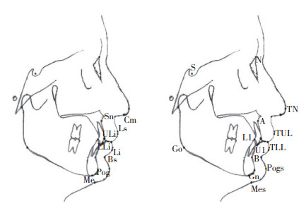
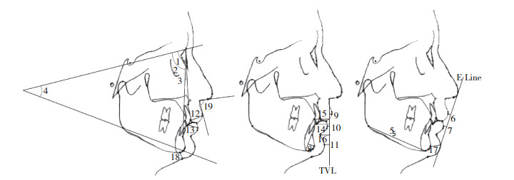
1.2 研究方法在一段连续的时间内,应用Winceph9.0软件(上海华景医疗器材有限公司)对所有研究对象矫治前后的头颅侧位X线进行定点测量。头影测量项目值均由研究者本人经3次测量取得平均值后纳入。对Ricketts、Arnett、Burstone分析法的24个测量项目进行测量。测量标志点及测量项目见图 1、2。

|
| S,sella;N,nasion;A,subspinale;B,supramental;Go,gonion;Gn,gnathion;Pog,pogonion;Me,menton;U1,upper incisor;L1,lower incisor;Prn,pronasale;Cm,nasal columella;Sn,subnasale;Ls,labrale superius;TUL,top of upper lip;ULi,interior of upper lip;TLL,top of lower lip;LLi,interior of lower lip;Li,labrale inferius;Bs,mentolabial sulces;Pogs,pogonion of soft tissue;Mes,menton of soft tissue. 图 1 测量标志点 Fig.1 Measurement landmarks |

|
| 1,the angle formed by sella,nasion and subspinale;2,the angle formed by sella,nasion and supramental;3,the angle formed by subspinale,nasion and supramentalS;4,mandibular plane angle;5,mandibular length;6,the distance from top of upper lip to E Line;7,the distance from top of lower lip to E Line;8,the protrusion in the anterior part of the chin of hard tissue;9,the protrusion of upper lip;10,the protrusion of lower lip;11,the protrusion of the chin;12,the thickness of upper lip;13,the thickness of lower lip;14,the protrusion of upper central incisor;15,the protrusion of lower central incisor;16,chin groove depth;17,the thickness in the anterior part of the chin of soft tissue;18,the thicSkness in the bottom part of the chin of soft tissue;19,nasolabial angle 图 2 测量项目 Fig.2 Measurement items |
1.3 统计学分析
采用SPSS 23.0软件进行统计分析,计量资料以x ±s表示。对性别的测量值进行独立样本t检验。将矫治前后头颅侧位片的测量结果进行配对样本t检验。对软组织变化进行回归相关分析。P < 0.05为差异有统计学意义。
2 结果 2.1 性别差异本研究对纳入的研究对象性别在矫治前后各项指标进行了独立样本t检验。结果显示性别无统计学差异(P > 0.05),因此将男女数据进行合并研究。
2.2 矫治前后面下部软组织变化矫治后鼻唇角呈减小趋势;上唇突度、下唇突度、颏突度呈增大趋势,差异均有统计学意义(P < 0.05)。见表 1。
| (x±s) | |||||||||||||||||||||||||||||
| Measurements | Before | After | SD | t | P | ||||||||||||||||||||||||
| ∠SNA(°) | 81.46±3.23 | 81.38±3.14 | -0.08±1.07 | -0.425 | 0.674 | ||||||||||||||||||||||||
| ∠SNB(°) | 75.29±2.85 | 75.99±3.02 | 0.70±1.50 | 2.653 | 0.012 | ||||||||||||||||||||||||
| ∠ANB(°) | 6.17±1.57 | 5.41±1.81 | -0.76±1.04 | -4.123 | < 0.001 | ||||||||||||||||||||||||
| GoGn to SN(°) | 34.83±3.08 | 35.51±3.18 | 0.68±0.81 | 4.780 | < 0.001 | ||||||||||||||||||||||||
| Go-Me(mm) | 66.39±5.80 | 67.90±5.56 | 1.51±3.29 | 2.595 | 0.014 | ||||||||||||||||||||||||
| B-Pog(MP)(mm) | 5.61±1.57 | 6.16±1.64 | 0.55±0.82 | 3.783 | 0.001 | ||||||||||||||||||||||||
| TUL-E Line(mm) | 1.03±1.91 | 0.54±1.81 | -0.49±1.52 | -1.828 | 0.077 | ||||||||||||||||||||||||
| TLL-E Line(mm) | 0.67±2.32 | 1.09±2.73 | 0.42±1.66 | 1.434 | 0.162 | ||||||||||||||||||||||||
| TUL-TVL(mm) | 4.26±1.84 | 4.70±1.68 | 0.45±1.09 | 2.313 | 0.028 | ||||||||||||||||||||||||
| TLL-TVL(mm) | -0.56±2.51 | 0.83±2.58 | 1.38±1.77 | 4.404 | < 0.001 | ||||||||||||||||||||||||
| Pogs-TVL(mm) | -7.87±3.80 | -7.10±3.68 | 0.77±0.29 | 2.659 | 0.012 | ||||||||||||||||||||||||
| ULi-TUL(mm) | 13.47±2.19 | 12.96±1.83 | -0.51±1.51 | -1.914 | 0.065 | ||||||||||||||||||||||||
| LLi-TLL(mm) | 11.41±1.79 | 11.53±1.57 | 0.11±1.53 | 0.404 | 0.689 | ||||||||||||||||||||||||
| U1-TVL(mm) | -12.19±2.96 | -11.03±2.79 | 1.16±1.53 | 4.267 | < 0.001 | ||||||||||||||||||||||||
| L1-TVL(mm) | -18.10±3.51 | -15.43±3.17 | 2.67±2.47 | 6.101 | < 0.001 | ||||||||||||||||||||||||
| Bs-TVL(mm) | -11.51±2.26 | -11.47±2.61 | 0.04±1.72 | 0.144 | 0.887 | ||||||||||||||||||||||||
| Pog-Pogs(mm) | 13.08±2.11 | 13.27±1.98 | 0.19±1.63 | 0.661 | 0.514 | ||||||||||||||||||||||||
| Me-Mes(mm) | 7.78±1.61 | 7.67±1.68 | -0.11±0.82 | -0.754 | 0.456 | ||||||||||||||||||||||||
| Cm-Sn-Ls(°) | 97.17±11.77 | 93.24±12.15 | -3.93±7.70 | -2.886 | 0.007 | ||||||||||||||||||||||||
2.3 矫治前后面下部硬组织变化
矫治后ANB角呈减小趋势;SNB角、下颌平面角呈增大趋势;下颌体长度、硬组织颏前突度、上下中切牙突度增加;上中切牙暴露量呈减小趋势;差异均有统计学意义(P < 0.05)。见表 1。
2.4 软组织变化间的相关性鼻唇角的改变量与上唇的前突量呈负相关(r = -0.466)。下唇突度的改变量与上唇突度的改变量呈正相关(r = 0.627)。上下唇突度的改变量与上下中切牙突度的改变量无相关性。见表 2。
| Dependent variable | Independent variable | r | P |
| Cm-Sn-Ls | TUL-TVL | -0.466 | 0.007 |
| TLL-TVL | TUL-TVL | 0.627 | < 0.001 |
| TUL-TVL | U1-TVL | 0.094 | 0.609 |
| TUL-TVL | L1-TVL | 0.185 | 0.311 |
| TLL-TVL | U1-TVL | 0.094 | 0.607 |
| TLL-TVL | L1-TVL | 0.075 | 0.684 |
3 讨论
研究[3]发现,女性唇部对牙齿移动的反应略大于男性,且男女软组织厚度之间存在差异[4]。然而本研究结果显示性别间并无统计学差异,因此将男女数据进行合并研究。
研究[5-6]显示,ANB角增大、下颌平面角增大,会使Ⅱ类患者的面型变差。对安氏Ⅱ类2分类错牙合的生长发育研究[7]表明,该类错牙合早期会抑制下颌骨矢状向上的生长,且不会随着生长发育自行纠正到正常,青少年可以通过矫治对骨畸形及软组织进行改善。本研究结果显示,下颌平面角变大,其原因可能为安氏Ⅱ类2分类的患者咬合打开后,后牙升高,下颌发生了顺时针旋转。而下颌的顺时针旋转会使面型变差[5],故应注意咬合打开的同时控制后牙高度。矫治后SNB角变大,可能为前牙的牙合障去除后,下颌牙合位改变所致。而下颌平面角增大,SNB角反而增大,这恰恰证明了下颌矢状向位置发生了改变。
矫治后下颌体长度及颏前部突度均增大,说明矫治前后青少年下颌及颏部向前生长。这与SUBRAMANIAM等[8]和AL-NIMRI等[9]的研究结果一致。有研究[10]表明青少年鼻部和颏部会一直向前生长。矫治前后SNA角的变化不显著,证明上颌并未发生矢状向的改变,即软组织鼻下点位置稳定。故考虑到青少年鼻部及颏部的生长,本研究同时选择了相对稳定的真垂线作为参考平面。
矫治后上下唇突度增加,可能为闭锁性深覆牙合解除后,上下切牙唇倾所致。有研究[11]显示,下唇的前移使安氏Ⅱ类2分类错牙合患者颏唇曲线趋于完美。通过相关性分析,下唇矢状向位置变化与上唇矢状向位置的变化呈正相关。如今,种植体支抗技术已经日渐成熟,可以通过种植体支抗在一定范围内有效的辅助回收上下牙列,改善侧貌突度[12]。
矫治后上切牙暴露量减少,其原因可能为上切牙唇倾带来的相对压低。未经正畸治疗的正常牙合人群中69%的人属于有吸引力的中位微笑[13]。而安氏Ⅱ类2分类错牙合的患者,由于上牙槽发育过度,通常表现为露龈笑,极度影响微笑时的美观[14]。因此上中切牙暴露量减少是相对愿意看到的,也是正畸医生可以调控的[15]。矫治后上下切牙切缘矢状向位置增加,其原因可能为内倾型深覆牙合打开,上下切牙唇倾。通过相关性分析发现,上下唇突度的改变量与上下中切牙矢状向位置的改变量无明显相关性。这与安氏Ⅱ类1分类错牙合的患者不同[16]。其原因可能为矫治前安氏Ⅱ类2分类错牙合患者上下前牙过于舌倾,上下唇张力较小。矫治结束后虽上下前牙轴倾度得到改善,但上下唇张力增大。上下唇突度的改变是张力所导致的软组织形变与上下前牙唇倾所导致的前移相互作用的结果。因此对于安氏Ⅱ类2分类错牙合患者不可简单的根据切牙改变量来直接估计唇突度的改变量,仍需考虑其他影响因素,如年龄、软组织厚度、唇肌的张力等。
鼻唇角是一个有争议的指标,不同学者对此角的判断标准不同。CHOI等[17]认为鼻唇角在95°~100°是和谐美观的。本研究结果显示矫治后鼻唇角减小。相关性分析结果显示,鼻唇角的变化与上唇矢状向位置的变化呈负相关。即随着上唇突度的增加鼻唇角有变小趋势。
综上所述,青少年患者矫治后上下唇突度虽有所增加,鼻唇角变小,但其各项测量值都仍处于侧貌理想范围内。故在正畸治疗过程中通过很好的控制,可以有效地避免Ⅱ类2分类错牙合患者面型变差。在矫治时通过预测患者面下部的改变,可提前与患者沟通。同时医生也可以通过适当的干预尽量不破坏安氏Ⅱ类2分类错牙合患者现有的较好侧貌外形。
| [1] |
傅民魁. 口腔正畸学[M]. 北京: 人民卫生出版社, 2000.
|
| [2] |
高辉, 赵志河, 陈扬熙, 等.安氏Ⅱ类2分类错  青少年侧貌的美学特征[J].中国美容医学, 2003, 12(1):71-73. DOI:10.15909/j.cnki.cn61-1347/r.2003.01.034. 青少年侧貌的美学特征[J].中国美容医学, 2003, 12(1):71-73. DOI:10.15909/j.cnki.cn61-1347/r.2003.01.034.
|
| [3] |
MAETEVORAKUL S, VITEPORN S. Factors influencing soft tissue profile changes following orthodontic treatment in patients with class Ⅱdivision 1 malocclusion[J]. Prog Orthod, 2016, 17(1): 13. DOI:10.1186/s40510-016-0125-1 |
| [4] |
PEROVIC T, BLAŽEJ Z. Male and female characteristics of facial soft tissue thickness in different orthodontic malocclusions evaluated by cephalometric radiography[J]. Med Sci Monit, 2018, 24: 3415-3424. DOI:10.12659/msm.907485 |
| [5] |
GUSTI AJU WAHJU ARDANI I, WILLYANTI I, NARMADA IB. Correlation between vertical components and skeletal classⅡmalocclusion in ethnic Javanese[J]. Clin Cosmet Investig Dent, 2018, 10: 297-302. DOI:10.2147/CCIDE.S188414 |
| [6] |
MEHTA P, SAGARKAR RM, MATHEW S. Photographic assessment of cephalometric measurements in skeletal classⅡcases:a comparative study[J]. J Clin Diagn Res:JCDR, 2017, 11(6): ZC60-ZC64. DOI:10.7860/JCDR/2017/25042.10075 |
| [7] |
FURQUIM BD, JANSON G, DE CASTRO CABRERA COPE L, et al. Comparative effects of the mandibular protraction appliance in adolescents and adults[J]. Dent Press J Orthod, 2018, 23(3): 63-72. DOI:10.1590/2177-6709.23.3.063-072.oar |
| [8] |
SUBRAMANIAM P, NAIDU P. Mandibular dimensional changes and skeletal maturity[J]. Contemp Clin Dent, 2010, 1(4): 218-222. DOI:10.4103/0976-237X.76387 |
| [9] |
AL-NIMRI K, ABO-ZOMOR M, ALOMARI S. Changes in mandibular position in treated classⅡdivision 2 malocclusions in growing and non-growing subjects[J]. Aust Orthod J, 2016, 32(1): 73-81. |
| [10] |
PRIMOZIC J, PERINETTI G, CONTARDO L, et al. Facial soft tissue changes during the pre-pubertal and pubertal growth phase:a mixed longitudinal laser-scanning study[J]. Eur J Orthod, 2017, 39(1): 52-60. DOI:10.1093/ejo/cjw008 |
| [11] |
韦佳黛, 刘建英, 莫水学. 非拔牙矫治成人安氏Ⅱ类2分类颏唇美学分析[J]. 口腔医学研究, 2015, 31(7): 695-698. DOI:10.13701/j.cnki.kqyxyj.2015.07.017 |
| [12] |
KATADA H. Esthetic improvement through orthodontic treatment involving extraction:use of orthodontic anchor screws[J]. Bull Tokyo Dent Coll, 2019, 60(2): 115-129. DOI:10.2209/tdcpublication.2018-0041 |
| [13] |
PARRINI S, ROSSINI G, CASTROFLORIO T, et al. Laypeople's perceptions of frontal smile esthetics:a systematic review[J]. Am J Orthod Dentofac Orthop, 2016, 150(5): 740-750. DOI:10.1016/j.ajodo.2016.06.022 |
| [14] |
CHENG HC, CHENG PC. Factors affecting smile esthetics in adults with different types of anterior overjet malocclusion[J]. Korean J Orthod, 2017, 47(1): 31-38. DOI:10.4041/kjod.2017.47.1.31 |
| [15] |
ISHIDA Y, ONO T. Nonsurgical treatment of an adult with a skeletal classⅡgummy smile using zygomatic temporary anchorage devices and improved superelastic nickel-titanium alloy wires[J]. Am J Orthod Dentofac Orthop, 2017, 152(5): 693-705. DOI:10.1016/j.ajodo.2016.09.030 |
| [16] |
虞菲, 房兵. 切牙内收与唇部位置变化相关研究[J]. 中国实用口腔科杂志, 2018, 11(8): 495-497. DOI:10.19538/j.kq.2018.08.012 |
| [17] |
CHOI SY, KIM SJ, LEE HY, et al. Esthetic nasolabial angle according to the degree of upper lip protrusion in an Asian population[J]. Am J Rhinol Allergy, 2018, 32(1): 66-70. DOI:10.2500/ajra.2018.32.4485 |
 2020, Vol. 49


