文章信息
- 杨耀博, 渠江帅, 毛馨族, 肖亮
- YANG Yaobo, QU Jiangshuai, MAO Xinzu, XIAO Liang
- 药物涂层球囊治疗股浅动脉支架内再狭窄
- Clinical efficacy of a drug-coated balloon in the treatment of superficial femoral artery in-stent restenosis
- 中国医科大学学报, 2020, 49(10): 882-886, 892
- Journal of China Medical University, 2020, 49(10): 882-886, 892
-
文章历史
- 收稿日期:2019-05-15
- 网络出版时间:2020-10-07 16:03
下肢动脉硬化闭塞症(lower extremity athero-sclerotic occlusive disease,LEAOD)是指下肢动脉发生粥样硬化性病变导致管腔狭窄或闭塞引起的肢体缺血性疾病。有报道[1-2]称LEAOD发病率可达3%~12%。支架置入目前广泛应用于LEAOD的治疗,但后期易出现支架内再狭窄(in-stent restenosis,ISR) [3]。股浅动脉支架术后1年发生ISR的比例约为18%~40%[4],严重影响患者的预后。药物涂层球囊(drug-coated balloon,DCB)是一种携带化学治疗药物至病变血管表面,从而治疗狭窄或闭塞性血管病变的新型装置,其表面的化学药物紫杉醇通过球囊扩张与病变血管壁充分接触后,迅速渗透到动脉壁中,抑制内膜增生,发挥预防血管成形术(percutaneous transluminal angioplas,PTA)后再狭窄的作用[5]。本研究拟分析比较股浅动脉ISR患者分别行DCB和普通球囊(conventional balloon,CB) PTA治疗后的临床效果。
1 材料与方法 1.1 研究对象选取2017年3月至2019年3月期间于中国医科大学附属第四医院介入科行股浅动脉支架置入后出现再狭窄的患者98例。其中,男74例(75.5%),女24例(24.5%),年龄37~90岁,平均(67.92±0.46)岁。糖尿病53例(54.1%),吸烟48例(49%),冠状动脉粥样硬化性心脏病30例(30.6%),高血压49例(50%),高血脂11例(11.2%)。根据治疗方式,将患者分为DCB组(n = 48)和CB组(n = 50),分别给予DCB治疗或CB治疗。
纳入标准:(1)股浅动脉闭塞经支架置入再次出现狭窄或闭塞症状,需入院治疗;(2)预期寿命 > 12个月;(3)造影提示股浅动脉严重狭窄或闭塞;(4)膝下动脉至少1条流出道,但如果膝下动脉存在轻度狭窄性病变却不影响血流的也可以入组;(5)不存在影响血流的主髂动脉流入道病变,或主髂动脉病变经过处理后通畅,但如果主髂动脉狭窄不影响血流的也可以入组。排除标准:(1)有出血性疾病或出血倾向(如血友病或血小板减少症);(2)急性下肢缺血;(3)造影剂或紫杉醇等临床试验药物过敏;(4)严重的肝肾功能障碍;(5)残余影响血流的流入道病变;(6)膝下动脉无流出道。本研究经中国医科大学附属第四医院伦理委员会批准,所有患者均签署知情同意书。
1.2 方法 1.2.1 手术及术后治疗患者仰卧位,术野区消毒铺单,局麻后应用Seldering技术行对侧股动脉拟行穿刺,置入5-6F动脉鞘管(长度11~45 cm)。给予肝素(0.5~0.6 mg/kg)完成肝素化,常规下肢动脉数字减影血管造影(digital subtraction angiography,DSA)。见患侧股浅动脉支架内中至重度狭窄,在0.035英寸超硬导丝引导下交换6F-90 cm长鞘做支撑用,然后用0.035英寸超滑导丝配合4F支持导管缓慢通过狭窄或闭塞段。导丝通过病变段进入远端真腔后,对DCB组患者先用CB (直径 < 参考血管0.5~1 mm)预扩张2 min,在0.035英寸超硬导丝引导下交换7F动脉鞘管。再用5或5.5 mm直径的DCB (长度 > 靶病变近远端各10 mm)扩张,扩张压力6~8个大气压(1个大气压=101.325 kPa),持续约3 min;对CB组患者,直接用6 mm CB扩张,压力和持续时间同DCB组。2组扩张完成后均行股浅动脉造影,显示股浅动脉血流恢复通畅,至少有1支直通足部的膝下动脉,如果存在膝下动脉病变,则至少开通1支膝下动脉,记录血管病最小管腔直径(minimal lumen diameter,MLD),穿刺点压迫器加压包扎,术毕。术后口服盐酸沙格雷酯(100 mg,3次/d) +阿司匹林(100 mg/d) 1年。严格戒烟,积极控制血压、血糖。
1.2.2 随访应用彩色多普勒超声(color doppler ultrasound,DUS)、计算机断层扫描血管造影(computed tomography angiography,CTA)或DSA,每3~6个月进行定期随访检查。记录患者的病变部位在术前、术后7 d及6、12个月的踝肱指数(ankle brachial index,ABI)、MLD、晚期管损失(late lumen loss,LLL)、相对正常管径(relative diameter,RD)、最窄部位收缩期流速峰值、相对正常段流速,计算收缩期流速峰值比(peak systolic velocity ratio,PSVR) =最窄部位收缩期流速峰值/相对正常段流速×100%。再狭窄定义为PSVR > 2.4[6],或支架内及支架近端和远端管腔狭窄 > 50%,或患者间歇性跛行距离 < 100 m[7]。根据Rutherford分类标准,确定临床症状分级。根据泛大西洋协作组于2007年修订的股动脉TASCⅡ分型[8],筛选出A、B、C型3类患者。
1.3 统计学分析采用SPSS 21.0软件进行统计学分析。计量资料用x±s表示,计数资料用百分比表示。2组间比较采用独立样本t检验,P < 0.05为差异有统计学意义。
2 结果本研究共纳入98例患者,股浅动脉TASCⅡ分型A型7例,B型47例,C型44例。2组一般情况及股浅动脉TASCⅡ分型情况比较,差异均无统计学意义(P > 0.05),见表 1。
| Clinical features | DCB group (n = 48) | CB group (n = 50) | P |
| Gender (male/female) | 39/9 | 35/15 | 0.20 |
| Age (year) | 67.2±11.3 | 68.6±9.6 | 0.53 |
| Smoking [n (%)] | 22(45.8) | 26(52.0) | 0.54 |
| Hypertension [n (%)] | 29(60.4) | 30(60.0) | 0.84 |
| Diabetes mellitus [n (%)] | 27(56.3) | 26(52.0) | 0.67 |
| CAD [n (%)] | 13(27.1) | 17(34.0) | 0.46 |
| Hyperlipidemia [n (%)] | 5(10.4) | 6(12.0) | 0.80 |
| TASCⅡtype [n (%)] | 0.89 | ||
| TASC A | 4(8.3) | 3(6.0) | |
| TASC B | 23(47.9) | 24(48.0) | |
| TASC C | 21(43.8) | 23(46.0) | |
| CAD,coronary artery disease. | |||
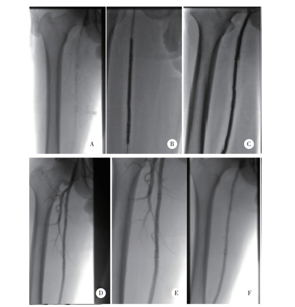
DCB治疗股浅动脉支架长段闭塞病变的影像学资料见图 1。所有患者随访期间均未观察到严重不良事件(死亡及严重的心、脑血管意外等)。

|
| A, DSA angiography showed that the stent length of the right superficial femoral artery was occluded; B, occlusion section of 5 mm×140 mm ordinary balloon pre-expansion stent; C, 5.5 mm×300 mm drug-coated balloon dilatation stent disease occlusion; D, postoperative angiography suggested smooth blood circulation in the superficial femoral artery; E, DSA review 6 months after the operation indicated smooth blood circulation in the superficial femoral artery without obvious stenosis or occlusion; F, DSA review 12 months after the operation indicated smooth blood circulation in the superficial femoral artery without obvious stenosis or occlusion. 图 1 药物涂层球囊治疗股浅动脉支架长段闭塞病变的影像 Fig.1 Image of long segment occlusion of superficial femoral artery stents treated with drug-coated balloon |
术后6、12个月,DCB组病变部位的再狭窄率(4.2%,18.0%)均低于CB组(26.0%,84.0%),差异均有统计学意义(均P < 0.05)。
术前和术后7 d内,2组患者MLD、ABI、PSVR以及Rutherford分级比较,差异无统计学意义(P > 0.05);术后6、12个月,DCB组ABI、MLD均高于CB组,LLL、PSVR均低于CB组(均P < 0.05),见表 2。
| Follow-up indicators | DCB group | CB group | t | P |
| ABI | ||||
| Pre-operation | 0.32±0.60 | 0.30±0.80 | 1.49 | 0.14 |
| 7 days after surgery | 0.93±0.34 | 0.88±0.82 | 1.22 | 0.23 |
| 6 months after surgery | 0.83±0.80 | 0.79±0.60 | 2.24 | 0.03 |
| 12 months after surgery | 0.78±0.67 | 0.45±0.76 | 20.06 | < 0.001 |
| MLD (mm) | ||||
| Pre-operation | 0.51±0.15 | 0.48±0.11 | 1.01 | 0.31 |
| Immediate postoperative measurement | 3.31±0.36 | 3.23±0.35 | 1.12 | 0.27 |
| 6 months after surgery | 3.07±0.41 | 2.89±0.42 | 2.18 | 0.03 |
| 12 months after surgery | 2.91±0.44 | 1.58±0.36 | 16.39 | < 0.001 |
| LLL (mm) | ||||
| 6 months after surgery | 0.24±0.16 | 0.34±0.30 | -2.09 | 0.04 |
| 12 months after surgery | 0.39±0.23 | 1.64±0.51 | -15.30 | < 0.001 |
| PSVR | ||||
| Pre-operation | 3.45±2.18 | 3.80±2.09 | -0.82 | 0.42 |
| 7 days after surgery | 1.15±0.12 | 1.17±0.65 | -1.10 | 0.27 |
| 6 months after surgery | 1.55±0.75 | 2.28±0.73 | -6.70 | < 0.001 |
| 12 months after surgery | 2.01±0.79 | 3.57±1.00 | -8.50 | < 0.001 |
| ABI,ankle brachial index;MLD,minimum lumen diameter;LLL,late lumen loss;PSVR,peak velocity ratio. | ||||
术后6个月,DCB组Rutherford分级1、2级略多于CB组,但2组之间无统计学差异(P > 0.05);术后12个月,DCB组Rutherford分级1、2级明显多于CB组(P < 0.05),见表 3。
| Group | 0 d | 1 d | 2 d | 3 d | 4 d | P |
| Pre-operation | 0.920 | |||||
| DCB group | 0 | 0 | 0 | 34 | 14 | |
| CB group | 0 | 0 | 0 | 35 | 15 | |
| 7 days after surgery | 0.860 | |||||
| DCB group | 40 | 8 | 0 | 0 | 0 | |
| CB group | 41 | 9 | 0 | 0 | 0 | |
| 6 months after operation | 0.191 | |||||
| DCB group | 0 | 35 | 11 | 2 | 0 | |
| CB group | 0 | 28 | 17 | 5 | 0 | |
| 12 months after operation | < 0.001 | |||||
| DCB group | 0 | 30 | 13 | 5 | 0 | |
| CB group | 0 | 5 | 4 | 19 | 22 |
3 讨论
目前关于ISR的机制尚未明确,血管内的超声波研究表明,血管支架的运用完全可以消除血管壁的回缩以及血管壁的负面重构,而支架内的再狭窄则完全是新生内膜的结果[9-11],而新生内膜主要是由增殖的平滑肌细胞[12]和细胞外基质[13]组成。有研究[4]指出,在股浅动脉支架术后的患者中,1年ISR的比例约为18%~40%。目前治疗ISR最常用的方式是单纯CB扩张,其操作简单,成功率高,但是远期疗效并不令人满意,1年的再狭窄率高达49.9%~84.8%[14]。
DCB作为一种新型腔内治疗方式,近年来在股腘动脉病变治疗中得到一定的应用,其表面涂有亲脂性抗增殖药物紫杉醇,该药物具有很高的渗透性,且组织吸收率较高,球囊扩张可使药物迅速渗透入血管壁,且作用持久[15]。DCB主要优点如下:(1)避免了由金属和聚合物诱导的再狭窄、血栓形成以及支架断裂的风险,术后无需持续双重抗血小板;(2)管壁接触面积更大,药物释放更均匀,生物利用度更高;(3)对于扭曲血管、分叉部血管和弥散性病变适应性更好[16];(4)能够在动脉血管壁的平滑肌细胞层和成纤维细胞层保持较高的药物浓度,有效地抑制病变血管内膜增生[17-19]。张文例等[20]的研究也证实了紫杉醇可持续抑制血管平滑肌的增殖。
KRANKENBERG等[21]进行了一项多中心、前瞻性、随机对照临床试验,共纳入119例患者,随机分配为DCB组(62例)和CB组(57例),6个月ISR发生率分别为15.4% (DCB组)和44.7% (CB组),差异有统计学意义(P = 0.002),12个月Rutherford分级至少提高1级。IN.PACT Global试验[22]纳入了131例ISR的患者(149条肢体),34%完全闭塞病变,59.1%钙化病变,药物球囊扩张术后12个月的通畅率为88.7%。国内AcoArtⅠ研究[23]亚组分析中,股腘动脉ISR 46例,DCB组26例,CB组20例,12个月再狭窄发生率分别为23.0%、95.0% (P < 0.01),证明了DCB治疗股腘动脉ISR的中短期疗效。本研究中,术后6个月DCB组和CB组病变部位的再狭窄率分别为4.2%和26.0%,术后12个月再狭窄率分别为18.0%和84.0%,均较前述研究结果略低,可能与本组病例术后口服的盐酸沙格雷酯药物有关。盐酸沙格雷酯对血管平滑肌的5-羟色胺2A受体具有特异性拮抗作用,沙格雷酯+阿司匹林可以显著降低外周血管支架术后的再狭窄发生率,同时显著减少支架内内膜增殖,此外,还可以改善侧支循环[24]。DCB作为一种抗癌药物,过量应用可引起骨髓抑制、过敏反应、消化道反应、神经毒性不良反应[25]。本研究记录了患者于术前后血常规、尿常规、肝功能、肾功能、凝血功能的变化,并观察了术后一般情况,结果并未发现有临床意义的指标变化,也未观察到明显的临床反应。
综上所述,本研究结果表明,DCB治疗股浅动脉ISR的临床疗效要明显优于CB。但本研究也存在不足之处,如样本量偏小,数据测量方面存在主观因素影响,随访时间较短。因此,仍需大样本、多中心对照试验研究进一步论证以上结果。
| [1] |
ABOYANS V, RICCO JB, BARTELINK MLEL, et al. Editor's choice-2017 ESC guidelines on the diagnosis and treatment of peripheral arterial diseases, in collaboration with the European society for vascular surgery (ESVS)[J]. Eur J Vasc Endovascular Surg, 2018, 55(3): 305-368. DOI:10.1016/j.ejvs.2017.07.018 |
| [2] |
CHICHARRO-LUNA E, GRACIA-VESGA MA, OROZCO-BELTRÁN D. Prevalence of peripheral arterial disease and associated factors in elderly patients using an automated oscillometric device[J]. Revista De Enfermeria Barcelona Spain, 2014, 37(5): 18-24. |
| [3] |
KINSTNER CM, LAMMER J, WILLFORT-EHRINGER A, et al. Paclitaxel-eluting balloon versus standard balloon angioplasty in in-stent restenosis of the superficial femoral and proximal popliteal artery:1-year results of the PACUBA trial[J]. JACC:Cardiovasc Interv, 2016, 9(13): 1386-1392. DOI:10.1016/j.jcin.2016.04.012 |
| [4] |
LAIRD JR, KATZEN BT, SCHEINERT D, et al. Nitinol stent implantation versus balloon angioplasty for lesions in the superficial femoral artery and proximal popliteal artery:twelve-month results from the RESILIENT randomized trial[J]. Circ Cardiovasc Interv, 2010, 3(3): 267-276. DOI:10.1161/CIRCINTERVENTIONS.109.903468 |
| [5] |
CREMERS B, BINYAMIN G, CLEVER YP, et al. A novel constrained, paclitaxel-coated angioplasty balloon catheter[J]. EuroIntervention, 2017, 12(17): 2140-2147. DOI:10.4244/EIJ-D-16-00093 |
| [6] |
HUMPHRIES MD, PEVEC WC, LAIRD JR, et al. Early duplex scanning after infrainguinal endovascular therapy[J]. J Vasc Surg, 2011, 53(2): 353-358. DOI:10.1016/j.jvs.2010.08.045 |
| [7] |
周辰光, 董巍巍, 赵晓静. 下肢动脉硬化闭塞症合并糖尿病足的介入治疗及其研究进展[J]. 医学综述, 2015, 21(1): 109-111. DOI:10.3969/j.issn.1006-2084.2015.01.043 |
| [8] |
NORGREN L, HIATT WR, DORMANDY JA, et al. Inter-society consensus for the management of peripheral arterial disease (TASC II)[J]. J Vasc Surg, 2007, 45(1): S5-S67. DOI:10.1016/j.jvs.2006.12.037 |
| [9] |
FINN AV, NAKAZAWA G, KOLODGIE FD, et al. Temporal course of neointimal formation after drug-eluting stent placement:is our understanding of restenosis changing?[J]. JACC:Cardiovasc Interv, 2009, 2(4): 300-302. DOI:10.1016/j.jcin.2009.01.004 |
| [10] |
SUZUMURA H, SUZUKI T, HOSOKAWA H, et al. Neointima in coronary stent does not increase during over 1-year in non-restenosed lesion at 6 months follow-up:serial volumetric intravascular ultrasound study[J]. Jpn Heart J, 2002, 43(6): 581-591. DOI:10.1536/jhj.43.581 |
| [11] |
YAMANAGA K, TSUJITA K, SHIMOMURA H, et al. Serial intravascular ultrasound assessment of very late stent thrombosis after sirolimus-eluting stent placement[J]. J Cardiol, 2014, 64(4): 279-284. DOI:10.1016/j.jjcc.2014.02.008 |
| [12] |
符伟国, 岳嘉宁. 股腘动脉段病变支架内再狭窄的腔内治疗策略分析[J]. 中华外科杂志, 2016, 54(8): 586-590. DOI:10.3760/cma.j.issn.0529-5815.2016.08.006 |
| [13] |
AKBOGA MK, YILMAZ S. Predictors of in-stent restenosis[J]. Angiology, 2019, 70(3): 279. DOI:10.1177/0003319718776796 |
| [14] |
TOSAKA A, SOGA Y, IIDA O, et al. Classification and clinical impact of restenosis after femoropopliteal stenting[J]. J Am Coll Cardiol, 2012, 59(1): 16-23. DOI:10.1016/j.jacc.2011.09.036 |
| [15] |
SCHELLER B, SPECK U, SCHMITT A, et al. Addition of paclitaxel to contrast media prevents restenosis after coronary stent implantation[J]. J Am Coll Cardiol, 2003, 42(8): 1415-1420. DOI:10.1016/S0735-1097(03)01056-8 |
| [16] |
SARODE K, SPELBER DA, BHATT DL, et al. Drug delivering technology for endovascular management of infrainguinal peripheral artery disease[J]. JACC:Cardiovasc Interv, 2014, 7(8): 827-839. DOI:10.1016/j.jcin.2014.05.008 |
| [17] |
NG VG, MENA C, PIETRAS C, et al. Local delivery of paclitaxel in the treatment of peripheral arterial disease[J]. Eur J Clin Investig, 2015, 45(3): 333-345. DOI:10.1111/eci.12407 |
| [18] |
COLLERAN R, HARADA Y, CASSESE S, et al. Drug coated balloon angioplasty in the treatment of peripheral artery disease[J]. Expert Rev Med Devices, 2016, 13(6): 569-582. DOI:10.1080/17434440.2016.1184969 |
| [19] |
TEPE G, SCHNORR B, ALBRECHT T, et al. Angioplasty of femoral-popliteal arteries with drug-coated balloons:5-year follow-up of the THUNDER trial[J]. JACC:Cardiovasc Interv, 2015, 8(1): 102-108. DOI:10.1016/j.jcin.2014.07.023 |
| [20] |
张文俐, 杜润, 朱政斌, 等. 药物洗脱球囊抑制下肢动脉狭窄性病变的实验研究[J]. 介入放射学杂志, 2014, 23(5): 423-426. DOI:10.3969/j.issn.1008-794X.2014.05.014 |
| [21] |
KRANKENBERG H, TÜBLER T, INGWERSEN M, et al. Drug-coated balloon versus standard balloon for superficial femoral artery in-stent restenosis:the randomized femoral artery in-stent restenosis (FAIR) trial[J]. Circulation, 2015, 132(23): 2230-2236. DOI:10.1161/CIRCULATIONAHA.115.017364 |
| [22] |
BRODMANN M, KEIRSE K, SCHEINERT D, et al. Drug-coated balloon treatment for femoropopliteal artery disease:the IN.PACT global study de novo in-stent restenosis imaging cohort[J]. JACC:Cardiovasc Interv, 2017, 10(20): 2113-2123. DOI:10.1016/j.jcin.2017.06.018 |
| [23] |
JIA X, ZHANG JW, ZHUANG BX, et al. Acotec drug-coated balloon catheter:randomized, multicenter, controlled clinical study in femoropopliteal arteries:evidence from the AcoArt I trial[J]. JACC:Cardiovasc Interv, 2016, 9(18): 1941-1949. DOI:10.1016/j.jcin.2016.06.055 |
| [24] |
刘丹, 陈忠, 翟梦瑶, 等. 药物治疗对外周动脉硬化闭塞症支架术后再狭窄的影响[J]. 中华普通外科杂志, 2012, 27(11): 896-899. DOI:10.3760/cma.j.issn.1007-631X.2012.11.012 |
| [25] |
CASADEI GARDINI A, TENTI E, MASINI C, et al. Multicentric survey on dose reduction/interruption of cancer drug therapy in 12.472 patients:indicators of suspected adverse reactions[J]. Oncotarget, 2016, 7(26): 40719-40724. DOI:10.18632/oncotarget.8942 |
 2020, Vol. 49


