文章信息
- 曹正飞, 王青青, 关慧娟, 冯翠娟
- CAO Zhengfei, WANG Qingqing, GUAN Huijuan, FENG Cuijuan
- 恒牙
 早期安氏Ⅱ类2分类患者牙弓及基骨弓宽度的特征分析
 - Analysis of the dental and alveolar arch widths in patients with early permanent classⅡ division 2 malocclusion
- 中国医科大学学报, 2020, 49(1): 35-38
- Journal of China Medical University, 2020, 49(1): 35-38
-
文章历史
- 收稿日期:2019-01-07
- 网络出版时间:2019-12-11 17:02
 早期安氏Ⅱ类2分类患者牙弓及基骨弓宽度的特征分析
 早期安氏Ⅱ类2分类错
 畸形牙弓及基骨弓宽度的特征。方法 选取处于恒牙
 早期的患者共57例,其中安氏Ⅰ类29例,安氏Ⅱ类2分类28例,年龄11~15岁。使用3shape扫描仪扫描患者正畸前记存模型获得数字化模型,对数字化模型进行测量分析。结果 与安氏Ⅰ类患者相比,安氏Ⅱ类2分类患者上颌前磨牙区及下颌尖牙区牙弓宽度较小(P < 0.05),其余牙弓测量项目无统计学差异(P>0.05)。2种错
 类型患者下颌牙槽骨宽度无统计学差异(P>0.05)。结论 安氏Ⅱ类2分类患者上颌前磨牙区及下颌尖牙区牙弓宽度小于安氏Ⅰ类患者,下颌基骨弓宽度与安氏Ⅰ类患者相比无差异。安氏Ⅱ类2分类患者在正畸治疗过程中应注意上下颌牙弓宽度的调整。
安氏Ⅱ类2分类是指磨牙关系主要表现为Ⅱ类关系、上切牙舌倾、下切牙代偿性伸长、覆盖小、覆


选取2016年3月至2018年6月间中国医科大学附属口腔医院正畸二科诊治的恒牙
纳入标准:(1)年龄11~15岁;(2)恒牙列完整(除第三磨牙),除第二磨牙外其他牙齿完全萌出建


排除标准:(1)存在颌骨偏斜等骨性畸形;(2)存在唇裂、腭裂等先天性颅颌面畸形;(3)存在全身系统性疾病;(4)就诊前接受过正畸治疗。
1.2 研究对象分组标准 1.2.1 安氏Ⅰ类(1)正中

(1)正中
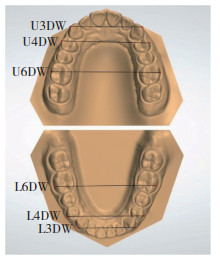
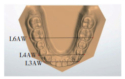
使用藻酸盐印模材料获取印模,随即用硬石膏材料灌制获得患者记存模型,要求模型完整、准确、清晰,包括所有牙列、基骨、唇舌系带,软组织皱襞及腭部,无明显气泡及石膏瘤等。而后使用3shape扫描仪扫描石膏模型,获得数字化模型。选取目标牙位临床冠颊侧中心点(facial-axis point,FA)作为牙弓宽度测量标志点;选取目标牙位临床冠长轴龈向延长线与WALA嵴的交点作为基骨测量标记点,分别以尖牙间、第一前磨牙间及第一磨牙间测量数据代表尖牙区、前磨牙区及磨牙区牙弓或基骨弓宽度(图 1、2)。所有定点与测量工作由同一研究者在一定连续时间内完成,每个项目重复测量3次,取平均值。

|
| U3DW, maxillary canine width; U4DW, maxillary premolar width; U6DW, maxillary molar width; L3DW, mandibular canine width; L4DW, mandibular premolar width; L6DW, mandibular molar width. 图 1 上下颌牙弓宽度测量 Fig.1 Measurement of the maxillary and mandibular dental arch width |

|
| L3AW, alveolar width of the mandibular canine; L4AW, alveolar width of the mandibular premolar; L6AW, alveolar width of the mandibular molar. 图 2 下颌牙槽骨宽度测量 Fig.2 Measurement of the mandibular alveolar arch width |
1.4 统计学分析
采用SPSS 20.0软件进行统计学分析,检验水准设定为α=0.05。采用独立样本t检验比较2组牙弓宽度及基骨弓宽度测量指标。
2 结果与安氏Ⅰ类患者相比,安氏Ⅱ类2分类患者上颌前磨牙区及下颌尖牙区牙弓宽度较小(P < 0.05),其余牙弓测量项目无统计学差异(P> 0.05)。2种错
| Item | ClassⅠ | Class Ⅱ division 2 | t | P |
| U3DW(mm) | 38.80±1.83 | 38.21±2.12 | 1.128 | 0.264 |
| U4DW(mm) | 47.30±1.92 | 46.04±2.19 | 2.302 | 0.025 |
| U6DW(mm) | 58.05±2.57 | 57.04±2.82 | 1.404 | 0.166 |
| L3DW(mm) | 29.80±1.44 | 28.81±1.74 | 2.336 | 0.023 |
| L4DW(mm) | 39.70±1.91 | 39.10±2.49 | 1.026 | 0.309 |
| L6DW(mm) | 52.31±2.26 | 51.90±2.76 | 0.610 | 0.544 |
| L3AW(mm) | 30.77±1.62 | 30.17±2.02 | 1.245 | 0.218 |
| L4AW(mm) | 41.62±1.63 | 41.07±2.54 | 0.958 | 0.343 |
| L6AW(mm) | 58.11±2.22 | 57.85±2.77 | 0.382 | 0.704 |
3 讨论
牙弓、基骨弓宽度是影响正畸治疗的重要因素,了解二者形态的差异可以帮助医生设计更为合理的治疗计划,从而达到理想的治疗效果。一直以来,基骨弓形态测量标记点的确定方法有很大差异,有学者使用根尖点及根中心点作为基骨测量点[7-8]。但是,牙根的变异性以及锥形束计算机断层扫描显像清晰程度的差异会造成一定的测量误差。2000年,ANDREWS提出的WALA嵴,即附着龈与牙槽黏膜交界软组织带上最凸点的连线。研究[9-12]表明,WALA嵴与下颌FA点间距离存在显著相关性,并可以代表下颌骨基骨的形态特征及宽度。因此,本研究应用“WALA嵴”概念并结合模型3D测量对安氏Ⅱ类2分类患者牙弓、基骨弓宽度进行分析,为此类患者的治疗提供指导。
本研究发现,安氏Ⅱ类2分类患者上颌前磨牙区牙弓宽度小于安氏Ⅰ类患者,上颌尖牙及磨牙区牙弓宽度的差异无统计学意义。有研究[13]认为安氏Ⅱ类2分类患者的上颌尖牙区和前磨牙区的宽度明显小于个别正常


安氏Ⅱ类2分类患者由于上颌切牙腭侧倾斜,使上颌尖牙区形成类似于卵圆形或方圆形牙弓的转折,因此上颌尖牙区牙弓宽度与安氏Ⅰ类相比无差异。其次,由于下颌牙弓矢状向发育受限,且下颌前牙不能形成与后牙区相似的尖窝接触咬合关系,在受到某些因素影响时更易出现移位,因此下颌前牙段出现了拥挤及尖牙区宽度不足[17]。另外,后缩的下颌使上下颌牙弓不能正常匹配,继而上颌前磨牙区牙弓横向发育受到下颌牙弓的限制而出现狭窄。同时舌体对下颌磨牙区的水平向力量增加,且此处牙弓宽度发育受到的限制较小,所以与安氏Ⅰ类相比下颌磨牙区宽度无差异[18]。
因此,对安氏Ⅱ类2分类青少年患者应采取早期治疗,解除前牙闭锁


综上所述,安氏Ⅱ类2分类患者在正畸治疗过程中,应考虑解决患者的牙弓及基骨弓宽度异常,并注意上下牙弓宽度的调整,尤其是青少年患者,这将有利于患者牙颌面部的正常生长发育及正畸治疗效果的稳定。本研究由于纳入研究对象的数量限制,未对不同垂直骨面性、性别等可能会影响牙弓宽度的因素进行分组分析,今后可以以此作为出发点,进行更大样本量的高质量研究。
| [1] |
陈扬熙. 口腔正畸学[M]. 北京: 人民卫生出版社, 2012: 611.
|
| [2] |
MARUO IT. Class Ⅱ division 2 subdivision left malocclusion associated with anterior deep overbite in an adult patient with temporomandibular disorder[J]. Dental Press J Orthod, 2017, 22(4): 102-112. DOI:10.1590/2177-6709.22.4.102-112.bbo |
| [3] |
ZHANG K, HUANG L, YANG L, et al. Effects of transverse relationships between maxillary arch, mouth, and face on smile esthetics[J]. Angle Orthod, 2016, 86(1): 135-141. DOI:10.2319/101514.1 |
| [4] |
OH H, MA N, FENG PP, et al. Evaluation of posttreatment stability after orthodontic treatment in the mixed and permanent dentitions[J]. Angle Orthod, 2016, 86(6): 1010-1018. DOI:10.2319/122315-881.1 |
| [5] |
PINHEIRO FH, GARIB DG, JANSON G, et al. Longitudinal stability of rapid and slow maxillary expansion[J]. Dental Press J Orthod, 2014, 19(6): 70-77. DOI:10.1590/2176-9451.19.6.070-077.oar |
| [6] |
MARINELLI A, MARIOTTI M, DEFRAIA E. Transverse dimensions of dental arches in subjects with class Ⅱ malocclusion in the early mixed dentition[J]. Prog Orthod, 2011, 12(1): 31-37. DOI:10.1016/j.pio.2011.02.006 |
| [7] |
刘荫, 李雪琦, 陈宇, 等. 安氏Ⅰ类错  患者下颌Bonwill-Hawley弓形与基骨弓形态相关性的三维测量研究[J]. 口腔疾病防治, 2017, 25(5): 300-304. DOI:10.12016/j.issn.2096-1456.2017.05.005 患者下颌Bonwill-Hawley弓形与基骨弓形态相关性的三维测量研究[J]. 口腔疾病防治, 2017, 25(5): 300-304. DOI:10.12016/j.issn.2096-1456.2017.05.005 |
| [8] |
SUK KE, PARK JH, BAYOME M, et al. Comparison between dental and basal arch forms in normal occlusion and class Ⅲ malocclusions utilizing cone-beam computed tomography[J]. Korean J Orthod, 2013, 43(1): 15-22. DOI:10.4041/kjod.2013.43.1.15 |
| [9] |
ZOU W, JIANG J, XU T, et al. Relationship between mandibular dental and basal bone arch forms for severe skeletal class Ⅲ patients[J]. Am J Orthod Dentofacial Orthop, 2015, 147(1): 37-44. DOI:10.1016/j.ajodo.2014.08.019 |
| [10] |
吴佳琪, 江久汇, 邹薇, 等. 骨性Ⅱ类错  下颌牙弓与基骨形态相关性的三维测量研究[J]. 华西口腔医学杂志, 2013, 31(6): 605-609. DOI:10.7518/hxkq.2013.06.015 下颌牙弓与基骨形态相关性的三维测量研究[J]. 华西口腔医学杂志, 2013, 31(6): 605-609. DOI:10.7518/hxkq.2013.06.015 |
| [11] |
郑洁, 韩梅, 贺红. 利用WALA嵴分析安氏Ⅱ类1分类患者牙弓与基骨形态[J]. 武汉大学学报(医学版), 2015, 36(4): 607-610, 615. DOI:10.14188/j.1671-8852.2015.04.026 |
| [12] |
李小兵. 牙弓/牙槽骨弓的塑形矫治:基于牙弓形态发育不良的儿童错  畸形诊断与阻断治疗[J]. 华西口腔医学杂志, 2016, 34(6): 556-563. DOI:10.7518/hxkq.2016.06.002 畸形诊断与阻断治疗[J]. 华西口腔医学杂志, 2016, 34(6): 556-563. DOI:10.7518/hxkq.2016.06.002 |
| [13] |
游新, 秦朴, 杜跃华, 等. 安氏Ⅱ类2分类错  颅颌面形态及牙弓宽度的特征分析[J]. 重庆医科大学学报, 2010, 35(12): 1863-1866. DOI:10.13406/j.cnki.cyxb.2010.12.035 颅颌面形态及牙弓宽度的特征分析[J]. 重庆医科大学学报, 2010, 35(12): 1863-1866. DOI:10.13406/j.cnki.cyxb.2010.12.035 |
| [14] |
HUTH J, STALEY RN, JACOBS R, et al. Arch widths in class Ⅱ-2 adults compared to adults with class Ⅱ-1 and normal occlusion[J]. Angle Orthod, 2007, 77(5): 837-844. DOI:10.2319/062305-209 |
| [15] |
PATEL D, MEHTA F, PATEL N, et al. Evaluation of arch width among class Ⅰ normal occlusion, class Ⅱ division 1, class Ⅱ division 2, and class Ⅲ malocclusion in Indian population[J]. Contemp Clin Dent, 2015, 6(Suppl 1): S202-S209. DOI:10.4103/0976-237X.166842 |
| [16] |
李琳, 张敏, 王桂子, 等. 安氏Ⅱ类2分类错  牙弓及牙槽骨弓宽度的特点分析[J]. 牙体牙髓牙周病学杂志, 2017, 27(1): 41-44, 52. DOI:10.15956/j.cnki.chin.j.conserv.dent.2017.01.009 牙弓及牙槽骨弓宽度的特点分析[J]. 牙体牙髓牙周病学杂志, 2017, 27(1): 41-44, 52. DOI:10.15956/j.cnki.chin.j.conserv.dent.2017.01.009 |
| [17] |
WALKOW TM, PECK S. Dental arch width in class Ⅱ division 2 deep-bite malocclusion[J]. Am J Orthod Dentofacial Orthop, 2002, 122(6): 608-613. DOI:10.1067/mod.2002.129189 |
| [18] |
武秀萍.恒牙期安氏Ⅱ类2分类错 |
| [19] |
SMITH DF, DALESIO NM, BENKE JR, et al. Anthropometric and dental measurements in children with obstructive sleep apnea[J]. J Clin Sleep Med, 2016, 12(9): 1279-1284. DOI:10.5664/jcsm.6132 |
| [20] |
PETRACCONE CAIXETA AC, ANDRADE I, BAHIA JUNQUEIRA PEREIRA T, et al. Dental arch dimensional changes after adenotonsillectomy in prepubertal children[J]. Am J Orthod Dentofacial Orthop, 2014, 145(4): 461-468. DOI:10.1016/j.ajodo.2013.12.018 |
| [21] |
IWASAKI T, SATO H, SUGA H, et al. Relationships among nasal resistance, adenoids, tonsils, and tongue posture and maxillofacial form in class Ⅱ and class Ⅲ children[J]. Am J Orthod Dentofacial Orthop, 2017, 151(5): 929-940. DOI:10.1016/j.ajodo.2016.10.027 |
| [22] |
MUTINELLI S, COZZANI M. Rapid maxillary expansion in early-mixed dentition:effectiveness of increasing arch dimension with anchorage on deciduous teeth[J]. Eur J Paediatr Dent, 2015, 16(2): 115-122. |
| [23] |
ESENLIK E, RÜBENDÜZ M. An evaluation of the dentoskeletal effects of slow maxillary expansion from the mixed to the permanent dentition[J]. Aust Orthod J, 2015, 31(1): 2-13. |
| [24] |
MERTENS B, ANGIONI C, ORTI V, et al. Collaboration between periodontics and orthodontics:interest of alveolar corticotomies and piezocision. Review of literature[J]. Orthod Fr, 2017, 88(2): 179-191. DOI:10.1051/orthodfr/2017010 |
| [25] |
YEN SL. A comparison between osteotomy and corticotomy-assisted tooth movement[J]. Front Oral Biol, 2016, 18: 124-129. DOI:10.1159/000351907 |
 2020, Vol. 49


