Fatty acids (FAs) play important roles in biological processes, and are essential molecular components of all organisms. As fundamental components of phospholipids in cell membranes, fatty acids affect the composition of cell membranes and influence organism health. Fatty acids are divided into saturated fatty acids, monounsaturated fatty acids (UFAs) and polyunsaturated fatty acids (PUFAs) depending on their degree of saturation. PUFAs are basic components of cell membranes that control cell configuration and permeability (Xiao et al., 2001). These fatty acids play essential roles in biological growth and lipid metabolism. Studies have shown that PUFA anabolism in fish is affected by external factors, such as nutrition and the environment, as well as genetic factors, which may influence the mRNA level of key enzymes in the high UFA synthesis pathway (Xie et al., 2013). Cook et al. (2000 and Barberá et al. (2011 studied the fatty acid composition of sea urchin gonads and showed that three PUFAs mainly contribute to gonad composition, namely, eicosapentaenoic acid (C20:5n-3), arachidonic acid (C20: 4n-6) and palmitoleic acid (C16:1n-7). PUFAs are also essential to sea urchin growth and development. Studies have shown that the growth rate of sea urchins rich in PUFAs is superior to that of control group (Ding, 2014).
Peroxisome proliferator-activated receptors (PPARs) are members of the nuclear receptor superfamily that regulates the expression of target genes, particularly those associated with lipid metabolism (Zhu et al., 2000). Issemann et al. (1990 discovered that this protein family is activated by fatty acid-like compounds called peroxisome proliferators; and the nuclear receptors came to be known as PPARs. The proteins can be divided into three structural type, namely, α, β (or δ), and γ, which are products of different genes (Issemann, 1990; Dreyer, 1992; Kliewer, 1994; Zhu, 1995). Mammalian PPARγ strongly influences lipid metabolism, cell proliferation, and inflammation. PPARγ signaling pathways control lipid uptake, transport, storage, and disposal (Walczak and Tontonoz, 2002). PPARγ also regulates the expression of gene networks required for cell proliferation, differentiation, morphogenesis, and metabolic homeostasis, thus highlighting its ability to activate lipogenic genes and adipocyte differentiation. Given its interesting properties, the functions of PPARγ have become an important research area in recent years.
The PPARγ gene sequences of several fish species, including Salmo salar (Ruyter et al., 1997), Pleuronectes platessa (Leaver et al., 1998), Danio rerio (Ibabe, 2002), Dicentrarchus labrax (Boukouvala et al., 2004), Mugil cephalus (Ibabe et al., 2004), Salmo trutta f. fario (Batista-Pinto, 2005), Carassius auratus (Mimeault et al., 2006), Rachycentron canadum (Tsai et al., 2008), and Ctenopharyngodon idella (He et al., 2012), have been cloned and functionally identified. However, no reports of PPARγ identification in Strongylocentrotus intermedius have yet been published.
Uncoupling protein-2 (UCP2) was discovered in 1997 as a homologue of the brown-fat UCP (UCP1). Similar to UCP1, which is able to dissipate caloric energy as heat by uncoupling mitochondrial respiration from ATP production, UCP2 can uncouple respiration, at least in vitro (Fleury, 1997). UCP2 is present in many tissues involved in fuel metabolism. Its expression is increased in fat and muscle in response to elevated circulating free fatty acids from fasting and high-fat feeding (Medvedev et al., 2001).
Medvedev et al. (2001 proposed a role for PPARγ as a mediator of physiological changes in UCP2, because thiazolidinediones increase UCP2 expression in the cells of tissues related to fuel metabolism (Aubert, 1997). The PPAR-dependent pathway might play a role in mediating the response of UCP2 to fatty acids because treatment with PPARγ agonists can increase UCP2 mRNA levels both in vivo and in vitro (Aubert, 1997; Camirand, 1998). However, the regulatory relationship between these two genes in S. intermedius remains unclear.
In 1989, S. intermedius was introduced from Japan by Dalian Ocean University and promoted in coastal areas, such as the Liaoning and Shandong Peninsulas, of China. Today, this species is one of the main sea urchin varieties cultivated in China (Chang et al., 2004). The only edible component of the animal is the gonad, and its nutritional value is mainly reflected by the medicinal effects of its fatty acids (Zuo et al., 2016). These fatty acids are extremely complex and contribute to the growth and reproduction of the organism (Russell, 1998; Palacios et al., 2007; Wei et al., 2016). Many types of fatty acids occur in sea urchins, and their total amount outweighs those found in fish species from both seawater and freshwater environments. Sea urchins feature much higher amounts of UFAs compared with marine fish and shrimp (Wang et al., 2006). UFAs from gonads are efficacious for preventing certain cardiovascular and cerebrovascular diseases and have high nutritional and medicinal value. Previous studies showed that UFAs are closely related to the growth, development, and reproduction of sea urchins (Kang and Leaf, 1996; Tavazzi et al., 2008; Carboni et al., 2013).
PPARγ is a fatty acid-related gene. To understand the function of PPARγ in S. intermedius, we used rapid amplification of cDNA ends (RACE) polymerase chain reaction (PCR) to obtain the full-length cDNA sequence encoding PPARγ in this species. Then, we investigated the expression patterns of PPARγ using fluorescence quantitative PCR and studied the relationship between PPARγ and UCP2.
2 Materials and Methods 2.1 Animals, Rearing Conditions, and Sample CollectionThe 2-year-old S. intermedius samples used in this study were bred at the Key Laboratory of Mariculture and Stock Enhancement in the North China Sea, Ministry of Agriculture and Rural Affairs, China. The healthy sea urchins were homogeneous in size with shell heights of (29.11±2.10) mm, shell lengths of (50.13±3.46) mm and weights of (41.53±5.05) g.
Tube feet, peristomial membrane, Aristotle's lantern, coelomocyte tissues, gonads, and intestines were dissected from nine sea urchins. Samples from the different developmental stages were also collected as follows. Three females and three males were induced to spawn by injection of 1 mL of KCl (40 μL per gram of body mass) into the coelom via the peristomial membrane (Kelly et al., 2000). The fertilized ovum develops from the multicell stage to the blastocyst stage through cleavage, enters the gastrula stage after blastocyst rotation, and then develops to the planktonic stage. The planktonic larvae are divided into two stages, including prismatic larvae and long-winged larvae. In the late stage of the long-winged larvae, a larva's wrist gradually disappears, and it eventually becomes a juvenile sea urchin after a series of metamorphic transformations. Samples were pooled and passed through a 400 μm sieve to remove impurities, taken up by pipettes, and placed in a 1.5 mL centrifuge tube. All tissue and developmental stage samples were frozen in liquid nitrogen immediately and stored at -80℃ for RNA extraction and fatty acid analyses.
2.2 Cloning of a Partial PPARγ cDNA FragmentTotal RNA was extracted from the intestines of three S. Intermedius individuals using Trizol reagent (Invitrogen) and digested by the RNase-free DNase I (TaKaRa, China) to remove any possible genomic DNA contaminants. Firststrand cDNA was synthesized with a reverse transcription system (TaKaRa) according to the manufacturer's protocol. A pair of degenerate primers, PPARγ-F/PPARγ-R (Table 1), was designed based on the conserved regions of PPARγ sequences from other species and used for PCR under following conditions: initial denaturation at 94℃ for 5 min, followed by 35 cycles of 94℃ for 30 s, 60℃ for 30 s, and 72℃ for 45 s, and a final extension at 72℃ for 10 min. The PCR product was cloned into the pMD18-T simple vector (TaKaRa) and sequenced.
|
|
Table 1 Primers used in this study |
The partial cDNA sequences of PPARγ were obtained from our results of cloning of a partial PPARγ cDNA fragment, and the gene-specific primers were designed according to these cDNA sequences by Primer Premier 5.0. The nucleotide sequences of all of the primers used in this study are shown in Table 1, and the primers were obtained from Sangon Biotech (Shanghai, China). RACE products were extracted and purified using the Quick Gel Extraction Kit (Transgen Biotech, China). The purified products were subcloned into PEASYⓇ-1 Cloning Vector (Transgen Biotech, China) and immediately transformed into Trans1-T1 phage-resistant chemically competent cells (Transgen Biotech, China). Positive recombinants were identified by colony PCR using M13 primers (Transgen Biotech, China) and finally sequenced at Sangon (Shanghai, China) for sequence confirmation.
2.4 Bioinformatics AnalysisThe nucleotide sequence of the full-length PPARγ cDNA sequence was analyzed using the Basic Local Alignment Search Tool (BLAST) program at the National Center for Biotechnology Information (https://www.ncbi.nlm.nih.gov/blast/), and the deduced AA sequence was determined using the Expert Protein Analysis System (http://www.expasy.org/). The open reading frame (ORF) was identified with ORF Finder (https://www.ncbi.nlm.nih.gov/orffinder/). The physical and chemical parameters of the deduced protein were computed using the ProtParam tool (http://web.expasy.org/protparam/). The computed parameters also included the molecular weight, theoretical isoelectric point (pI), and grand average of hydropathicity (GRAVY). The presence and location of the signal peptide cleavage sites in the AA sequence were predicted by SignalP 4.1 Server (http://www.cbs.dtu.dk/services/SignalP/). The functional domains and important sites of the protein were predicted by InterPro (http://www.ebi.ac.uk/interpro/). Multiple sequence alignment of the deduced AA sequences derived from PPARγ was performed using the ClustalW program (http://www.ch.embnet.org/software/ClustalW.html). A phylogenetic tree was constructed according to the AA sequences of the selected PPARγ using the neighbor-joining (NJ) method in MEGA 6.0 software.
2.5 Real-Time PCR Analyses of PPARγ ExpressionThe relative expression levels of PPARγ mRNA transcripts in different tissues and at different developmental stages (i.e., eggs, 2 cells, 16 cells, 32 cells, 64 cells, blastula, gastrula, four-armed arm larvae, and six-armed larvae) were analyzed by quantitative real-time PCR (qRT-PCR), which was conducted on an Applied Biosystems 7500 real-time PCR system (Foster City, CA, USA) following the manufacturer's instructions on the use of SYBR Premix Ex Taq (SYBR PrimeScriptTM RT-PCR Kit II, Ta-KaRa, Japan). The 18S rRNA gene was used as the internal reference gene. Amplification was performed in a total volume of 20 μL containing 2 μL of 1:5 diluted original cDNA, 10 µL of 2×SYBR Green Master mix (TaKaRa, Japan), 0.4 μL of ROX Reference Dye II, 6 μL of PCR-grade water and 0.8 μL (10 mmol L-1) of each primer. The reaction was performed as follows: 40 cycles of 94℃ for 5 min, 94℃ for 30 s, 60℃ for 30 s, and 72℃ for 30 s. The final extension step was conducted at 72℃ for 5 min. Three independent biological replicates and three technical repetitions of each group were carried out. At the end of the PCR reactions, melting curve analysis of the amplification products confirmed that a single PCR product was present in these reactions. The relative expression levels of the target gene were calculated by the 2-ΔΔCT method described by Livak and Schmittgen (2002.
2.6 RNAi in S. intermedius and Identification of a Relationship Between PPARγ and UCP2Small interfering RNAs (siRNAs) targeting PPARγ (Table 1) were designed and synthesized by GenePharma (Shanghai, China). Six healthy sea urchins with consistent growth, an average shell diameter of (43.25±1.19) mm and an average wet weight of (25.36±2.25) g were randomly divided into the negative control and experimental groups with three individuals in each group. The first group was pipetted with 5 μL of NC (negative control), 5 μL of Lipo6000TM transfection reagent (Beyotime, Shanghai), and 40 μL PBS buffer; the second group was pipetted with 5 μL of siRNA, 5 μL of Lipo6000TM transfection reagent (Beyotime), and 40 μL of PBS buffer. These components were mixed evenly, allowed to stand for 5 min, and then injected into the body cavity through the sea urchin's mouth. After 48 h, total RNA was extracted from the coelomocyte tissues, intestines, and gonads and reversetranscribed. Finally, we detected the relative expression levels of PPARγ and UCP2 genes using qRT-PCR and detected changes of fatty acid composition before and after interference by capillary gas chromatography.
2.7 Statistical AnalysisAll data were expressed as mean±SD (standard deviation). All analyses were performed using one-way ANOVA (Analysis of variance) with SPSS software (version 16.0). The P value was adjusted for multiple tests using a false discovery rate (Benjamini-Hochberg), and P < 0.05 was considered statistically significant
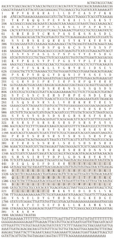
3 Results 3.1 Sequence Analysis of PPARγThe full-length sequence of PPARγ comprised 2286 base pairs (bp), including a 5' untranslated region (UTR) of 145 bp, a 3' UTR of 386 bp and a putative ORF of 1755 bp, The gene encoded a protein with 584 AA residues. The predicted molecular mass of PPARγ is 67.27 kDa, and its theoretical pI is 10.07. The protein sequence included an RRM (RNA recognition motif) domain (Fig. 1).
|
Fig. 1 Nucleotide and deduced amino acid (AA) sequences of PPARγ. The initiation codon is shown in italics. The asterisk (*) indicates a terminal codon. RRM (RNA recognition motif) combined with its domain is shown in the shaded area. |
The AA hydrophilicity/hydrophobicity of the PPARγ enzyme was analyzed using the online ProtScale database, which showed that the aliphatic index is 30.97, the maximum hydrophilic coefficient is 1.389, the minimum hydrophilic coefficient is -3.500, and the GRAVY is 0.835. These data suggest the protein is hydrophobic in nature.
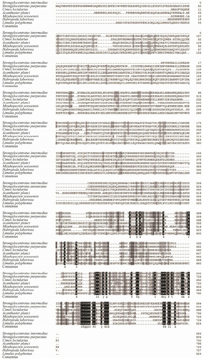
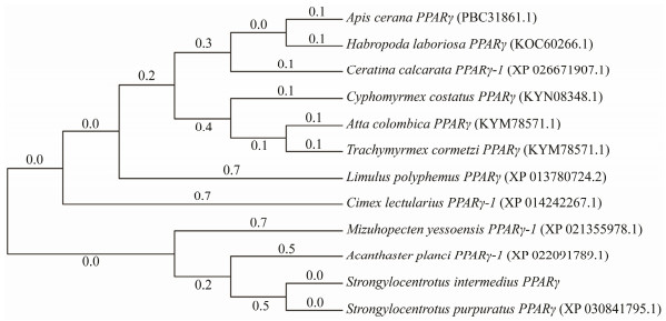
3.3 Multiple Alignment and Phylogenetic Analyses of PPARγThe deduced AA sequence of PPARγ in S. intermedius was analyzed using BLAST. The results showed that PPARγ shares high homology with PPARγ from other vertebrates and invertebrates, including Strongylocentrotus purpuratus (97%), Cimex lectularius (50%), Acanthaster planci (47%), Mizuhopecten yessoensis (52%), Habropoda laboriosa (46%), and Limulus polyphemus (53%). We constructed a phylogenetic tree using the NJ algorithm to reveal the phylogeny of PPARγ, and the results are shown in Figs. 2 and 3.
|
Fig. 2 ClustalX alignment of S. intermedius PPARγ. Amino acid (AA) sequences. The shaded regions indicate identical residues. Other conserved but dissimilar AAs are shaded in gray. |
|
Fig. 3 Consensus neighbor-joining (NJ) tree based on the amino acid sequences of PPARγ genes from other species. The evolutionary history was inferred using the NJ method. Evolutionary analyses were conducted in MEGA7.0. |
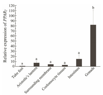
Tissue distributions of PPARγ mRNA were detected by qRT-PCR. The PPARγ gene was expressed in all tissues tested. Expression levels were the highest in the gonads and the lowest in the tube feet. The coelomocyte tissues also showed low PPARγ expression levels (Fig. 4).
|
Fig. 4 Relative expression levels of PPARγ in different tissues. Each vertical bar represents the mean±SD (n = 3). 18S rRNA was used as the reference gene. Different Roman letters above the bars provided indicate significant differences in different tissues at P < 0.05. |
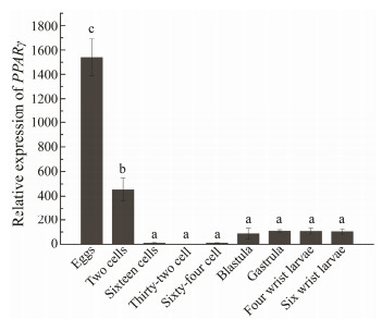
To understand the role of PPARγ at different developmental stages of sea urchin and the synthesis of fatty acids during development, we examined PPARγ expression in the egg, 2-cells, 16-cells, 32-cells, blastula, gastrula, fourarmed larva and six-armed larva stages of S. intermedius. PPARγ was expressed at all nine stages of development. The relative expression level of PPARγ was highest during the egg stage and lowest in the 32-cell stage. PPARγ expression initially decreased and then increased over the entire developmental process of sea urchin (Fig. 5).
|
Fig. 5 Relative expression of PPARγ in S. intermedius at the different developmental stages. 18S rRNA was used as the reference gene. Different Roman letters above the bars provided indicate significant differences in different tissues at P < 0.05. |
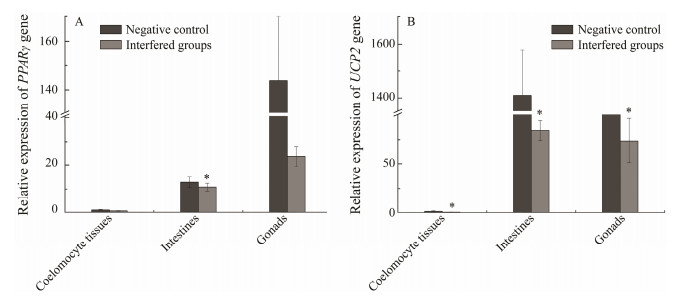
PPARγ siRNA experiments were performed to understand the regulatory relationship between PPARγ and UCP2. Compared with that in the negative control, 48 h of transfection of PPARγ siRNA reduced PPARγ expression in the coelomocyte tissue, intestines and gonads by 43.17%, 16.16%, 83.56%, respectively. Compared with that in the negative control, 48 h of transfection of PPARγ siRNA reduced the expression of UCP2 mRNA in the coelomocyte tissue, intestines, and gonads by 5.96%, 16.29%, and 33.75%, respectively (Fig. 6).
|
Fig. 6 (A) PPARγ mRNA levels of RNAi-PPARγ. (B) UCP2 mRNA levels of RNAi-PPARγ. Values are presented as mean±SD (n = 3). Asterisks indicate significant differences, P < 0.05. |
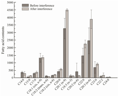
To determine the role of PPARγ in fatty acid synthesis, we examined the changes of fatty acid levels in the gonads of sea urchins, before and after PPARγ siRNA interference. The results showed that C4:0, C18:2(trans, n-6) and C20:3(n-6) levels were decreased after PPARγ siRNA interference (Fig. 7).
|
Fig. 7 Changes in fatty acid contents before and after PPARγ siRNA interference (n = 3). Asterisks indicate significant differences, P < 0.05. |
Sea urchin is a delicacy in many countries, including China, Japan, and France. Essential nutrients such as lipids and PUFAs are stored in the gonads. They not only determine the nutritional value of this delicacy but also ensure the normal growth and reproduction of sea urchins in culture (Han et al., 2019). S. intermedius gonads are rich in fatty acids, especially UFAs. PPARγ can activate lipogenic genes and adipocyte differentiation. In this study, we characterized and analyzed the expression of a PPARγ cDNA in S. intermedius. The full-length cDNA of PPARγ, which includes 1755 bp was obtained. Phylogenetic analysis revealed that S. intermedius and S. purpuratus are clustered together, thus indicatingthat these two species are closely related and have a long relationship with other vertebrates. Similarities in the sequence, conserved domains, and phylogenetic analyses of these species provide supporting evidence that the cloned geneis indeed S. intermedius PPARγ.
Many studies have investigated the relationship between the tissue expression of fish PPARs and their functions. Tsai et al. (2008 cloned PPARα, PPARβ, and PPARγ from R. canadum, and found that PPARγ is expressed in all tissues, particularly in visceral fat deposits. He et al. (2012 cloned the three PPAR genes from C. idellaa and determined that PPARγ is highly expressed in the liver and micro-expressed in other tissues. Because liver is one of the most important organs regulating glucose and lipid metabolism, these results confirm that PPARγ is an important transcriptional regulator of glucose and lipid metabolism. In this study, qRT-PCR analysis showed that PPARγ is expressed in all tissues of S. intermedius, especially in the gonads, followed by the intestines and Aristotle's lantern. The high expression of PPARγ observed in the gonads agrees with the hypothesis that the UFA content of the gonads of sea urchins is high (Chang et al., 2004). Our data partly agree with the tissue expression patterns of PPARγ mRNA determined in the loach, where the highest levels was found in the brain, followed by the male gonads and then the female gonads (Cui et al., 2018). During gonadal development in sea urchin, fatty acids contribute to the accumulation of ovarian lipids before spawning to ensure nutrient accumulation of yolk and maintain normal embryo development and early larvae development. Unlike other animals, both female and male sea urchins can accumulate high levels of yolk protein, which can reach 10%-15% of their total protein content (Chang, 2014). Yolk protein is a lipoprotein with high lipid contents and it is also an important nutrient and energy source for larval development (Chang et al., 2004). In the loach, the expressions of fads2 and elovl5 coincide with PPARγ regulation, implying that PPARγ may be involved in long-chain LC-PUFA biosynthetic pathways (Cui et al., 2018).
The normal accumulation of total lipids, triglycerides, phospholipids, and UFAs in aquatic animals is critical for gonadal and embryonic development, as well as early larval growth (Palacios et al., 2007; Barberá et al., 2011). In previous studies, the utilization of different fatty acids in embryos varied with the developmental stage (Yao and Zhao, 2006). During the early development of the stingray embryo, for example, PUFAs are present at higher proportions in the blastocyst stage, the gastrula stage, and during organogenesis, indicating increased PUFA levels are involved in metabolism, decomposition, and energy preparation for embryonic development in the yolk during these periods (Yao et al., 2009). In the present study, PPARγ was expressed during all developmental stages of sea urchin. PPARγ expression was the highest in the eggs because the protein is directly involved in the maturation of germ cells in the testes and oocytes (Huang, 2008). During the transition period from eggs to 32-cells embryos, PPARγ expression gradually decreases because of cell division. PPARγ expression gradually increases from the 32-cells stage to the gastrula stages because the sea urchin differentiates out of the intestine in the latter stage and ingests nutrients outside this organ. After the gastrula stage, PPARγ expression stabilizes. Because rich UFAs are available in the food, marine animals are not required to synthesize their own UFAs; however, lower invertebrates appear to retain an in vivo capacity for UFA synthesis (Zuo et al., 2016). The results indicate that PPARγ expression is closely related to UFA synthesis during the early developmental stages of sea urchin. This inference provides a basis for investigating fatty acid synthesis at the different developmental stages of sea urchins.
UCP2 is an important mitochondrial inner membrane protein associated with glucose and lipid metabolism (Zhou et al., 2016). Adipose-specific increases in expression of UCP2 have been observed in obesity-resistant mouse strains in response to high-fat diets (Fleury, 1997). Fasting-induced increases of circulating free fatty acids lead to a stimulation of UCP2 expression in adipose tissue and muscle, whereas subsequent refeeding suppresses UCP2 expression (Boss, 1997; Samec, 1998). These research findings have provide compelling evidence of the involvement of fatty acids in UCP2 regulation, though the molecular mechanisms still unknown. It is possible that UCP2 mediates its response to fatty acids because some fatty acids through a PPAR-dependent pathway, considering some fatty acids can specifically bind PPARγ, that acts as a natural ligand for this transcription factor (Forman et al., 1997; Kliewer et al., 1997). To understand the relationship between PPARγ and UCP2, we targeted the siRNA of PPARγ and detected PPARγ and UCP2 expression using qRT-PCR. The results showed that PPARγ expression in S. intermedius was significantly down-regulated after 48 h of interference (P < 0.05); and the interference efficiency was the highest in the gonads followed by the intestines. Similar to the results of PPARγ, the expression of UCP2 also significantly decreased in the gonads and intestines. This result may be due to the fact that PPARγ activates UCP2 indirectly by altering the activity or expression of other transcription factors that bind to the UCP2 promoter (Medvedev et al., 2001). Thus, we speculate that UCP2 is located downstream of PPARγ and a positive regulatory relationship exists between these two genes.
The gonads are important tissues for reproduction and energy storage in sea urchins. Fat is an important source of energy and fatty acids for marine animals (Tong et al., 1998; Zuo et al., 2016). To determine the role of PPARγ in fatty acid synthesis, we examined the changes of fatty acid levels in the gonads of S. intermedius before and after PPARγ siRNA interference. PPARγ was sufficiently inhibited after 48 h of interference while fatty acid C18:2(trans, n-6) and C20:3(n-6) synthesis contents also decreased. So PPARγ can promotes fatty acid synthesis and affect fatty acid metabolism, which might be realized through the regulation of UCP2.
In conclusion, PPARγ cDNA was cloned from S. intermedius for the first time, and bioinformatics analysis was conducted on the obtained sequences. The tissue distribution and time-course expression of PPARγ at different developmental stages were detected, while the highest expression level of PPARγ was observed in the gonads. Our results showed that PPARγ expression gradually increases and then decreases during the embryonic development. PPARγ and UCP2 have a positive regulatory effect. PPARγ expression can directly affect C18:2(trans, n-6) and C20: 3(n-6) levels. Future studies should elucidate what kinds of PUFAs are regulated by PPARγ, which is helpful to determine the categories and functions of key enzymes involved in PUFA biosynthesis and reveal the enzymatic mechanisms of nutritional regulation in S. intermedius.
AcknowledgementsThis work was supported by the National Natural Science Foundation of China (No. 31772849), the Liaoning Higher School Innovation Team Support Program (No. LT 2019003), and the Doctoral Startup Foundation of Liaoning Province (No. 20170520095).
Aubert, J., Champigny, O., Saint-Marc, P., Negrel, R., Collins, S., Ricquier, D. and Ailhaud, G., 1997. Up-regulation of UCP-2 gene expression by PPAR agonists in preadipose and adipose cells. Biochemical and Biophysical Research Communications, 238(2): 606-11. DOI:10.1006/bbrc.1997.7348 (  0) 0) |
Barberá, C., Fernández-Jover, D., López Jiménez, J. A., González, Silvera D., Hinz, H. and Moranta, J., 2011. Trophic ecology of the sea urchin Spatangus purpureus elucidated from gonad fatty acids composition analysis. Marine Environmental Research, 71(4): 235-246. DOI:10.1016/j.marenvres.2011.01.008 (  0) 0) |
Batista-Pinto, C., Rodrigues, P., Rocha, E. and Lobo-da-Cunha, A., 2005. Identification and organ expression of peroxisome proliferator activated receptors in brown trout (Salmo trutta f. fario). Biochimica et Biophysica Acta, 1731(2): 88-94. DOI:10.1016/j.bbaexp.2005.09.001 (  0) 0) |
Boss, O., Samec, S., Dulloo, A., Seydoux, J., Muzzin, P. and Giacobino, J. P., 1997. Tissue-dependent upregulation of rat uncoupling protein-2 expression in response to fasting or cold. FEBS Letters, 412(1): 111-4. DOI:10.1016/S0014-5793(97)00755-2 (  0) 0) |
Boukouvala, E., Antonopoulou, E., Favre-Krey, L., Diez, A., Bautista, J. M., Leaver, M. J., Tocher, D. R. and Krey, G., 2004. Molecular characterization of three peroxisome proliferatoractivated receptors from the sea bass (Dicentrarchus labrax). Lipids, 39(11): 1085-1092. DOI:10.1007/s11745-004-1334-z (  0) 0) |
Camirand, A., Marie, V., Rabelo, R. and Silva, J. E., 1998. Thiazolidinediones stimulate uncoupling protein-2 expression in cell lines representing white and brown adipose tissues and skeletal muscle. Endocrinology, 139(1): 428-431. DOI:10.1210/endo.139.1.5808 (  0) 0) |
Carboni, S., Hughes, A. D., Atack, T., Tocher, D. R. and Migaud, H., 2013. Fatty acid profiles during gametogenesis in sea urchin (Paracentrotus lividus): Effects of dietary inputs on gonad, egg and embryo profiles. Comparative Biochemistry & Physiology Part A Molecular & Integrative Physiology, 164(2): 376-382. (  0) 0) |
Chang, Y. Q., Ding, J., and Song, J., 2004. Sea Cucumber, Sea Urchin Biology Research and Culture. Ocean Press, Beijing, 364pp (in Chinese).
(  0) 0) |
Chen, W. C., Yang, C. C., Sheu, H. M., Seltmann, H. and Zouboulis, C. C., 2003. Expression of peroxisome proliferator activated receptor and CCAAT/enhancer binding protein transcription factors in cultured human sebocytes. The Journal of Investigative Dermatology, 121(3): 441-447. DOI:10.1046/j.1523-1747.2003.12411.x (  0) 0) |
Cook, E. J., Bell, M. V., Black, K. D. and Kelly, M. S., 2000. Fatty acid compositions of gonadal material and diets of the sea urchin, Psammechinus miliaris: Trophic and nutritional implications. Journal of Experimental Marine Biology & Ecology, 255(2): 261. (  0) 0) |
Cui, Y., Liang, X., Cao, X. J. and Gao, J., 2018. Molecular characterization of peroxisome proliferator activated receptor gamma (PPARγ) in loach Misgurnus anguillicaudatus and its potential roles in fatty acid metabolism in vitro. Process Biochemistry, 66: 205-211. DOI:10.1016/j.procbio.2018.01.008 (  0) 0) |
Ding, Y. L., 2014. Construction and preliminary study of sea urchin family with different tube foot color and high unsaturated fatty acid. PhD thesis. Dalian Ocean University (in Chinese).
(  0) 0) |
Di-Poi, N., Michalik, L., Desvergen, B. and Wahli, W., 2004. Functions of peroxisome proliferator activated receptors (PPAR) in skin homeostasis. Lipids, 39(11): 1093-1099. DOI:10.1007/s11745-004-1335-y (  0) 0) |
Downie, M. M. T., Sanders, D., Maier, L., Stock, D. M. and Kealey, T., 2004. Peroxisome proliferator-activated receptor and farnesoid X receptor ligands differentially regulate sebaceous differentiation in human sebaceous gland organ cultures in vitro. The British Journal of Dermatology, 151(4): 766-775. DOI:10.1111/j.1365-2133.2004.06171.x (  0) 0) |
Dreyer, C., Krey, G., Keller, H., Givel, F., Helftenbein, G. and Walter, W., 1992. Control of the peroxisomal β-oxidation pathway by a novel family of nuclear hormone receptors. Cell, 68(5): 879-887. DOI:10.1016/0092-8674(92)90031-7 (  0) 0) |
Fleury, C., Neverova, M., Collins, S., Raimbault, S., Champigny, O., Levi-Meyrueis, C., Bouillaud, F., Seldin, M. F., Surwit, R. S., Ricquier, D. and Warden, C. H., 1997. Uncoupling protein-2: A novel gene linked to obesity and hyperinsulinemia. Nature Genetics, 15(3): 269-272. DOI:10.1038/ng0397-269 (  0) 0) |
Forman, B. M., Chen, J. and Evans, R. M., 1997. Hypolipidemic drugs, polyunsaturated fatty acids, and eicosanoids are ligands for peroxisome proliferator-activated receptors α and δ. Proceedings of the National Academy of Sciences of the United States of America, 94: 4312-4317. DOI:10.1073/pnas.94.9.4312 (  0) 0) |
Göttlicher, M., Widmark, E., Li, Q. and Gustafsson, J. A., 1992. Fatty acids activate a chimera of the clofibric acid-activated receptor and the glucocorticoid receptor. Proceedings of the National Academy of Sciences of the United States of America, 89(10): 4653-4657. DOI:10.1073/pnas.89.10.4653 (  0) 0) |
Han, L. S., Ding, J., Wang, H., Zuo, R. T., Quan, Z. J., Fan, Z. H., Liu, Q. D. and Chang, Y. Q., 2019. Molecular characterization and expression of SiFad1 in the sea urchin (Strongylocentrotus intermedius). Gene, 705: 133-141. DOI:10.1016/j.gene.2019.04.043 (  0) 0) |
He, S., Liang, X. F., Qu, C. M., Huang, W., Shen, D., Zhang, W. B. and Mai, K. S., 2012. Identification, organ expression and ligand-dependent expression levels of peroxisome proliferator activated receptors in grass carp (Ctenopharyngodon idella). Comparative Biochemistry and Physiology. Toxicology & Pharmacology: CBP, 155(2): 381-388. (  0) 0) |
Huang, J. C., 2008. The role of peroxisome proliferator-activated receptors in the development and physiology of gametes and preimplantation embryos. PPAR Research, 2008: 732303. DOI:10.1155/2008/732303 (  0) 0) |
Ibabe, A., Grabenbauer, M., Baumgart, E., Fahimi, H. D. and Cajaravillel, M. P., 2001. Expression of peroxisome proliferatoractivated receptors (PPARs) in zebrafish (Danio rerio). Histochemie, 118(3): 231-239. (  0) 0) |
Ibabe, A., Grabenbauer, M., Baumgart, E., Völkl, A., Fahimi, H. D. and Cajaraville, M. P., 2004. Expression of peroxisome proliferator-activated receptors in the liver of gray mullet (Mugil cephalus). Acta Histochem, 106(1): 11-19. DOI:10.1016/j.acthis.2003.09.002 (  0) 0) |
Issemann, I. and Green, S., 1990. Activation of a member of the steroid hormone receptor superfamily by peroxisome proliferators. Nature, 347: 645-650. DOI:10.1038/347645a0 (  0) 0) |
Juge-Aubry, C. E., Gorla-Bajszczak, A., Pernin, A., Lemberger, T., Wahli, W., Burger, A. G. and Meier, C. A., 1995. Peroxisome proliferator-activated receptor mediates cross-talk with thyroid hormone receptor by competition for retinoid X receptor. Possible role of a leucine zipper-like heptad repeat. The Journal of Biological Chemistry, 270(30): 18117-18122. DOI:10.1074/jbc.270.30.18117 (  0) 0) |
Kang, J. X. and Leaf, A., 1996. The cardiac antiarrhythmic effects of polyunsaturated fatty acid. Lipids, 31: S41-S44. DOI:10.1007/BF02637049 (  0) 0) |
Karen, L. H., Bridget, M. C. and Pamela, J. S., 2002. Peroxisome proliferator-activated receptor gamma (PPARγ) and its ligands: A review. Domestic Animal Endocrinology, 22: 1-23. DOI:10.1016/S0739-7240(01)00117-5 (  0) 0) |
Kelly, M. S., Hunter, A. J., Claire, L. S. and McKenzie, J. D., 2000. Morphology and survivorship of larval Psammechinus miliaris (Gmelin) (Echinodermata: Echinoidea) in response to varying food quantity and quality. Aquaculture, 183(3-4): 223-240. DOI:10.1016/S0044-8486(99)00296-3 (  0) 0) |
Kim, M. J., Deplewski, D., Ciletti, N., Michel, S. and Rosenfield, R. L., 2001. Limited cooperation between peroxisome proliferator-activated receptors and retinoid X receptor agonists in sebocyte growth and development. Molecular Genetics & Metabolism, 74(3): 362-369. (  0) 0) |
Kliewer, S. A., Forman, B. M., Blumberg, B., Ong, E. S., Borgmeyer, U., Mangelsdorf, D. J., Umesono, K. and Evans, R. M., 1994. Differential expression and activation of a family of murine peroxisome proliferator-activated receptors. Proceedings of the National Academy of Sciences of the United States of America, 91(15): 7355-9. DOI:10.1073/pnas.91.15.7355 (  0) 0) |
Kliewer, S. A., Sundseth, S. S., Jones, S. A., Brown, P. J., Wisely, G. B., Koble, C. S., Devchand, P., Wahli, W., Willson, T. M., Lenhard, J. M. and Lehmann, J. M., 1997. Fatty acids and eicosanoids regulate gene expression through direct interactions with peroxisome proliferator-activated receptors α and γ. Proceedings of the National Academy of Sciences of the United States of America, 94: 4318-4323. DOI:10.1073/pnas.94.9.4318 (  0) 0) |
Lapsys, N. M., Kriketos, A. D., Limfraser, M., Poynten, A. M., Lowy, A., Furler, S. M., Chisholm, D. J. and Cooney, G. J., 2000. Expression of genes involved in lipid metabolism correlate with peroxisome proliferator-activated receptor gamma expression in human skeletal muscle. Journal of Clinical Endocrinology & Metabolism, 85(11): 4293-4297. (  0) 0) |
Leaver, M. J., Wright, J. and George, S. G., 1998. A peroxisomal proliferator-activated receptor gene from the marine flatfish, the plaice (Pleuronectes platessa). Marine Environmental Research, 46(1-5): 75-79. DOI:10.1016/S0141-1136(97)00070-6 (  0) 0) |
Li, S., Gul, Y., Wang, W., Qian, X. Q. and Zhao, Y. H., 2013. PPARγ, an important gene related to lipid metabolism and immunity in Megalobrama amblycephala: Cloning, characterization and transcription analysis by GeNorm. Gene, 512(2): 321-330. DOI:10.1016/j.gene.2012.10.003 (  0) 0) |
Livak, K. J. and Schmittgen, T. D., 2001. Analysis of relative gene expression data using real-time quantitative PCR and the 2-ΔΔCT method. Methods, 25: 402-408. DOI:10.1006/meth.2001.1262 (  0) 0) |
Medvedev, A. V., Snedden, S. K., Raimbault, S., Ricquier, D. and Collins, S., 2001. Transcriptional regulation of the mouse uncoupling protein-2 gene. Journal of Biological Chemistry, 276(14): 10817-10823. DOI:10.1074/jbc.M010587200 (  0) 0) |
Mimeault, C., Trudeau, V. L. and Moon, T. W., 2006. Waterborne gemfibrozil challenges the hepatic antioxidant defense system and down-regulates peroxisome proliferator activated receptor beta (PPARbeta) mRNA levels in male goldfish (Carassius auratus). Toxicology, 228(2-3): 140-150. DOI:10.1016/j.tox.2006.08.025 (  0) 0) |
Muller, E., Drori, S., Aiyer, A., Yie, J., Sarraf, P., Chen, H., Hauser, S., Rosen, E. D., Ge, K., Roeder, R. G. and Spiegelman, B. M., 2002. Genetic analysis of adipogenesis through peroxisome proliferator activated receptor γ isoforms. Journal of Biological Chemistry, 277(44): 41925-41930. DOI:10.1074/jbc.M206950200 (  0) 0) |
Palacios, E., Racotta, I. S., Arjona, O., Marty, Y., Le Coz, J. R., Moal, J. and Samain, J. F., 2007. Lipid composition of the Pacific lion-paw scallop, Nodipecten subnodosus, in relation to gametogenesis. Aquaculture, 266(1-4): 266-273. DOI:10.1016/j.aquaculture.2007.02.030 (  0) 0) |
Ren, D., Collingwood, T. N., Rebar, E. J., Wolffe, A. P., Camp, H. S., Ren, D., Collingwood, T. N., Rebar, E. J., Wolffe, A. P. and Camp, H. S., 2002. PPARγ knockdown by engineered transcription fators: Exogenous PPARγ2 but not PPARγ1 reactivates adipogenesis. Genes & Development, 16(1): 27-32. (  0) 0) |
Rosenfield, R. L., Deplewski, D., Kentsis, A. and Ciletti, N., 1998. Mechanisms of androgen induction of sebocyte differentiation. Dermatology, 196(1): 43-46. DOI:10.1159/000017864 (  0) 0) |
Russell, M. P., 1998. Resource allocation plasticity in sea urchins: Rapid, diet induced, phenotypic changes in the green sea urchin, Strongylocentrotus droebachiensis (Müller). Journal of Experimental Marine Biology & Ecology, 220(1): 1-14. (  0) 0) |
Ruyter, B, Andersen, O., Dehli, A, Farrants, A. O., Gjøen, T. and Thomassen, M. S., 1997. Peroxisome proliferator activated receptors in Atlantic salmon (Salmo salar): Effects on PPAR transcription and acyl-CoA oxidase activity in hepatocytes by peroxisome proliferators and fatty acids. Biochimica et Biophysica Acta (BBA) - Lipids and Lipid Metabolism, 1348(3): 331-338. DOI:10.1016/S0005-2760(97)00080-5 (  0) 0) |
Samec, S., Seydoux, J. and Dulloo, A. G., 1998. Role of UCP homologues in skeletal muscles and brown adipose tissue: Mediators of thermogenesis or regulators of lipids as fuel substrate. Faseb Journal, 12(9): 715. DOI:10.1096/fasebj.12.9.715 (  0) 0) |
Singh, R., Artaza, J. N., Taylor, W. E., Gonzalez-Cadavid, N. F. and Bhasin, S., 2003. Androgens stimulate myogenic differentiation and inhibit adipogenesis in C3H 10T1/2 pluripotent cells through an androgen receptor-mediated pathway. Endocrinology, 144(11): 5081-5088. DOI:10.1210/en.2003-0741 (  0) 0) |
Tavazzi, L., Maggioni, A. P., Marchioli, R., Barlera, S., Franzosi, M. G., Latini, R., Lucci, D., Nicolosi, G. L., Porcu, M. and Tognoni, G., 2008. Effect of n-3 polyunsaturated fatty acids in patients with chronic heart failure (the GISSI-HF trial): A randomised, double-blind, placebo-controlled trial. Lancet, 372(9645): 1223-1230. DOI:10.1016/S0140-6736(08)61239-8 (  0) 0) |
Tong, S. Y., Chen, W., Yu, X. and Liu, H. L., 1998. Study on lipid and fatty acids composition of three kinds of Echinoidea's gonad. Journal of Fisheries of China, 22(03): 247-252 (in Chinese with English abstract). (  0) 0) |
Tontonoz, P., Hu, E. and Spiegelman, B. M., 1994. Stimulation of adipogenesis in fibroblasts by PPAR gamma 2, a lipid-activated transcription factor. Cell, 79(7): 1147-1156. DOI:10.1016/0092-8674(94)90006-X (  0) 0) |
Tsai, M. L., Chen, H. Y., Tseng, M. C. and Chang, R. C., 2008. Cloning of peroxisome proliferators activated receptors in the cobia (Rachycentron canadum) and their expression at different life-cycle stages under cage aquaculture. Gene, 425(1-2): 69-78. DOI:10.1016/j.gene.2008.08.004 (  0) 0) |
Walczak, R. and Tontonoz, P., 2002. PPARadigms and PPARadoxes: Expanding roles for PPARgamma in the control of lipid metabolism. Journal of Lipid Research, 43(2): 177. DOI:10.1016/S0022-2275(20)30159-0 (  0) 0) |
Wang, L. M., Wei, C. and Ying, S., 2006. Analysis of general nutritional components and fatty acid composition of hybrid sea urchins and amphibians. Journal of Dalian Ocean University, 21(3): 255-258 (in Chinese with English abstract). (  0) 0) |
Wei, J., Zhao, C., Zhang, L., Yang, L. M., Zuo, R. T., Hou, S. Q. and Chang, Y. Q., 2016. Effects of short-term continuous and intermittent feeding regimes on food consumption, growth, gonad production and quality of sea urchin Strongylocentrotus intermedius fed a formulated feed. Journal of the Marine Biological Association of the United Kingdom, 97(2): 359-367. (  0) 0) |
Xiao, Y. F., Ke, Q. and Wang, S. Y., 2001. Single point mutations affect fatty acid block of human myocardial sodium channel alpha subunit Na+ channels. Proceedings of the National Academy of Sciences, 98(6): 3606-3611. DOI:10.1073/pnas.061003798 (  0) 0) |
Xie, D. Z., Wang, S. Q., You, C. H., Chen, F., Zhang, Q. H and Li, Y. Y., 2013. Influencing factors and mechanisms on HUFA biosynthesis in teleosts. Journal of Fishery Sciences of China, 20(2): 456-466. DOI:10.3724/SP.J.1118.2013.00456 (  0) 0) |
Yao, J. J. and Zhao, Y. L., 2006. Lipid changes during the embryonic development of Macrobrachium rosenbergii. Journal of East China Normal University, 4: 103-109 (in Chinese with English abstract). (  0) 0) |
Yao, J. J., Zhao, Y. L., Li, C., He, D. and Hu, C. Y., 2009. Study on the changes of fatty acids in the early development of yellow catfish Pelteobagrus fiulvidraco. Journal of Fisheries Science, 28(11): 644-647 (in Chinese with English abstract). (  0) 0) |
Zhou, M. C., Yu, P., Sun, Q. and Li, Y. X., 2016. Expression profiling analysis: Uncoupling protein 2 deficiency improves hepatic glucose, lipid profiles and insulin sensitivity in high-fat diet-fed mice by modulating expression of genes in peroxisome proliferator-activated receptor signaling pathway. Journal of Diabetes Iinvestigation, 7(2): 179-189. DOI:10.1111/jdi.12402 (  0) 0) |
Zhu, Y., 2000. Isolation and characterization of peroxisome proliferator-activated receptor (PPAR) interacting protein (PRIP) as a coactivator for PPAR. Journal of Biological Chemistry, 275(18): 13510-13516. DOI:10.1074/jbc.275.18.13510 (  0) 0) |
Zhu, Y., Qi, C., Korenberg, J. R., Chen, X. N., Noya, D., Rao, M. S. and Reddy, J. K., 1995. Structural organization of mouse peroxisome proliferator-activated receptor gamma (mPPAR gamma) gene: Alternative promoter use and different splicing yield two mPPAR gamma isoforms. Proceedings of the National Academy of Sciences, 92(17): 7921-7925. DOI:10.1073/pnas.92.17.7921 (  0) 0) |
Zuo, R. T., Hou, C. Q., Chang, Y. Q., Ding, J., Song, J., Zhao, C. and Zhang, W. J., 2016. Progress in nutritional physiology of sea urchin. Journal of Dalian Ocean University, 31(4): 463-468. (  0) 0) |
 2021, Vol. 20


