2) Laboratory for Fisheries Science and Food Production Processes, Qingdao National Laboratory for Marine Science and Technology, Qingdao 266237, China;
3) Rizhao Ocean and Fisheries Research Institute, Rizhao 276826, China;
4) College of Fisheries and Life Science, Shanghai Ocean University, Shanghai 201306, China
The pen shell, Atrina pectinata, is a large size bivalve that lives naturally in small clusters in the muddy or sandy sediment of tidal flats and subtidal zones. It widely distri butes in the IndoWest Pacific from southeastern Africa to Melanesia and New Zealand, north to Japan and south to New South Wales.A. pectinata exhibits extensive morphological variations and a rich genetic diversity (Liu et al., 2011). A. pectinata is one of the important edible bivalves in east Asian countries including China, Japan, Korea among others. However, the standing stock of the species has been dramatically reducing in recent years due to reckless overharvesting (Chung et al., 2006). Meanwhile, there are multiple difficulties in rearing A. pectinata larvae and juveniles due to their death by suffocation (Ohashi et al., 2008). The main sources of pen shell cultures are from natural spat collection (Son et al., 2005). The spawning of this species takes place once a year from June to August while its spawning concentrates in a short period from June to July when the seawater temperature is around 20℃. Its spawning season sustains from June to August in the Boryeong coastal waters of Korea (Chung et al., 2006); however, in the coastal waters of Rizhao, Shandong, China, June is the later breeding time.
Several previous studies focused on the reproduction, physiology and genetic diversity of A. pectinata. Some researchers have tried to understand the mass mortality of pen shell by observing the histology and nutritional conditions (Yurimoto et al., 2008) and investigating the density-dependent growth and survival rates (Kim et al., 2008), little is known about its karyotypic variation. The oocyte diameter, early and late apoptosis, embryo development and chromosomal ploidy of blastocysts can be used to assess the parameters of karyotypic variation of prepubertal goat oocytes with different follicle diameters (Romaguera et al., 2010) and compare the effectiveness of different treatments on the activation and subsequent development of bovine oocytes (Wang et al., 2008). Therefore, the knowledge of the karyotype of this species will provide information necessary for resolving the high mor tality at the early embryonic development, juvenile settlement and metamorphosis stages, which may help to improve the survival of A. pectinata larvae and juveniles.
In our earlier studies, we have found that the primary sex-determination type of this species is XX/XY. In the present study, we analyzed the karyotypes of A. pectinata parents and their early embryos in late breeding season. Our aim was to determine whether the abnormal karyotype of early embryos caused high mortality. Our findings should enrich the current knowledge of juvenile shellfish aquaculture.
2 Materials and Methods 2.1 Sample Collection, Phenotype Measurement and PretreatmentAll early embryos and their parents were obtained from Rizhao Ocean and Fisheries Research Institute. These parents matured naturally and were induced to spawn with airdrying method. Approximately 100 million larvae were obtained in later breeding season in, and seven male and five female A. pectinata were selected randomly from parent population. The average adult shell length was 21.77 cm ± 3.72 cm and average body weight was 291.46 g ± 53.06 g. Early embryos were at blastula stage (5 h after insemination) and trochophore stage (20 h after insemination). The slides were washed in a strong acid lotion and then immersed in pure ethanol. The postspawning adult pen shell was placed on corner ground and shaded for 10 – 30 min with their small front of shell leaning against wall. The shell wide back end will actively open. The gill tissue was then clipped quickly to generate a wound which will yield a mass group of proliferating cells at the metaphase stage of mitosis. Thirty minutes later, the animal was placed back into rearing water.
2.2 Gonad Histological AnalysisFive female and five male parents were newly selected for gonad histological analysis. Their gonads were fixed immediately in Bouin's solution upon spawning. Gonad tissue sections were stained with hematoxylin and eosin to identify the state of gonad development and gametogenesis under a Leica Microsystem (TYPE DM4000B; Leica Microsystems Wetzlar GmbH, Wetzlar, Germany).
2.3 Karyotype Slide PreparationThe gill proliferating cells and early embryos enriched with sieve cloth were immediately placed into 0.04% colchicine containing 50% sterile seawater for about 35 min. All cells and embryos were hypotony treated in 0.075 mol L−1 potassium chloride at room temperature (23℃ ± 3℃) for 45 min and transferred to precooled Carnoy's fixer (3 methanol:1 acetic acid) with the fixer changed every 15 min. The fixed tissue was dissociated by 50% acetic acid. Dissociated suspension were spattered onto clean hot slides at approximately 55℃, 3 drops each, to prepare the metaphase karyotypes. The slides were airdried, stained with 10% Giemsa solution in phosphate buffer, pH 6.8, for 30 min and rinsed with distilled water.
2.4 Nuclear Ploidy AnalysisPhotomicrographs of metaphase plates were obtained using a Leica Microsystem (TYPE DM4000B; Leica Microsystems Wetzlar GmbH, Wetzlar, Germany). Each plate, 15 – 30 metaphases were analyzed to determine the mode number of chromosomes. The normal diploid chromosomal morphometries were obtained using Adobe Photoshop CS5 software. For each chromosome, the lengths of short and long arms, average total length, relative length (RL, the percentage of the length of an chromosome to the sum of the total) and arm ratio (AR, the length ratio of long to short arms) were measured and calculated. The chromosomal pairs were classified following the conventions suggested by Levan et al. (1964) into metacentric (m), submetacentric (sm), subtelocentric (st) and telocentric (t) with AR varied between 1 and 1.7, between 1.7 and 3, between 3 and 7 and between 7 and ∞, respectively. The numbers of sex chromosomes and total chromosomes were used to ploidy analysis.
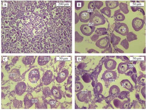
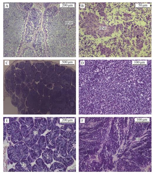
3 Results 3.1 Degeneration of Pparent Gonads in Later Breeding SeasonWe met success in artificially promoting maturation but failed to inducing spawning in proper breeding season indoor in May, 2017. We fetched the parents matured naturally in sea, induced them to spawn with airdrying method, observed their gonad tissue sections post spawning, and characterized the gonad histology of parents postspawning in later breeding season (Figs. 1 and 2). The orange female gonads looked plump; however, there were still a large number of unspawned eggs in the follicles (Fig. 1A). Many oocytes appeared to have a heteronucleus (HN) outside cell nucleus (CN) (Figs. 1B, D). Lots of oocytes showed degeneration clues: a condensed cell nucleus and an irregular or lack of nucleolus. These degenerated oocytes (DO) degraded into fragments gradually (Figs. 1C, D). White male gonads showed some transparent blocks although they still looked plump and their follicles were full of spermatids (Figs. 2A, C, E). Degeneration also appeared in the testes. Degenerated spermatids were clustered in some individuals and mitochondria were rarely seen or not present in spermatids (Figs. 2A, B). In some individuals, sperm development fully synchronized with mitochondrion development. However, Sperms lost their flagella and evenly dispersed in follicles (Figs. 2C, D). In some individuals, sperm development did not synchronized as the normal sperms did. They did not discharge seminal fluid even when spermiation was induced (Figs. 2E, F).
|
Fig. 1 Gonad histological characteristics of maternal clams in the late breeding season. A, A large number of eggs in the gonad follicles of maternal clams after spawning, ×100; B, Heteronuclear (HN) outside the cell nucleus (CN) in some eggs remaining in follicles, ×400; C, Degenerated oocytes (DO) degraded into fragments gradually, ×400; D, Different egg degradation states in the same follicle, ×400. HN, heteronuclear cell; N, nucleolus; CN, cell nucleus; DO, degenerated oocyte. |
|
Fig. 2 Gonad histological characteristics of paternal clams in the late breeding season. A, Degenerated spermatids were clustered in some follicles, the morphology of mitochondrion and flagellum of sperm is not clear, ×100; B, Degenerated spermatids were clustered in some follicles, the morphology of mitochondrion and flagellum of sperm is not clear, ×400; C, Sperms developed fully-synchronized with mitochondria but without flagella, ×40; D, Sperms developed fully-synchronized with mitochondria but without flagella, ×200; E, Sperm development was not synchronized and bundles of flagella of mature sperms was in the center of follicles, ×100; F, Sperm development was not synchronized and bundles of flagella of mature sperms was in the center of follicles, ×200. DSC, degenerated spermatid cluster; FB, flagella bunch. |
A total of 200 sets of well-spread mitotic metaphase chromosomes were examined to determine the mode diploid chromosome number (Cn) (Table 1). Fifty metaphases were from blastula and trochophore larval cells; 50 were from maternal gill cells; and 50 were from paternal gill cells. The modal diploid number of chromosomes (2n) was 34, which varied between 31 and 92 with a mode of 34. The chromosome length varied in size between 1.69 and 8.70 μm.
|
|
Table 1 Mitotic metaphase cell number, chromosome number and percentage ofAtrina pectinata |
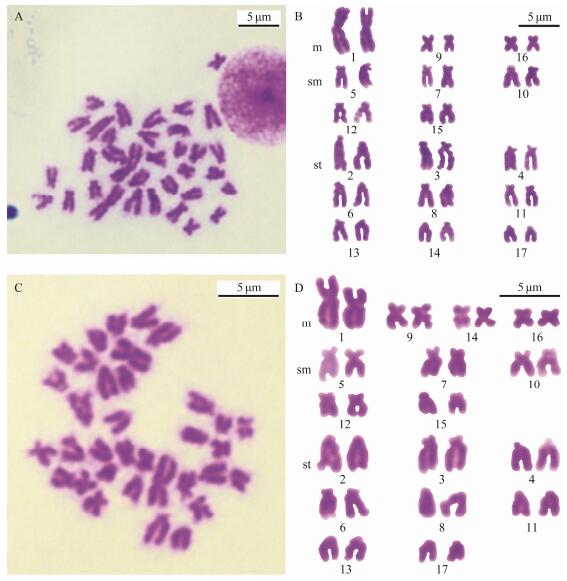
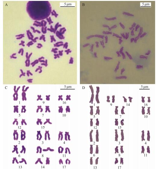
The well-spread metaphase plate, and the karyogram of the 34 chromosomes from a maternal gill cell with the largest pair of homomorphic chromosomes are shown in Figs. 3A and B. The well-spread metaphase plate and the karyogram of the 34 chromosomes from a paternal gill cell with the largest pair of heteromorphic chromosomes are shown in Figs. 3C and D. The metaphase plate and the karyogram from a trochophore-stage cell with the largest pair of homomorphic chromosomes are show in Figs. 4A and C. The metaphase plate and the karyogram from another trochophore-stage cell with the largest pair of heteromorphic chromosomes are shown in Figs. 4B and D. There is a primary XX/XY type of sex-determination in A. pectinata, and the largest pair of chromosomes present a lower-level sex differentiation; they were heterotypic in male somatic cells, but the difference between X and Y chromosomes was sometimes not obvious. The 14th pair of chromosomes were metacentric in male but telocentric in female metaphase somatic cells; therefore, the primary sex-determination mechanism manifests as an XX/XY type which we have found before (Zhou et al., 2018).
|
Fig. 3 The metaphase chromosome and karyotype of female and male Atrina pectinata. A, B, The metaphase chromosomes and karyotype of female A. pectinata. C, D, Metaphase chromosomes and karyotype of male A. pectinata. Scale indicates 5 μm. |
|
Fig. 4 The metaphase chromosome and karyotype of trochophore-stage Atrina pectinata. A, C, Metaphase chromosomes and karyotype of trochophore-stage cells with the largest pairs of homomorphic chromosomes; B, D, Metaphase chromosomes and karyotype of a trochophore-stage cell with the largest pairs of heteromorphic chromosomes. Scale indicates 5 μm. |
Both male and female metaphase chromosomes have almost the same sorting order; the male karyogram consisted of four m, five sm and eight st pairs while the female karyogram consisted of three m, five sm, and nine st pairs. The 14th pair was m in male and st in female metaphase cells. The result is consistent with our previous results (Zhou et al., 2018).
3.3 Ploidy of Early EmbryosBy using the largest pair of chromosomes as the classification standard, various multipleploidy embryos were found at the blastula stage, which included triploidy with three of the largest homomorphic or heteromorphic chromosomes (Fig. 5), pentaploidy with five of the largest homomorphic or heteromorphic chromosomes (Fig. 6) and aneuploidy with different numbers of the largest homomorphic or heteromorphic chromosomes (Fig. 7). At the trochophore stage, the 34 chromosomes can be distinguished as female and male groups via the largest pair of chromosomes length; only few abnormal embryo cells, such as 34 chromosomes together with three of the largest chromosomes, were observed (Fig. 8). Ten with the largest pairs of homomorphic chromosomes and 10 with the largest pairs of heteromorphic chromosome trochophorestage metaphase cells were used for karyotype analysis. The chromosome set was similar to that of their parents. The two largest chromosomes were also homomorphic X/X and heteromorphic X/Y (Fig. 4, Table 2).
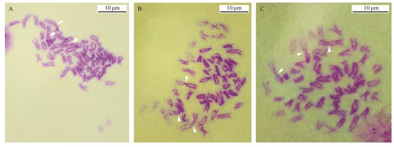
|
Fig. 5 Three triploid metaphase chromosomes at the blastula stage. 3n = 51. Arrows show the largest chromosomes. Scale indicates 10 μm. |
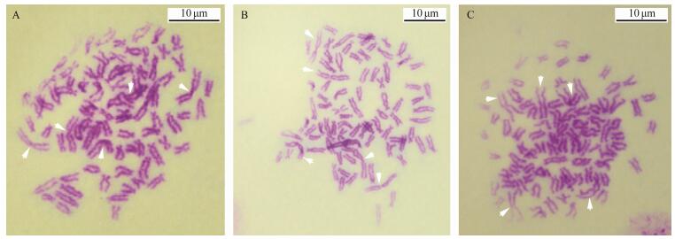
|
Fig. 6 Three triploid metaphase chromosomes at the blastula stage. 5n = 85. Arrows show the largest chromosomes. Scale indicates 10 μm. |
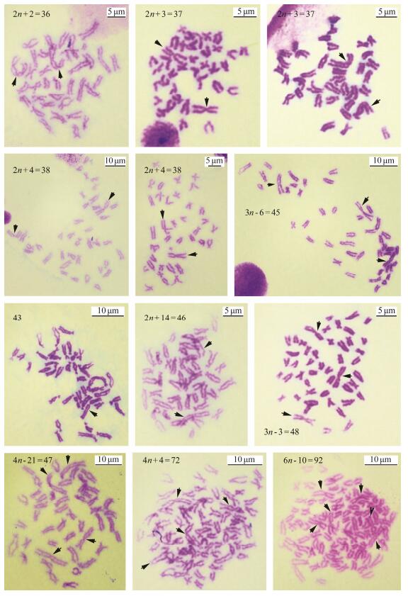
|
Fig. 7 Aneuploid metaphase chromosomes at the blastula stage. Arrows show the largest chromosomes. Scale indicates 5 μm or 10 μm. |
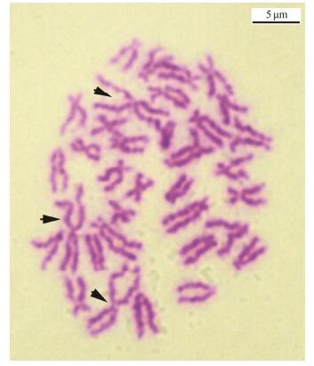
|
Fig. 8 An abnormal chromosome set of trochophore-stage cells with 34 chromosomes but three of the largest chromosomes in one metaphase cell. Arrows show the largest chromosomes. Scale indicates 5 μm. |
|
|
Table 2 Relative length and arm ratio of metaphase chromosomes of trochophore-stage A. pectinata |
Owing to the degeneration of overmature gonads, the eggs and sperms may have subtle genetic deficient in quality although they fertilized and developed successfully at the beginning time. The embryo development and chromosomal ploidy of the blastula stage was abnormal. A large number of deformative and asynchronous embryos died and degraded on the first hatching day till trocho phorestage and all embryos died and degraded at the D-shaped larval stage 2 days after insemination.
4 DiscussionAll the observed A. pectinata in this study belong to the same geographical group. Their genetic diversity should be low; however, there were many ploidy and abnormal embryos that eventually died, especially early embryos in their later breeding season. It was presumed that the overmature gametes underwent degeneration via apoptosis or programmed cell death (PCD). The cattle embryo cleavage rates were clearly influenced by the oocyte quality, and the apoptosis happened regardless of the oocyte quality to maintain tissue homeostasis (Bakri et al., 2016). As an experimental model, Artemia sinica has a diapause process during embryo development because of apoptosis, which is induced to resist adverse conditions such as high salinity, low temperature and anoxic conditions (Zhang et al., 2017). Shellfish species, the calyptraeid gastropods Crepidula navicella and Calyptraea lichen, produce viable and nutritive embryos, where nutritive embryos arrest their development after gastrulation by apoptosis and are ingested by their viable developing siblings as a form of intracapsular nutrition. Therefore, the role of MAPK (mitogen-activated protein kinase) signaling and apoptosis were examined as potential players in the early developmental switch between viable and nutritive embryos (Lesoway et al., 2017). Embryos of the freshwater prawn Macrobrachium olfersii were found to be able to activate the cell function to protect against environmental changes such as UVB (ultraviolet radiation b) (Schramm et al., 2017). Overall, the degeneration of parent gonads and deformative embryos were possibly underlined by apoptosis. We presumed that A. pectinata did not propagate naturally in an inappropriate season to ensure the fitness and survival of their descendants in the most economical way.
The number of goat embryos developing to term was lower in adolescent than in adult oocytes after they were transfered to receptor females. It is presumed that those deficiencies could be attributed to cytoplasmic, ultrastructural, metabolic and/or chromosomal abnormalities in oocytes and embryos from adolescent females (Romaguera et al., 2010). According to our results, in the late breeding season, degeneration took place in the gonads of overmature parents as well as in early embryos. Degeneration could not produce a strong generation surviving in worse growth conditions such as colder water temperature and a lack of food; otherwise, the fate of spats is still death in the early development stage, which is a waste of resources. Genetic deficiency may take place in overmature oocytes and earlier embryos in our observation such as condensed cell nuclei, irregular or lack of nucleoli in the oocytes, and rich triploid, pentaploid and aneuploidy embryos with different numbers of the largest homomorphic or heteromorphic chromosomes in earlier embryo cells while the chromosome composition in parents were normal diploid. Though overmature eggs and sperms with degeneration can fertilize successfully, the embryo development and chromosomal ploidy of the blastula stage was abnormal. Most embryos died and degraded, and all embryos died and degraded at the D-shaped larvae stage 2 days after insemination. All of them are called abortion spats. The genetic quality of eggs and sperms is vital. Degeneration in the overmature gonad and abnormal chromosome com position would cause a high mortality rate in early embryos in the later breeding season. The aneuploids Pacific oyster Crassostrea gigas were produced by 2n ♀ × 3n ♂ (DT group), 3n ♀ × 2n ♂ (TD group), 3n ♀ × 3n ♂ (TT group). These three groups had more abnormal embryos than normal DD group. Their embryo ploidy mainly was 2.5n in 12 h and 30 h, and became 2n in 5 d. Mass death of larvae with chromosomes pairing disorder was observed (Gong and Zhang, 2003). The survival rate of D-shaped larvae was only 14.7% when gynogenesis diploids were induced by 6-dimethylaminopurine (60 mg L-1; 6-DMAP) which disrupted the spindle at mitosis and inhibited chromosome movement, resulting in the abnormal chromosome composition (Yang et al., 2009). It is important to raise the parental pen shells in advance, and then cultivate seedlings in an appropriate season to ensure the fitness and survival of their descendants.
It is well known that the growth rate will decrease and even stagnate with increasing age, and the proliferation capacity of somatic cells is limited. By adjusting the water temperature of temporary rearing and using the hyperplasia-healing tissue of the gill for chromosomal investigation, we improved the methods of chromosome preparation, and obtained many well-spread mitotic chromosomal plates of A. pectinata, a largescale bivalve (Zhou et al., 2018). Few study on bivalve wound hyperplasiahealing tissue for chromosome preparation was found. In one example, A technique was reported for the preparation of metaphase chromosomes from the mantle tissue of adult pearl oyster Pinctada martensii, in which they cut off the adductor and removed the shell of one side to expose the mantle and visceral mass, and made shearing cuts on the mantle to culture hyperplasia tissue. The experimen tal animal did not survive, and the researchers suggested that young animals could get better results (Shen et al., 1993). In the present study, it is simple, practical, and re peatable to prepare chromosomes of adult A. pectinata. By adjusting the temperature in temporary cultures, and using the hyperplasia-healing tissue of the gill wound, the experimental animal can be kept alive and healthy after the procedure.
In this study, two pairs of chromosomes with sex difference in adults and larvae A. pectinata were chosen to study. The two largest chromosomes were female homomorphic as X and X chromosomes and male heteromorphic as X and Y chromosomes, respectively. The male 14th pair chro mosomes were metacentric and telocentric in female metaphase somatic cells, but it was not certain whether they were sex differentiated. In most sexually dimorphic animals, the determination of primary sex characteristics is controlled autonomously by sex chromosomes, secondary sex characteristics may be determined by sex chromosomes or alternatively by hormones released from the developing gonad (Twyman, 2002). The results of the induced gynogenetic and triploid analyses indirectly suggested that the dwarf surf clam may have an XX-female, XY-male sex determination with Y-domination (Guo et al., 1994). However, no more direct information on chromosomes for sex determination in bivalves was found at present.
5 ConclusionsThe gonad tissue slices showed the degeneration of eggs and sperms in later breeding season. The largest chromosomes could be used for sex and ploidy identification of embryos. Abnormal chromosome composition and genetic deficiency would cause a high mortality rate in early embryos in the late breeding season. These data will enrich the current knowledge of juvenile pen shell aquaculture.
AcknowledgementsThe authors are grateful to Mr. Yuzhong Sun, Director of Rizhao Ocean and Fisheries Research Institute, and engineer Shengnong Zhang at the Yellow Sea Fisheries Institute, Chinese Academy of Fishery Sciences, for their aids in cytogenetic experiments and chromosome preparations. This study was supported in part by the Central PublicInterest Scientific Institution Basal Research Fund, YSFRI, CAFS, China (No. 20603022018004), the National Natural Science Foundation of China (No. 31672637), the National Key R & D Program of China (No. 2018YFD0900800), and the Key Research and Development Plan of Shandong Province, China (No. 2016GSF115012).
Bakri, N. M., Ibrahim, S. F., Osman, N. A., Hasan, N., Jaffar, F. H. F., Rahman, Z. A. and Osman, K., 2016. Embryo apoptosis identification: Oocyte grade or cleavage stage?. Saudi Journal of Biology Science, 23: S50-S55. DOI:10.1016/j.sjbs.2015.10.023 (  0) 0) |
Chung, E. Y., Baik, S. H. and Ryu, D. K., 2006. Reproductive biology of the pen shell, Atrina (Servatrina) pectinata on the Boryeong coastal waters of Korea. Korean Journal of Malacology, 22(2): 143-150. (  0) 0) |
Gong, N. and Zhang, G. F., 2003. Embryo development and survival of aneuploid Pacific oyster Crassostrea gigas produced by mating its triploids and diploids. Journal of Fishery Sciences of China, 10(1): 5-9. (  0) 0) |
Guo, X. M., Allen, Jr. and S., K., 1994. Sex determination and polyploid gigantism in the dwarf surf clam (Mulinia lateralis Say). Genetics, 138(3): 1199-1206. (  0) 0) |
Kim, D. H., Yoon, H. S., An, Y. K., Lee, S. D. and Choi, S. D., 2008. Density dependent growth and survival rates of Atrina pectinata in Duekryang Bay, Korea. Korean Journal Malacology, 24(2): 137-142. (  0) 0) |
Lesoway, M. P., Collin, R. and Abouheif, E., 2017. Early activation of MAPK and apoptosis in nutritive embryos of Calyptraeid gastropods. Journal of Experimental Zoology, 328: 449-461. DOI:10.1002/jez.b.22745 (  0) 0) |
Levan, A., Fredga, K. and Sandberg, A. A., 1964. Nomenclature for centromeric position on chromosomes. Hereditas, 52(2): 201-220. (  0) 0) |
Liu, J., Li, Q., Kong, L. F. and Zheng, X. D., 2011. Cryptic diversity in the pen shell Atrina pectinata (Bivalvia: Pinnidae): High divergence and hybridization revealed by molecular and morphological data. Molecular Ecology, 20(20): 4332-4345. DOI:10.1111/j.1365-294X.2011.05275.x (  0) 0) |
Ohashi, S., Fujii, A., Oniki, H., Osako, K., Maeno, Y. and Yoshikoshi, K., 2008. The rearing of the pen shell Atrina pectinata larvae and juveniles (Preliminary note). Aquaculture Science, 56(2): 181-191. (  0) 0) |
Romaguera, R., Casanovas, A., Morató,, R, ., Izquierdo, D., Catalá,, M, ., Jimenez-Macedo, A. R., Mogas, T. and Paramio, M. T., 2010. Effect of follicle diameter on oocyte apoptosis, embryo development and chromosomal ploidy in prepubertal goats. Theriogenology, 74(3): 364-373. DOI:10.1016/j.theriogenology.2010.02.019 (  0) 0) |
Schramm, H., Jaramillo, M. L., Quadros, T., Zeni, E. C., Müller,, Y., M. R., Ammar, D. and Nazari, E. M., 2017. Effect of UVB radiation exposure in the expression of genes and proteins related to apoptosis in freshwater prawn embryos. Aquatic Toxicology, 191: 25-33. DOI:10.1016/j.aquatox.2017.07.014 (  0) 0) |
Shen, Y. P., Wang, X. J., Ma, W. T. and Zhang, X. Y., 1993. A technique for preparation of metaphase chromosome from mantle tissue of adult pearl oyster Pinctada martensii Dunker. Journal of Wuhan University (Nature Science Edition), 5: 121-122. (  0) 0) |
Son, P. W., Ha, D. S., Lee, C. H., Jang, D. S. and Kim, D. K., 2005. Study on the natural spat collection of the pen shell, Atrina pectinata. Korean Journal Malacology, 21(2): 113-120. (  0) 0) |
Twyman, R. M., 2002. Instant Notes in Developmental Biology. Bios Scientific Publishers Limited, Norwich: 151-159. (  0) 0) |
Wang, Z. G., Wang, W., Yu, S. D. and Xu, Z. R., 2008. Effects of different activation protocols on preimplantation development, apoptosis and ploidy of bovine parthenogenetic embryos. Animal Reproduction Science, 105(3-4): 292-301. DOI:10.1016/j.anireprosci.2007.03.017 (  0) 0) |
Yang, Q., Li, Q., Yu, R. H., Kong, L. F. and Zheng, X. D., 2009. Artificial induction of gynogenetic diploids by suppression of the first cleavage in Atrina pectinata. Marine Sciences, 33(10): 63-67. (  0) 0) |
Yurimoto, T., Yoshida, M. and Maeno, M., 2008. Histology and nutritional condition of the pen shells Atrina pectinata protruded above the sediment surface during summer in the tidal flat of Ariake Bay. Aquaculture Science, 56(4): 587-594. (  0) 0) |
Zhang, S., Yao, F., Jing, T., Zhang, M. C., Zhao, W., Zou, X. Y., Sui, L. L. and Hou, L., 2017. Cloning, expression pattern, and potential role of apoptosis inhibitor 5 in the termination of embryonic diapause and early embryo development of Artemia sinica. Gene, 628: 170-179. DOI:10.1016/j.gene.2017.07.021 (  0) 0) |
Zhou, L. Q., Wang, X. M., Wu, B., Sun, X. J., Chen, S. Q., Liu, Z. H., Yang, A. G., Zhang, S. N., Zhao, Q. and Zhang, G. W., 2018. Chromosome preparation and karyotypes analysis of both male and female Atrina pectinata. Progress in Fishery Science, 39(5): 66-72. (  0) 0) |
 2020, Vol. 19


