 2012, Vol. 28


2. 兰州市中医医院;
3. 中国人民解放军第四军医大学药物研究所;
4. 兰州军区第一医院
动物实验证实,过量的锰透过血脑屏障沉积于广泛脑区,导致神经元的毁灭和神经髓鞘的空泡状变性等改变〔1, 2〕,锰中毒和帕金森氏病都与基底节多巴胺能神经元的退行性变性有关〔3〕。丝裂原活化蛋白激酶( mitogen-activated-proteinkinase,MAPKs) 参与细胞增殖、分化、转化及凋亡反应,其中胞外信号调节激酶( extracellular-signal-regulated kinase,Erk) 主要参与细胞增殖与分化的调控〔4〕。本研究以PC12 细胞为模型,观察锰对神经细胞增殖抑制作用特点及此过程中Erk 活化表达的特点,探讨帕金森氏病( Parkinson's disease,PD) 相关环境危险因子锰对神经细胞增殖抑制的分子机制。
1 材料和方法 1. 1 细胞培养〔5〕及实验分组RPMI 1640 培养液; 10% 小牛血清( 杭州四季青公司) 5% CO2 培养箱( 英国Forma Scientif 公司) 去离子水将MnCl2 ( 美国Sigma 公司) 配制成10 mol /L 的储存液,4 ℃保存。实验分为对照组,不同氯化锰浓度作用的实验组,二甲基亚砜组。
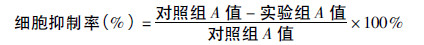
1. 2 细胞毒试验采用四甲基偶氮噻唑蓝( 3-( 4-,5-dimethylthiazol- 2Vyl) -2,5 -diphenyl tetrasolium bromide,MTT 法)〔5〕取对数生长期的细胞( 细胞密度为1 × 10⁵ ~ 2 × 10⁵ /L) ,接种于 96 孔培养板( 200 μl /孔) 中。MnCl2 对细胞增殖的毒性作用以抑制率来反映,其抑制率的计算公式:
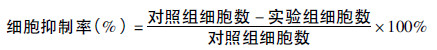
取对数生长期的细胞以每孔2 × 10⁴ 个接种于24 孔板中以每天活细胞数均值绘制生长曲线,及计算MnCl2 对神经细胞生长抑制率。
分别收集实验组和对照组细胞按透 射电镜要求制样并观察。
1. 5 凋亡细胞的基因组DNA 电泳〔6〕分别取对照组及实验 组细胞提取DNA,在加有溴化乙锭( ethidium bromide,EB) 的 1%琼脂糖凝胶上电泳过夜,DL-2000 用作分子标志。
1. 6 免疫印迹法( western blot) 测定蛋白磷酸化水平〔7〕 1. 6. 1 样品处理裂解细胞,分离提取蛋白〔7〕。 1. 6. 2 蛋白质定量分析用改进的Folin-酚法定量蛋白。 1. 6. 3 免疫印迹法( western blot) 测定蛋白磷酸化水平〔7〕一抗Erk1 /2( 兔多抗,美国Santa Cruz 公司) ; 二抗( Erk1 /2 二抗,辣根过氧化物酶标记,羊抗兔,美国Santa Cruz 公司) ; p - Erk 一抗( 鼠单抗,美国Santa Cruz 公司; 二抗( 辣根过氧化物酶标记,羊抗鼠,美国Santa Cruz 公司) 压片成像: 硝酸纤维素膜稍干后,滤膜滴加1 mL 化学荧光显迹液化学荧光发光剂 ( 美国Pierce 公司) ,反应1 min 保鲜膜覆盖,X 光胶片压片,凝胶成像系统测定免疫印迹区带的光密度,预染标准分子量蛋白Marker( 美国Bio-Rad 实验室) ; 标准分子量蛋白Marker ( 美国Santa Cruz 公司) ; Erk 特异性阻断剂PD98059 ( promega) 。
1. 7 统计分析采用SPSS13. 0 软件进行统计分析。组间率 的比较采用χ2 检验,吸光度用x± s 表示组间比较采用t 检 验。免疫印迹法( western blot) 测定蛋白磷酸化水平采用IMAGINGQUANT 数据处理系统分析结果。
2 结果 2. 1 不同浓度MnCl2 对PC12 细胞增殖的抑制作用( 表 1)| 表 1 表1 不同浓度MnCl2 对PC12 细胞的抑制率( %) |
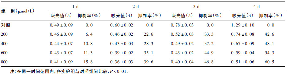
600、800 μmol /L MnCl2 作用4 d 细胞的抑制率分别是 54. 3%、60. 5%,随作用时间的延长和浓度的上升而增强,呈 现时间依赖效应和浓度依赖效应,表现慢性毒作用。实验组 与对照组相比差异有统计学意义( 均P < 0. 01) 。二甲基亚砜 对PC12 细胞抑制作用与对照组比较差异无统计学意义。
2. 2 台盼蓝拒染法绘制细胞生长曲线各剂量组在24 h 的抑 制率分别为32. 67%、56. 32%和74. 14%,与MTT 法结果相符。
2. 3 透射电镜下PC12 细胞观察600 μmol /L MnCl2 作用4 d 透射电镜下可见细胞体积明显缩小,细胞膜表面突起消 失,细胞质中有空泡形成,核膜完整但皱缩,核体积缩小,染色 质致密且边集于细胞核周边,表现为细胞凋亡的形态学改变。
2. 4 MnCl2 对PC12 细胞基因组DNA 的影响( 图 1) | 注: 1: 对照; 2: 600 μmol /L MnCl2 作用于细胞3 d; 3: DL - 2000。 图 1 600 μmol /L MnCl2 作用于细胞3 d PC12 细胞 基因组DNA 凝胶电泳 |
600 μmol /L MnCl2 作用3 d 细胞基因组DNA 琼脂糖凝胶电泳出 现明显梯状条带。
2. 5 MnCl2 作用下PC12 细胞Erk1 /2 的活化表达( 图 2) | 图 2 不同浓度MnCl2 treated 作用PC12 4 d 时p - Erk 的表达 |
600 μmol /L MnCl2 作用1、2、3、4 d 可见p-Erk1 /2 逐渐降低, 其中2 d 时较对照降低了75%( n = 3,P < 0. 05) ,图 4 中200、 400、600 μmol /L MnCl2 分别作用4 d 时,p-Erk1 /2 亦逐渐降 低,400 μmol /L MnCl2 作用4 d 时较对照明降低了78% ( n = 3,P < 0. 05) ,MnCl2 对PC12 细胞p-Erk 的逐渐抑制作用表 现出浓度和时间依赖效应。使用MEK( Erk1 /2 上游分子) 特 异性阻制剂PD98059 实验结果表明锰通过MEK 磷酸化下游 的Erk,下调p-Erk1 /2( 图 5) ,使细胞增殖抑制。
3 讨论对锰神经毒作用机理的探讨一直与帕金森氏病的研究相 关〔8, 9〕,提示锰的神经毒作用机制与多巴胺能神经元的损伤 有一定关系〔10〕。因此我们以多巴胺能的鼠嗜铬神经瘤细胞 PC12 为模型,筛选锰对神经细胞增殖抑制作用的时间及剂量 效应关系。结果表明,MnCl2 对PC12 细胞的增殖抑制作用在 低剂量、较长时间段中表现出剂量和时间效应关系。
本研究结果显示,600 μmol /L MnCl2 作用4 d 电镜可见细胞凋亡的形态学改变。同样条件下细胞DNA 碎片化,进一步证实MnCl2 能够诱导PC12 细胞凋亡。使用MEK1 /2 特异性阻断剂PD98059 后,可以阻断Erk( p44 /42) 的磷酸化,增强锰的毒性,导致细胞死亡,推断此类激酶是一种细胞存活因子〔11〕。本实验发现600 μmol /L MnCl2 作用1、2、3、4 d Erk 磷酸化水平呈逐渐降低趋势,作用2 d 时较对照明显降低了 75%,200、400、600 μmol /L MnCl2 分别作用4 d 时,Erk 磷酸化水平亦逐渐降低,400 μmol /L MnCl2 作用4 d 时较对照明显降低了78%,表明MnCl2 对PC12 细胞ERK 激活的抑制作用呈浓度和时间依赖效应。结果提示锰致PC12 细胞增殖抑制为细胞凋亡,可能与Erk 磷酸化水平的降低有关。
| 〔1〕 | Bornhorst J,Ebert F,Hartwig A,et al.Manganese inhibits poly (ADP-ribosyl)ation in human cells:a possible mechanism behind manganese-induced toxicity[J].Environ Monit,2010,12(11): 2062-2069. |
| 〔2〕 | 黄波,吴训伟,王清海.PAS-Na治疗锰中毒大鼠中枢神经系统病变的观察[J].中华劳动卫生职业病杂志,1994,12(4): 205. |
| 〔3〕 | Verhoeven WM,Egger JI,Kuijpers HJ.Manganese and acute paranoid psychosis:a case report[J].Med Case Reports,2011,(12), 5:146. |
| 〔4〕 | 于燕,张瑞娟,张敬华.姜黄素对香烟主流烟气凝聚物诱变性的拮抗作用[J].中国公共卫生,2003,19(5):538-540. |
| 〔5〕 | 司徒镇强,吴军正.细胞培养[M].北京:世界图书出版公司, 1996:127-143. |
| 〔6〕 | Williams BB,Kwakye GF,Wegrzynowicz M,et al.Altered manganese homeostasis and manganese toxicity in a Huntington's disease striatal cell model are not explained by defects in the iron transport system[J].Toxicol Sci,2010,117(1):169-179. |
| 〔7〕 | 萨姆布鲁克.分子克隆实验指南[M].2版.北京:科学出版社, 1999:898-900. |
| 〔8〕 | Pohl HR,Roney N,Abadin HG.Metal ions affecting the neurological system[J].Met Ions Life Sci,2011,8:247-262. |
| 〔9〕 | Martin WR.Fuming over Parkinson disease:are welders at risk? [J].Neurology,2011,76(15):1286-1287. |
| 〔10〕 | Prabhakaran K,Chapman GD,Gunasekar PG.α-Synuclein overexpression enhances manganese-induced neurotoxicity through the NF-κB-mediated pathway[J].Toxicol Mech Methods,2011,21 (6):435-443. |
| 〔11〕 | Molina RM,Phattanarudee S,Kim J,et al.Ingestion of Mn and Pb by rats during and after pregnancy alters iron metabolism and behavior in offspring[J].Neurotoxicology,2011,32(4):413-422. |