宫颈癌是严重威胁女性健康的严重疾病,发展中国家尤为严重[1-3]。放射治疗是其重要的根治性治疗选择之一。近距离放射治疗是根治性放射治疗中的重要组成部分[4]。目前,国内绝大部分医院采用高剂量率近距离治疗方式,它通过单一近似点源模拟线源来实现体积照射。这种采用单一近似点源模拟线源的近距离治疗使得治疗计划中的驻留位置和驻留时间可以进行一定的优化。然而,驻留位置的选择不是随心所欲的。本文通过文献综述介绍了宫颈癌腔内近距离治疗(intracavitary brachytherapy,IC-BT)以及腔内联合组织间插植近距离治疗(intracavitary combined with interstitial brachytherapy,IC/IS-BT)的标准载源模型(standard loading pattern,SLP),并对计划优化模型和优化过程中的限制条件等做适当阐述,以期为宫颈癌近距离治疗计划设计提供重要参考。
1 IC-BT的SLP的来源宫颈癌的近距离治疗在临床中的应用已超过110年[5]。在漫长的应用历史上经历了基于经验性的镭疗时代,后发展了经典的剂量学系统。经典的剂量学系统已被大量实践证实具有较好的疗效。目前宫颈癌IC-BT施源器的设计绝大部分来源于经典剂量学系统。因此可以说IC-BT的SLP来源于经典剂量学系统,主要有曼彻斯特系统、巴黎系统和斯德哥尔摩系统[6]。
虽然各剂量学系统在放射源构造等方面有所不同,但他们有一定的共通点:将施源器分为2部分,即宫腔源和阴道源。宫腔源为一线源,位于子宫中轴线上,根据子宫体长短不同设置1~3个线源相互连接。阴道源均位于宫颈口和/或阴道穹窿处,分为两类,一类是类球形放射源(曼彻斯特系统和巴黎系统),另一类是平板状的放射源(斯德哥尔摩系统)。在斯德哥尔摩系统和曼彻斯特系统的典型应用中,宫腔源与阴道源的载源比值分别为1.06(74 mgRa:70 mgRa)和0.875(35 mgRa:40 mgRa),而巴黎系统中,这一比值的典型值为1(范围为0.66~1.5)。因此,总体而言,一般情况下经典的剂量学系统中,宫腔源的载源量与阴道源的载源量相近。这一比值在高剂量率近距离治疗阶段的SLP中有着重要的意义。
2 IC-BT的SLP近年来,宫颈癌高剂量率近距离治疗在施源器方面有着重要进展[7]。在IC-BT中,根据经典剂量学系统的原理与放射源结构设计出的施源器主要分为2类:一类是基于曼彻斯特系统研制的宫腔管 + 卵圆体模式;另一类是匹配斯德哥尔摩系统剂量学分布而设计的宫腔管 + 环形模式。
对于宫腔管 + 卵圆体模式的施源器,SLP为:宫腔源末端位于阴道源水平,卵圆体中的载源以与宫腔管侧向投影位置的交点为中点。乌德勒支施源器是宫腔管 + 卵圆体模式的IC-BT施源器的典型代表,其SLP和剂量分布见图1[8-11]。

|
图 1 乌德勒支腔内施源器的标准载源模式的剂量分布示例 Figure 1 An example of dose distribution of standard loading pattern for Utrecht intracavitary applicator |
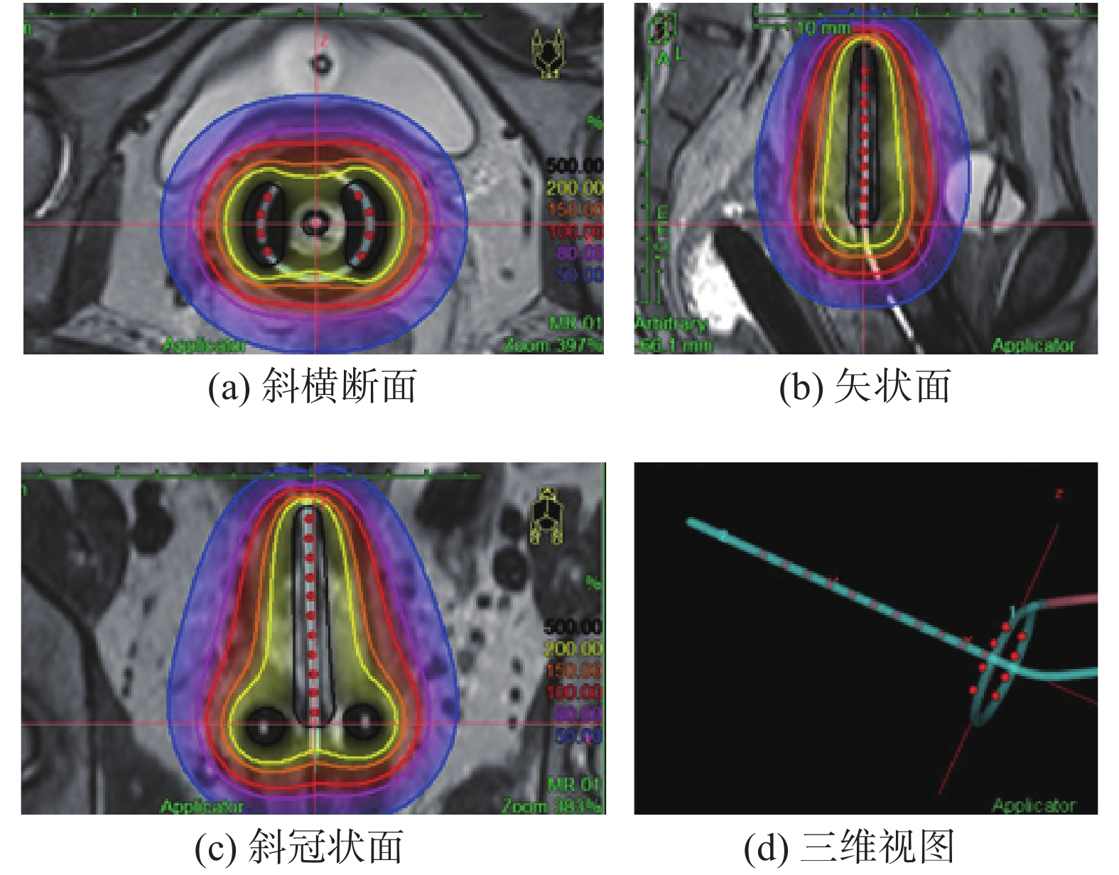
对于宫腔管 + 环形模式施源器的SLP,根据环形尺寸和宫腔管长度不同,有不同的设置,见表1[12-13]。总的来说环形上左右两侧各4个驻留点,相邻驻留点间隔为5 mm。环形施源器是宫腔管 + 环形模式的IC-BT施源器的典型代表,其SLP和剂量分布见图2[10-11]。在IC-BT的SLP下,其等剂量曲线形似一个压扁了的啤梨,通常称之为“梨形剂量分布”[14-15]。
|
|
表 1 环形施源器的最常用标准载源模式 Table 1 Standard loading pattern for mostly used Ring applicator |

|
图 2 环形腔内施源器标准载源模式的剂量分布示例 Figure 2 An example of dose distribution of standard loading pattern for Ring intracavitary applicator |
在很多临床应用中,SLP不一定适合当时的患者解剖,需要进行一定的优化。以环形施源器为例,有几种简单的优化模型[13]。该优化模型是以手动调整驻留点或驻留时间的方式实现,并且它仍然是变形的SLP。
当肿瘤位置偏心时,可调整左右两侧驻留点的驻留时间,当一侧驻留权重减小30%时,同侧A点剂量可下降15% ~20%。当直肠位置偏移中线并向腹侧突出时,可将同侧环形的驻留点向腹侧平移,必要时取消背侧的最后一个驻留点以使高剂量区有效回避直肠。当环形水平处的直肠向腹侧突出时,可将环形内的驻留点整体向腹侧移动优化剂量分布。宫腔管内的驻留时间可根据高危CTV失状位的形态变化调整各驻留点的驻留时间,最低可调整驻留权重到30%。
4 IC/IS-BT的SLP日本的一项研究[16]分析了2008年12月—2014年10月治疗的209例宫颈癌初次放射治疗患者数据。其中142例(67.9%)患者采用单纯腔内近距离治疗;25例(12.0%)患者采用单纯插植近距离治疗;其余42例(20.1%)患者采用IC/IS-BT。这表明,在宫颈癌的近距离放射治疗中,大部分病例采用IC-BT。然而,即使IC-BT具有一定的优化能力,但其在曼彻斯特A点水平层面,处方剂量的等剂量线不能超过宫腔管外25 mm[17]。对于高危CTV体积过大、宫旁侵犯较大、外照射后肿瘤消退不良,以及肿瘤与危及器官关系密切时,单纯采用IC-BT无法在满足危及器官剂量要求的前提下达到高剂量的靶区覆盖率[8,18-20]。宫颈癌根治性放射治疗中有着明确的剂量效应关系,即肿瘤剂量越高预期能获得更高的局部控制率[21-26]。因此,上述情况下,在IC-BT基础上增加插植针,即实施IC/IS-BT,可有效提高靶区的高剂量覆盖率,从而显著提高局部控制率[27-28]。
IC/IS-BT的SLP是基于IC-BT的SLP,仅在标准梨形剂量无法包绕靶区的部分设置驻留点,插植针驻留点的驻留时间仅为腔内部分的10%[13,18]。使得IC/IS-BT的等剂量曲线大体上仍保持梨形,仅在插植针驻留点区域将处方剂量等剂量线向外推移以覆盖高危CTV。优化后的计划中,插植针中的总驻留时间一般限定在腔内部分的10% ~20%[18]。
Nomden等[8]分析了乌德勒支IC/IS-BT施源器的20例患者的数据,患者给予2次脉冲剂量率治疗,首次治疗采用IC-BT,第二次采用IC/IS-BT。与首次IC-BT相比,IC/IS-BT在危及器官剂量方面有较大优势。最常用的插植针位置为7和10号,依次为6和11号,以及8号和9号。IC-BT的权重占绝大部分,平均插植针权重为19%,单插植针平均权重为7%。Fokdal等[19]研究了47例应用环形IC/IS-BT施源器的连续病人,基于预计划(BT0)剂量,有24例(41%)患者在近距离治疗(BT1或BT2)中采用IC/IS-BT。2次实际近距离治疗剂量与预计划剂量具有较高的一致性。BT1和BT2中有效插植针的数量分别为(4.7 ± 2.4)和(5.0 ± 2.7)根,平均插植深度分别为(3.0 ± 1.0)和(2.9 ± 1.1)cm。与预计划单纯IC-BT相比,预计划IC/IS-BT计划和实际近距离剂量均显著提高高危CTV D90和D100剂量,中危CTV D90、膀胱和直肠的D2cc未见显著改变,结肠和小肠的D2cc均显著减小。这是2种典型的IC/IS施源器的早期应用,为IC/IS-BT的SLP奠定了基础,具体的应用特征见表2。
|
|
表 2 两种腔内联合组织间插植施源器临床应用特征对比 Table 2 Comparison of clinical application characteristics of two kinds of intracavitary combined with interstitial applicators |
Zhao等[31]人分析了65分次局部晚期宫颈癌患者应用环形IC/IC-BT施源器的数据,应用最多的引导孔依次是5号、8号、6号和9号(26 mm环形),应用比例超过75%。对于宫旁进一步浸润的患者,通过宫腔管联合卵圆体或环形及基于此的宫旁插植难以包绕靶区,因此允许更多自由度的包括平行插植针和斜向插植针的Venezia施源器应运而生。Walter等[32]报到了首个该施源器应用于人体的临床结果。10例局部晚期宫颈癌患者,FIGO分期为IIB-IVB期,高危CTV体积中位值为42 cc(19 ~76 cc),每分次应用1 ~6根插植针,所有危及器官的计划目标均达到,有一例患者未达到任何的靶区计划目标,另一例患者达到4个靶区计划目标中的3个。相比于基于传统的A点计划和单纯腔内的计划,IC/IS-BT具有显著的剂量学优势。
5 小 结在宫颈癌近距离放射治疗中,应严格评估患者外照射后的残余肿瘤情况,对于能采用单纯IC-BT的,应采用IC-BT施源器。如外照射后残余肿瘤较大、位置偏离宫腔管、或不利于保护危及器官等情况,应考虑采用IC/IS-BT施源器。无论在IC-BT,还是在IC/IS-BT中,治疗计划的初始条件均应从SLP出发进行优化。IC/IS-BT的等剂量曲线总体上应仍保持梨形,即绝大部分剂量权重来自腔内施源器。
| [1] |
Bray F, Ferlay J, Soerjomataram I, et al. Global cancer statistics 2018: GLOBOCAN estimates of incidence and mortality worldwide for 36 cancers in 185 countries[J]. CA Cancer J Clin, 2018, 68(6): 394-424. DOI:10.3322/caac.21492 |
| [2] |
李钰, 高岩, 刘世龙, 等. 宫颈癌患者的膀胱充盈度一致性对放疗摆位误差的影响[J]. 中国辐射卫生, 2020, 29(3): 305-308. Li Y, Gao Y, Liu SL, et al. Effect of consistency of bladder filling volume on set-up errors in radiotherapy for the patients with cervical cancer[J]. Chin J Radiol Heal, 2020, 29(3): 305-308. DOI:10.13491/j.issn.1004-714X.2020.03.027 |
| [3] |
刘静雯, 任洪荣, 周冲, 等. 盆腔活性骨髓与宫颈癌放疗血液学毒性的关系[J]. 中国辐射卫生, 2020, 29(6): 696-699. Liu JW, Ren HR, Zhou C, et al. Correlation between pelvic active bone marrow and hematological toxicity in radiotherapy of cervical cancer[J]. Chin J Radiol Heal, 2020, 29(6): 696-699. DOI:10.13491/j.issn.1004-714X.2020.06.030 |
| [4] |
Han K, Milosevic M, Fyles A, et al. Trends in the utilization of brachytherapy in cervical cancer in the United States[J]. Int J Radiat Oncol Biol Phys, 2013, 87(1): 111-119. DOI:10.1016/j.ijrobp.2013.05.033 |
| [5] |
Chargari C, Deutsch E, Blanchard P, et al. Brachytherapy: an overview for clinicians[J]. CA Cancer J Clin, 2019, 69(5): 386-401. DOI:10.3322/caac.21578 |
| [6] |
Prescribing, recording, and reporting brachytherapy for cancer of the cervix[J]. J ICRU, 2013, 13(1/2): NP. DOI: 10.1093/jicru/ndw027.
|
| [7] |
唐笑迪, 岳海振, 赵红福. 宫颈癌高剂量率近距离治疗施源器进展[J]. 中国医疗设备, 2021, 36(4): 153-156, 161. Tang XD, Yue HZ, Zhao HF. The progress of source applicators in high-dose-rate brachytherapy for cervical cancer[J]. China Med Devices, 2021, 36(4): 153-156, 161. DOI:10.3969/j.issn.1674-1633.2021.04.035 |
| [8] |
Nomden CN, de Leeuw AA, Moerland MA, et al. Clinical use of the Utrecht applicator for combined intracavitary/interstitial brachytherapy treatment in locally advanced cervical cancer[J]. Int J Radiat Oncol Biol Phys, 2012, 82(4): 1424-1430. DOI:10.1016/j.ijrobp.2011.04.044 |
| [9] |
Tambas M, Tavli B, Bilici N, et al. Computed tomography-guided optimization of needle insertion for combined intracavitary/interstitial brachytherapy with Utrecht applicator in locally advanced cervical cancer[J]. Pract Radiat Oncol, 2021, 11(4): 272-281. DOI:10.1016/j.prro.2021.01.008 |
| [10] |
Serban M, Kirisits C, de Leeuw A, et al. Ring versus ovoids and intracavitary versus intracavitary-interstitial applicators in cervical cancer brachytherapy: results from the EMBRACE I study[J]. Int J Radiat Oncol Biol Phys, 2020, 106(5): 1052-1062. DOI:10.1016/j.ijrobp.2019.12.019 |
| [11] |
Gursel SB, Serarslan A, Meydan AD, et al. A comparison of tandem ring and tandem ovoid treatment as a curative brachytherapy component for cervical cancer[J]. J Contemp Brachytherapy, 2020, 12(2): 111-117. DOI:10.5114/jcb.2020.94308 |
| [12] |
Jürgenliemk-Schulz IM, Lang S, Tanderup K, et al. Variation of treatment planning parameters (D90 HR-CTV, D2cc for OAR) for cervical cancer tandem ring brachytherapy in a multicentre setting: Comparison of standard planning and 3D image guided optimisation based on a joint protocol for dose-volume constraints[J]. Radiother Oncol, 2010, 94(3): 339-345. DOI:10.1016/j.radonc.2009.10.011 |
| [13] |
Kirisits C, Pötter R, Lang S, et al. Dose and volume parameters for MRI-based treatment planning in intracavitary brachytherapy for cervical cancer[J]. Int J Radiat Oncol Biol Phys, 2005, 62(3): 901-911. DOI:10.1016/j.ijrobp.2005.02.040 |
| [14] |
Tanderup K, Nielsen SK, Nyvang GB, et al. From point A to the sculpted pear: MR image guidance significantly improves tumour dose and sparing of organs at risk in brachytherapy of cervical cancer[J]. Radiother Oncol, 2010, 94(2): 173-180. DOI:10.1016/j.radonc.2010.01.001 |
| [15] |
Shen S, Kim R, Duan J, et al. SU-E-T-433: pear-shaped based dose optimization for HDR intracavitary brachytherapy for cervical cancer patients with small uterus[J]. Med Phys, 2012, 39(6Part16): 3804. DOI:10.1118/1.4735522 |
| [16] |
Murakami N, Kobayashi K, Kato T, et al. The role of interstitial brachytherapy in the management of primary radiation therapy for uterine cervical cancer[J]. J Contemp Brachytherapy, 2016, 8(5): 391-398. DOI:10.5114/jcb.2016.62938 |
| [17] |
Kirisits C, Lang S, Dimopoulos J, et al. Uncertainties when using only one MRI-based treatment plan for subsequent high-dose-rate tandem and ring applications in brachytherapy of cervix cancer[J]. Radiother Oncol, 2006, 81(3): 269-275. DOI:10.1016/j.radonc.2006.10.016 |
| [18] |
Kirisits C, Lang S, Dimopoulos J, et al. The Vienna applicator for combined intracavitary and interstitial brachytherapy of cervical cancer: design, application, treatment planning, and dosimetric results[J]. Int J Radiat Oncol Biol Phys, 2006, 65(2): 624-630. DOI:10.1016/j.ijrobp.2006.01.036 |
| [19] |
Fokdal L, Tanderup K, Hokland SB, et al. Clinical feasibility of combined intracavitary/interstitial brachytherapy in locally advanced cervical cancer employing MRI with a tandem/ring applicator in situ and virtual preplanning of the interstitial component
[J]. Radiother Oncol, 2013, 107(1): 63-68. DOI:10.1016/j.radonc.2013.01.010 |
| [20] |
Palhares DMF, Marconi DG, Azevedo TL, et al. Predicting the necessity of adding catheters to intracavitary brachytherapy for women undergoing definitive chemoradiation for locally advanced cervical cancer[J]. Brachytherapy, 2018, 17(6): 935-943. DOI:10.1016/j.brachy.2018.07.003 |
| [21] |
Dimopoulos JC, Pötter R, Lang S, et al. Dose-effect relationship for local control of cervical cancer by magnetic resonance image-guided brachytherapy[J]. Radiother Oncol, 2009, 93(2): 311-315. DOI:10.1016/j.radonc.2009.07.001 |
| [22] |
Zhang N, Tang YH, Guo X, et al. Analysis of dose-effect relationship between DVH parameters and clinical prognosis of definitive radio (chemo) therapy combined with intracavitary/interstitial brachytherapy in patients with locally advanced cervical cancer: a single-center retrospective study[J]. Brachytherapy, 2020, 19(2): 194-200. DOI:10.1016/j.brachy.2019.09.008 |
| [23] |
Tanderup K, Fokdal LU, Sturdza A, et al. Effect of tumor dose, volume and overall treatment time on local control after radiochemotherapy including MRI guided brachytherapy of locally advanced cervical cancer[J]. Radiother Oncol, 2016, 120(3): 441-446. DOI:10.1016/j.radonc.2016.05.014 |
| [24] |
Mazeron R, Castelnau-Marchand P, Escande A, et al. Tumor dose-volume response in image-guided adaptive brachytherapy for cervical cancer: a meta-regression analysis[J]. Brachytherapy, 2016, 15(5): 537-542. DOI:10.1016/j.brachy.2016.05.009 |
| [25] |
Tang XD, Mu X, Zhao ZP, et al. Dose-effect response in image-guided adaptive brachytherapy for cervical cancer: a systematic review and meta-regression analysis[J]. Brachytherapy, 2020, 19(4): 438-446. DOI:10.1016/j.brachy.2020.02.012 |
| [26] |
Li F, Lu SC, Zhao HF, et al. Three-dimensional image-guided combined intracavitary and interstitial high-dose-rate brachytherapy in cervical cancer: a systematic review[J]. Brachytherapy, 2021, 20(1): 85-94. DOI:10.1016/j.brachy.2020.08.007 |
| [27] |
Gonzalez Y, Giap F, Klages P, et al. Predicting which patients may benefit from the hybrid intracavitary+interstitial needle (IC/IS) applicator for advanced cervical cancer: a dosimetric comparison and toxicity benefit analysis[J]. Brachytherapy, 2021, 20(1): 136-145. DOI:10.1016/j.brachy.2020.09.004 |
| [28] |
Fokdal L, Sturdza A, Mazeron R, et al. Image guided adaptive brachytherapy with combined intracavitary and interstitial technique improves the therapeutic ratio in locally advanced cervical cancer: Analysis from the retroEMBRACE study[J]. Radiother Oncol, 2016, 120(3): 434-440. DOI:10.1016/j.radonc.2016.03.020 |
| [29] |
赵红福, 程光惠, 韩东梅. 宫颈癌高剂量率三维近距离治疗计划设计[J]. 中华放射肿瘤学杂志, 2018, 27(5): 527-532. Zhao HF, Cheng GH, Han DM. Treatment plan design of three-dimensional high-dose-rate brachytherapy for cervical cancer[J]. Chin J Radiat Oncol, 2018, 27(5): 527-532. DOI:10.3760/cma.j.issn.1004-4221.2018.05.019 |
| [30] |
Tang B, Liu XY, Wang XL, et al. Dosimetric comparison of graphical optimization and inverse planning simulated annealing for brachytherapy of cervical cancer[J]. J Contemp Brachytherapy, 2019, 11(4): 379-383. DOI:10.5114/jcb.2019.87145 |
| [31] |
Zhao ZP, Zhang N, Liu Y, et al. Analysis of clinical utilization of ring applicator for combined intracavitary/interstitial image-guided brachytherapy treatment in Chinese patients with locally advanced cervical cancer[J]. J Contemp Brachytherapy, 2020, 12(3): 252-259. DOI:10.5114/jcb.2020.96866 |
| [32] |
Walter F, Maihöfer C, Schüttrumpf L, et al. Combined intracavitary and interstitial brachytherapy of cervical cancer using the novel hybrid applicator Venezia: Clinical feasibility and initial results[J]. Brachytherapy, 2018, 17(5): 775-781. DOI:10.1016/j.brachy.2018.05.009 |


