Translational potential of medicinal plants for Alzheimer’s disease: integrated evidence from molecular mechanisms to preclinical and clinical findings

Abstract
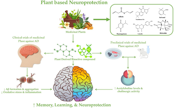
Alzheimer’s disease (AD) is a progressive neurodegenerative disorder driven by amyloid-β accumulation, tau hyperphosphorylation, oxidative stress, neuroinflammation, mitochondrial dysfunction, and cholinergic deficits. Despite extensive research, current FDA-approved therapies provide only modest symptomatic relief, underscoring the urgent need for safe and multi-targeted alternatives. This comprehensive review synthesizes evidence from in vitro, in vivo, and clinical studies on 15 medicinal plants and their 21 active compounds with therapeutic relevance to AD. Studies were included based on defined screening criteria: reported neuroprotective activity in at least one AD-related pathway, mechanistic evidence from preclinical or clinical investigations, and publication between 2015 and 2025. Analysis demonstrates that numerous plant extracts and isolated compounds including those from Allium sativum, Bacopa monnieri, Centella asiatica, Crocus sativus, Curcuma longa, Ginkgo biloba, Hericium erinaceus, Melissa officinalis, Nigella sativa, Salvia miltiorrhiza, Vitis vinifera, and others exert neuroprotective actions through convergent mechanisms. These include attenuation of oxidative and inflammatory pathways, inhibition of acetylcholinesterase and BACE-1, suppression of amyloid and tau pathology, enhancement of synaptic plasticity, mitochondrial support, and activation of neurotrophic signaling such as BDNF, TrkB and NRF2. Several compounds, including crocin, crocetin, curcumin, bilobalide, tanshinone IIA, bacosides, thymoquinone, and salvianolic acid B, demonstrate strong mechanistic activity across multiple AD-related pathways. Clinical trials further validate translational potential, with improvements reported in cognitive performance, behavioral symptoms, biomarker regulation, and daily functioning in individuals with mild cognitive impairment, AD, or age-related cognitive decline. Together, the evidence highlights medicinal plants as promising complementary or alternative strategies for modifying disease-relevant mechanisms and supporting cognitive health. Continued high-quality clinical investigations and optimized formulations are essential to advance these candidates toward therapeutic application.Graphical Abstract
Keywords
Medicinal plants Alzheimer’s disease Neuroprotection Phytochemicals Cognitive impairment Amyloid and tau pathology1 Introduction
Alzheimer’s disease (AD) is biologically characterized by extracellular β-amyloid (Aβ) plaque deposition and intracellular neurofibrillary tangles composed of hyperphosphorylated tau, which together drive a cascade of progressive neurodegeneration. Clinically, AD manifests in both genetic and sporadic forms and typically presents with amnestic impairments, although several less common variants demonstrate prominent non-amnestic cognitive decline [1, 2]. A broad set of pathological disturbances accompany these hallmark lesions, including mitochondrial dysfunction, cerebral hypometabolism, oxidative stress, neuroinflammation, and disruption of the blood–brain barrier. Early structural damage typically follows the Papez circuit affecting regions such as the hippocampal formation, entorhinal cortex, anterior thalamic nuclei, and posterior cingulate cortex. As AD advances, atrophy extends across multiple cortical and subcortical areas, with the posterior cingulate cortex, precuneus, and medial orbitofrontal regions showing disproportionate vulnerability. In contrast, auditory, sensorimotor, and premotor areas remain relatively preserved. Clinically, AD manifests as a gradual decline in memory, executive functioning, visuospatial processing, language, and speech production [3, 4]. These cognitive deficits impair daily activities and are often accompanied by behavioral and psychological symptoms of dementia (BPSD), which tend to emerge as the disease progresses [5]. Although the precise initiating mechanisms are still debated, two pathological signatures remain central to AD: extracellular Aβ fibrils forming senile plaques and intracellular neurofibrillary tangles composed of hyperphosphorylated tau. Current evidence suggests that Aβ accumulation represents an early and critical event in disease pathogenesis, with fibrillar deposits appearing years often decades before visible clinical symptoms [6] The global health burden of AD continues to rise sharply. Data from the Global Burden of Disease Study place AD among the fastest-growing causes of mortality. Projections estimate that the number of individuals living with AD will increase from 26.6 million in 2006 to approximately 107 million by 2050 [7], with about 16.5 million cases expected in Europe. Importantly, nearly 68% of this increase is anticipated in low- and middle-income countries, reflecting demographic shifts and expanding life expectancy [8]. Most dementia cases occur in adults over 60 years of age. As human lifespan continues to lengthen, the number of people affected by AD is growing rapidly, driving substantial research efforts to understand disease mechanisms and identify effective treatments. Despite these advances, current therapeutic strategies remain limited and do not halt or reverse the underlying neurodegenerative process [9].
Currently approved therapies for AD provide only modest symptomatic relief and do not stop disease progression. FDA-approved acetylcholinesterase inhibitors (donepezil, galantamine, rivastigmine) and the NMDA receptor antagonist memantine improve cognition and daily functioning to a limited extent and are often associated with side effects and adherence challenges. These limitations underscore the urgent need for alternative strategies, particularly those derived from medicinal plants, which offer multi-targeted neuroprotective mechanisms and favorable safety profiles [10]. Because of these limitations, increasing attention has turned toward herbal and traditional medical systems, including Ayurveda and traditional Chinese medicine (TCM), which are valued for their natural origin, favorable safety profiles, and generally lower incidence of adverse effects compared with many modern pharmacological agents [11, 12]. Over the past decades, natural products have been widely examined for their neuroprotective potential. Crude plant extracts, isolated phytochemicals, and standardized herbal formulations have shown meaningful promise in experimental models and early clinical evaluations for managing or delaying AD pathology [13, 14]. Many of these natural compounds demonstrate mechanisms relevant to AD such as antioxidant effects, anti-inflammatory actions, AChE inhibition, modulation of amyloid and tau pathways, and protection against synaptic dysfunction supporting their potential therapeutic role. Numerous phytochemicals with neuroactive properties continue to be evaluated for AD, adding to a growing body of evidence supporting their translational relevance [15]. Historically, many modern medicines trace their origins to plant derived compounds, and large portions of early pharmacopeias were built around herbal preparations. While the rise of modern pharmacology led to a substantial decline in the clinical use of traditional remedies, interest in herbal therapeutics has re-emerged, particularly for chronic, psychiatric, and neurological disorders. Several factors contribute to this trend: dissatisfaction with the limited effectiveness of conventional drugs, the appeal of treatments aligned with cultural or personal beliefs, and a growing preference for greater autonomy in healthcare decisions [16]. Importantly, many plant-derived constituents, including alkaloids, flavonoids, terpenoids, and diverse phenolic compounds, have demonstrated activity on key brain receptors and enzymes. A significant number exhibit robust AChE inhibitory activity, suggesting that herbal medicines may hold meaningful value in neurological disease management [17]. Both preclinical and clinical studies further support the scientific basis for using medicinal plants in treating cognitive disorders, reinforcing their potential as complementary or alternative options for AD therapy [16]. The aim of this study is to comprehensively evaluate the therapeutic potential of medicinal plant and their bioactive phytoconstituents in the prevention and management of AD. Specifically, the study seeks to explore their neuroprotective mechanisms such as antioxidant activity, anti-inflammatory effects, acetylcholinesterase inhibition, and modulation of amyloid-β and tau pathology through evidence derived from in vitro, in vivo, and clinical research. By integrating current findings on whole plant extracts, isolated phytochemicals, and dietary applications, the study aims to highlight the translational value of medicinal plants as complementary or alternative strategies for combating AD-related cognitive decline.
2 Methodology
2.1 Literature search strategy
A comprehensive literature search was conducted using Google Scholar, PubMed, Cochrane Library, ScienceDirect, and Scopus databases. The search utilized Boolean combinations of keywords to maximize relevance, including: “Alzheimer’s disease” OR “dementia” AND (“medicinal plant” OR “plant extract” OR “phytochemicals”) AND (“memory enhancement” OR “cognitive improvement” OR “neuroprotective”) AND (“in vitro” OR “in vivo” OR “preclinical” OR “clinical trials”). Additional keywords targeting specific plants and their bioactive compounds (e.g., Allium sativum, Bacopa monnieri, Centella asiatica, Crocus sativus, Curcuma longa, Ginkgo biloba) AND (“isolated compound” OR “phytochemical”) were applied to focus on studies investigating both plant extracts and isolated active compounds. The review included studies published between 2015 and 2025, with a total of 166 relevant studies identified. of these, 66 studies were published between 2015 and 2020, while 100 studies were published between 2021 and 2025, indicating an increasing research focus on plant-based neuroprotective interventions in recent years.
The selection criteria included in vitro studies, in vivo preclinical studies using animal models of AD or cognitive impairment, and clinical trials assessing cognitive outcomes. Specific inclusion criteria encompassed studies evaluating plant extracts or isolated compounds for: memory enhancement, antioxidant effects, inhibition of cholinesterase activity, anti-neuroinflammatory effects, or other molecular mechanisms relevant to Alzheimer’s pathology. Key data extracted from each study included the year of publication, plant species, plant part used, type of extract or compound, experimental model, dosage, duration, observed outcomes, and mechanistic insights.
Following full-text screening, studies were excluded for predefined reasons: studies were excluded due to lack of direct relevance to Alzheimer’s disease or cognitive impairment, studies did not report mechanistic or efficacy-related outcomes, 28 studies focused on non-plant-derived interventions, studies were review articles, conference abstracts, or commentaries, and studies were excluded due to non-English language or insufficient methodological detail. This systematic screening process ensured transparency and methodological rigor, resulting in the inclusion of high-quality studies that formed the basis for evaluating the therapeutic potential of medicinal plants and their bioactive compounds in AD.
3 Epidemiology
The global burden of AD continues to rise at an alarming pace. Recent estimates indicate that 52.56 million people aged 60 years or older were living with AD in 2021, with an age-standardized prevalence of 5146.66 cases per 100,000 population. Projections suggest that this number may triple by 2050 as the world’s population grows older. These estimates are based on data collected from 204 countries and classified into five socio-demographic index levels and 56 regions, including South and East Asia, Australasia, the Americas, and Eastern and Western Europe [18]. Variations in prevalence across these regions reflect interactions between genetic risk factors and environmental and lifestyle influences such as diet, education, physical activity, and vascular health. The interplay between genetics and lifestyle has been highlighted in recent work demonstrating that the APOE ε4 allele interacts with modifiable factors such as exercise, dietary habits, and smoking to influence AD risk [19]. Evidence from national and regional studies reveals striking demographic differences. In one region of China, AD affected 3.20% of individuals aged 60 years and above, with a markedly higher prevalence in women (4.17%) than in men (2.30%). Illiteracy emerged as an important determinant, with rates more than double those of individuals who had some level of education [20]. Across Asian populations generally, differences in education, urbanization, and cardiometabolic risk factors (e.g., hypertension, diabetes) contribute substantively to regional disparities in AD prevalence, as socioeconomic and lifestyle factors influence cognitive reserve and vascular health [21]. Data from 18 Indian studies report an overall prevalence ranging from 2 to 3.5%, though some subgroups showed rates as high as 5.1%. Women again bore a disproportionate burden (7.2% vs. 3.8% in men), with risk heightened by aging, low education, unmarried status, heavy alcohol use, cardiovascular and neurological conditions, depression, and nuclear living arrangements [22]. These findings echo broader evidence that lower education and vascular risk factors are consistently ranked among the most prominent modifiable AD risk factors (e.g., hypertension, diabetes) globally, with low education often outweighing smoking and inactivity among diverse populations [23]. In England, the prevalence of clinically diagnosed AD reached 378.39 per 100,000, compared with 292.81 per 100,000 for early-stage AD. The growing elderly population accounted for much of this increase, and prevalence was substantially higher in women. Socioeconomic deprivation also played a critical role, with the highest rates observed in the most disadvantaged communities [24]. Socioeconomic deprivation is linked with higher cardiovascular risk, reduced healthcare access, and lower lifetime cognitive stimulation factors that together accelerate AD pathology. Disparities by socioeconomic status persist even after adjusting for genetics, underscoring the role of environmental influences [25]. In the United States, some of the world’s highest AD prevalence levels have been documented, reaching 12.9% in states such as Maryland and New York, and 16.6% in Miami-Dade and the Bronx. Women had a 13% higher risk than men, while Black and Hispanic populations showed a markedly higher prevalence compared with White individuals. Lower educational attainment was also closely linked with increased risk [26]. This aligns with large cohort findings showing that racial and ethnic disparities in AD and dementia risk reflect both genetic risk, including higher APOE ε4 prevalence in some groups, and greater burdens of comorbidities like obesity, hypertension, and diabetes that modulate risk [25, 27]. Canada has experienced a steady rise in AD prevalence as well, increasing from 0.14% in 1994 to 0.80% in 2014, with a peak of 1.06% in 2013. Although women historically had higher rates, men have shown a sharper increase in recent years. Contributing factors include aging, lower education levels, diabetes, hypertension, and overweight [28]. These trends are consistent with evidence that vascular risk factors and lifestyle behaviors contribute substantially to AD risk, particularly in aging Western populations where cardiometabolic disease prevalence is rising [23]. In Australia, estimates of dementia prevalence range from 123,818 to 487,500 cases, with age- and sex-standardized rates between 21.4 and 65.9 per 1000 people aged 60 and above. These figures may underestimate the true burden, owing to misdiagnosis, late diagnosis, and limited data on institutionalized older adults particularly older women [29]. Socioeconomic and geographic disparities within Australia, including access to healthcare and preventive services, likely contribute to regional variation, while lifestyle determinants such as obesity and physical inactivity also influence prevalence [23]. Similarly, a New Zealand study comparing two population groups reported an age-adjusted dementia prevalence of 5.4% among non-Māori and 7.4% among Māori. The highest prevalence was seen in Māori men aged 85 years and older (27.2%). These disparities reflect the combined influence of chronic illnesses, aging, unequal access to dementia-related healthcare, and broader ethnic inequities [30]. Ethnic disparities in AD prevalence are influenced by differential exposure to modifiable risk factors, lifetime socioeconomic disadvantage, and healthcare access, which amplify risk beyond genetics alone [25]. In North Africa and the Middle East, the age-standardized AD prevalence reached 772.7 per 100,000, affecting an estimated 2.68 million people in 2021. Women were again disproportionately affected, a pattern shaped by factors such as older age, limited education, obesity, diabetes, hypertension, heart disease, smoking, sedentary lifestyles, dietary patterns, genetic susceptibility including ApoE4, and disruptions to healthcare during the COVID-19 pandemic [31]. Regional reviews highlight that rising cardiometabolic diseases, urbanization, and lifestyle changes including diet and physical inactivity are major contributors to the increasing burden of AD in the Middle East and North Africa (MENA) region [32]. Overall, Aging, gender, lack of education, socioeconomic deprivation, inactive lifestyle, disparities, chronic diseases conditions, psychosocial factors, and healthcare system limitations like misdiagnosis and a lack of access to healthcare are the most common factors impacting the prevalence of Alzheimer's disease across all regions. These findings demonstrated the importance of broad, focused public health strategies across all regions [18, 20, 22, 24, 29–31].
4 Therapeutic applications
4.1 Medicinal plant and their benefits for AD
The integration of medicinal plants into diets offers significant potential for improving cognitive health, particularly in preventing and managing AD. Table 1 highlights a range of plant, their therapeutic benefits, and modes of consumption, illustrating their profound effects on brain health. Medicinal Plant, in particular, have been recognized as a valuable source of anti-AD effects, including the reduction of specific enzyme activity, oxidative stress, Tau and Aβ protein aggregation, and neuroinflammation, as well as enhancing cellular protection and repair mechanisms (Fig. 1).
Medicinal plant beneficial for AD
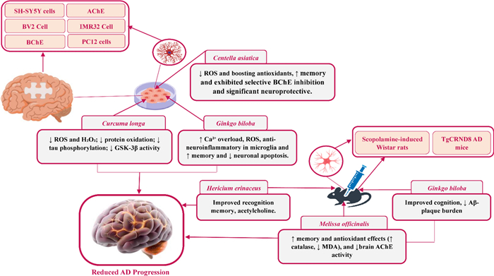
Proposed in vivo and in vitro mechanisms of action of Medicinal Plant in Alzheimer. (↑) indicates increased; (↓) indicates reduced; ROS, reactive oxygen species; GSK-3β, glycogen synthase kinase-3 beta; AChE, acetylcholinesterase; BChE, butyrylcholinesterase
4.2 Bioactive phytoconstituents of medicinal plant
Numerous in vivo, and in vitro studies have highlighted the potential of various commonly consumed medicinal plant in combating AD. These plants are rich in bioactive compounds that exhibit significant pharmacological activities, including AChE inhibition, antioxidant and anti-inflammatory effects, neuroprotection, modulation of Aβ toxicity, and regulation of neuronal pathways (Fig. 2). These phytoconstituents found in various medicinal plant that show benefits against AD are detailed in Table 2, with their chemical structures represented in Fig. 3.
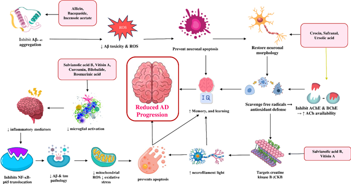
Schematic diagram illustrating the anti-Alzheimer’s mechanisms of action of various phytoconstituents found in medicinal plant. (↑) indicates increased; (↓) indicates reduced; ROS, reactive oxygen species; Nuclear factor kappa-light-chain-enhancer of activated B cells (NF-κB); Creatine Kinase B (CKB); AChE, acetylcholinesterase; BChE, butyrylcholinesterase
Pharmacological effects of the active phytoconstituents of selected medicinal plant against AD
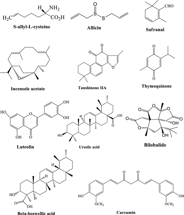
Chemical structure of bioactive phytoconstituent of medicinal plant
In addition to pharmacodynamic activity, the pharmacokinetic characteristics of key bioactive constituents critically influence their translational potential. Curcumin exhibits extremely low oral bioavailability due to poor aqueous solubility, rapid intestinal metabolism, and extensive first-pass hepatic clearance; however, nanoparticle, liposomal, phospholipid-complex, and bioenhancer-based formulations significantly improve systemic exposure and brain distribution [69–74]. Crocin displays limited gastrointestinal absorption and is rapidly hydrolyzed to crocetin, which shows superior membrane permeability, measurable blood–brain barrier penetration, and a longer elimination half-life, supporting its role as the principal bioavailable neuroactive metabolite of saffron [46, 47, 75–77]. Emerging pharmacokinetic studies of asiaticoside, bacosides, bilobalide, huperzine A, and erinacines further indicate variable absorption, metabolism, and brain exposure, emphasizing the importance of formulation strategies for optimized central nervous system delivery [71, 75, 78–81].
4.3 Allium sativum L.
A. sativum L. (garlic), a member of the Amaryllidaceae family, has long been used as both a culinary ingredient and a medicinal agent, with traditional applications ranging from respiratory and gastrointestinal ailments to hypertension, hyperlipidemia and general cognitive vitality [98]. Its historical use across Asia, Europe and the Middle East for inflammatory and circulatory disorders is particularly notable, as these conditions are now recognized contributors to neurodegenerative disease risk [98]. The pharmacological actions of garlic are primarily attributed to its organosulfur constituents including allicin and S-allyl-L-cysteine alongside flavonoids and other antioxidant molecules, which together exert antioxidative, anti-inflammatory, lipid-regulating and anti-amyloidogenic effects that support a mechanistic rationale for neuroprotection [98]. Evidence from preclinical studies shows that garlic extracts and isolated compounds reduce oxidative stress, inhibit acetylcholinesterase (AChE), modulate apoptosis-related markers (Bax/Bcl-2, caspase-3), suppress neuroinflammation and decrease amyloid-β burden, accompanied by improvements in learning and memory in rodent models [99, 100]. Aged garlic extract (AGE) and stabilized allicin formulations, developed to improve bioavailability and tolerability, have demonstrated the ability to preserve mitochondrial function and maintain synaptic proteins under Aβ-induced stress in both in vitro and in vivo assays [99, 100]. Consistent with these findings, essential oil derived from garlic cloves showed moderate anti-cholinesterase activity in vitro, inhibiting AChE (65.4%) and BChE (31.5%), with dose-dependent effects on cholinergic markers [33]. In Aβ(25–35)-induced AD rodent models, garlic administration increased hippocampal brain-derived neurotrophic factor (BDNF) and reduced AChE activity, supporting its neurotrophic potential [34]. S-allyl-L-cysteine further enhanced neurite outgrowth in primary mouse hippocampal neurons at 10–1000 ng/mL and improved cognitive performance in senescence-accelerated SAMP10 mice while preserving AMPA/NMDA receptor expression and p-CaMKII signaling [82]. Allicin supplementation (10–20 mg/kg) attenuated aluminum- and copper-induced cognitive deficits in Wistar rats, lowered oxidative and inflammatory markers (IL-1β, IL-6, TNF-α), restored neurotransmitter balance, and reduced Aβ(1–42) accumulation in a dose-dependent manner [83].
4.4 Bacopa monnier (L.) Wettst.
B. monnieri (brahmi, water hyssop) is a perennial creeping herb of the Plantaginaceae family, widely valued in Ayurvedic medicine as a cognitive enhancer and nervine. Traditionally, it has been used to improve memory, reduce anxiety, support sleep, and manage epilepsy, with additional ethnobotanical reports noting benefits for asthma, digestive discomfort, and wound healing [101, 102]. Contemporary ethnopharmacological research supports these long-standing uses and highlights growing interest in bacosides and related constituents for their antioxidant, anti-inflammatory, cholinergic, and synaptic-supportive effects, all of which are relevant to neurodegenerative disorders [101, 102]. Pharmaceutical development has focused on standardized bacoside-rich extracts used as nootropic supplements, isolation of bacoside A and other saponins as potential lead molecules, and formulation strategies including nanoparticle and liposomal carriers designed to enhance oral absorption and overcome poor solubility and first-pass metabolism. Combination products with other neuroprotective agents have also been explored to assess possible synergistic activity [38, 103]. Alzheimer’s-focused preclinical studies consistently show that B. monnieri exerts broad neuroprotective actions. In rodent and cell-based AD models, extracts reduce amyloid-β–induced toxicity, limit oxidative stress and neuroinflammation, attenuate tau-related pathology, and improve learning and memory performance in behavioural assays such as the Morris water maze [38, 102]. Mechanistic investigations report enhanced expression of synaptic markers, modulation of acetylcholinesterase activity, and reduced apoptotic signalling, indicating complementary actions that align well with key features of Alzheimer’s pathology [38, 101]. Individual studies further demonstrate that ethanolic extracts of the aerial parts protect against Aβ42-induced hippocampal damage at concentrations ranging from 0.25 to 400 μM, improving cognition, lowering oxidative and inflammatory markers, reducing cholinesterase activity, decreasing tau and amyloid pathology, and regulating the GSK-3β/Wnt/β-catenin pathway [36]. In colchicine-induced dementia models, a 50 mg/kg dose improved cognitive outcomes, decreased ROS and NO levels, reduced IL-6, TNF-α, and MCP-1, and lowered BACE-1 activity and amyloid production [37]. An aqueous extract also showed protective effects in IMR-32 neuroblastoma cells exposed to Aβ42 at 100 μg/mL [38]. Among isolated constituents, bacopaside Ⅰ (15–50 mg/kg) reduced amyloid plaque burden and improved memory in APP/PS1 transgenic mice [78], while bacoside A (1–100 μM) demonstrated acetylcholinesterase inhibitory activity and neuroprotective potential in vitro [79].
4.5 Centella asiatica (L.) Urb.
C. asiatica, widely known as gotu kola and pennywort and belonging to the Apiaceae family, is a perennial herb valued for centuries in Ayurvedic, Traditional Chinese and Southeast Asian systems of medicine for its wound-healing, anxiolytic and cognition-enhancing effects [81]. Traditional uses include whole leaves and aqueous preparations for the management of skin wounds and ulcers, gastrointestinal discomfort, anxiety, age-related memory complaints and general vitality, and ethnobotanical studies continue to report its recognition as a “memory herb” across regions of India, China and Indonesia [104, 105]. Its therapeutic profile is linked mainly to triterpenoid saponins such as asiaticoside, madecassoside, asiatic acid and madecassic acid, together with polyphenolic constituents, which act through antioxidant, anti-inflammatory, neuroprotective and tissue-repair mechanisms that support both external and internal applications [106]. Modern pharmaceutical development has expanded these traditional uses into standardized aqueous extracts designed for cognitive support, topical preparations for wound healing and scar reduction, nutraceutical formulations enriched with triterpenoid fractions and emerging pharmacokinetic studies aimed at defining exposure, safety and dosage for human use [107, 108]. Extensive preclinical evidence has examined the potential of C. asiatica in Alzheimer’s disease models. Aqueous extracts and defined constituents have been shown to regulate oxidative stress pathways, enhance mitochondrial biogenesis, activate Nrf2-dependent antioxidant gene networks and shift metabolic pathways associated with amyloid burden in transgenic models such as 5×FAD mice, leading to improved learning and memory across behavioural tasks [106]. Studies isolating fractions enriched in caffeoylquinic acids or specific triterpenoid components report reversal of cognitive deficits and reduction of neuropathological features in mouse systems, highlighting both symptomatic and potential disease-modifying actions [109, 110]. These findings are supported by validated analytical methods and an early-phase human pharmacokinetic and safety study conducted in older adults, marking important steps toward clinical translation and reinforcing the need for extract standardization and dose selection guided by animal data [107]. Additional experimental work using water extracts of C. asiatica leaves and stems at 200 and 1000 mg/kg in aged transgenic AD mice (Tg2576) and wild-type controls has demonstrated improvements in novel object recognition and contextual fear memory, changes in hippocampal metabolomic profiles, enhanced mitochondrial respiration and reductions in oxidative stress markers [40]. Ethanol extracts tested in neuronal cell lines such as PC12 and IMR32 at concentrations of 25, 50 and 100 μg/mL protected against Aβ1-40–induced toxicity, lowered reactive oxygen species and strengthened endogenous antioxidant defences including superoxide dismutase, catalase and glutathione systems [41]. In 5×FAD mice, daily treatment with water extracts at 200, 500 and 1000 mg/kg improved object recognition and contextual fear performance, increased expression of Nrf2-regulated antioxidant genes and reduced oxidative stress indicators [42]. Asiaticoside at 40 mg/kg in Aβ1-42-induced C57/BL6 mice alleviated memory impairment by inhibiting p38 MAPK activation and promoting synaptic repair, with evidence of reduced neuroinflammation and improved synaptic function, suggesting strong potential for therapeutic development [86]
4.6 Crocus sativus L.
C. sativus widely known as saffron and belonging to the family Iridaceae, is a perennial autumn-flowering bulb whose dried stigmas have been valued for millennia as a spice, dye, and medicinal substance throughout Mediterranean, Persian, Indian, and East Asian medical traditions. Across these long-standing records, saffron has been used for diverse conditions including mood disturbances, insomnia, menstrual irregularities, digestive problems and respiratory complaints, and historical writings together with ethnobotanical research also describe its application for memory and age-related cognitive decline [111]. Current pharmacological research attributes its central nervous system activity to major constituents such as crocin, crocetin, picrocrocin and safranal, which exhibit antioxidant, anti-inflammatory, anti-apoptotic and neurotransmitter-modulating actions that are mechanistically consistent with neuroprotection [112, 113]. Pharmaceutical development efforts increasingly focus on these compounds: crocin and crocetin are being evaluated as neuroprotective nutraceuticals and standardized botanical preparations for cognitive disorders; safranal is under investigation as a small-molecule modulator of GABAergic and monoaminergic signalling with potential antidepressant and anxiolytic benefits; standardized saffron extracts are being formulated for oral delivery to target mild cognitive impairment and reduce dementia risk in clinical settings; and derivative formulations such as by-products and encapsulated crocin complexes are being explored to improve stability, bioavailability and blood brain-barrier permeability for neurological indications [77, 113]. Converging preclinical evidence highlights robust multi-target effects relevant to AD. Studies in rodents and cell systems show that saffron extracts and isolated crocin attenuate amyloid-β toxicity, limit oxidative and inflammatory responses, inhibit cholinesterase activity, regulate autophagy and apoptosis pathways, and strengthen synaptic plasticity, leading to improvements in learning and memory across behavioral tasks such as the Morris water maze and passive avoidance tests [47, 77]. Importantly, these mechanisms do not act independently; rather, antioxidant activity appears to synergize with anti-inflammatory signaling by reducing reactive oxygen species–driven activation of inflammatory pathways such as NF-κB and NLRP3, thereby amplifying neuroprotection. This coordinated suppression of oxidative stress and neuroinflammation contributes to downstream stabilization of mitochondrial function, preservation of synaptic integrity, and reduced amyloidogenic processing, illustrating a tightly interconnected multi-target therapeutic network [47, 77]. In transgenic 5×FAD mice, a hydroalcoholic extract of C. sativus given at 50 mg/kg per day improved blood–brain-barrier integrity, increased amyloid-β clearance through P-gp and LRP1, reduced amyloid burden, up-regulated synaptic proteins and lowered neuroinflammation [46]. The enhancement of amyloid clearance is mechanistically linked to concurrent reductions in oxidative and inflammatory stress, which are known to impair transporter function at the blood–brain barrier, suggesting that antioxidant and anti-inflammatory actions indirectly facilitate amyloid efflux [46]. Complementary in vitro work using the same extract demonstrates enhanced amyloid-β clearance through P-gp induction, inhibition of the NLRP3 inflammasome with reduced IL-1β release, and protection of SH-SY5Y cells against amyloid-β and glutamate toxicity, alongside behavioral improvement in streptozotocin and scopolamine models [47]. Additional in vivo studies in streptozotocin-induced dementia in rats and scopolamine-induced amnesia in mice further support these effects at doses of 50 mg/kg and 100 mg/kg [47], while chronic scopolamine-treated rats show significant recovery of memory at 10, 15 and 20 mg/kg with reduced acetylcholinesterase activity, lower IL-6 and oxidative stress, and a reduction in amyloid plaques and neurofibrillary tangles [49]. Here, cholinesterase inhibition may act synergistically with anti-inflammatory and antioxidant effects to sustain cholinergic neurotransmission while minimizing cytokine- and oxidative-mediated synaptic damage [49] Crocin administered at 40 mg/kg in a hippocampal amyloid-β25–35 mouse model suppresses neuroinflammation through activation of the PI3K, AKT pathway, improving cognitive outcomes [75]. Crocetin at concentrations of 3.12–50 µM enhances amyloid clearance in vitro, lowers brain amyloid load and gliosis, and improves memory in 5XFAD mice, indicating autophagy-promoting and neuroprotective effects at doses of 5, 10 and 20 mg/kg in vivo [76]. Safranal given at 0.025, 0.1 and 0.2 mL/kg in an amyloid-β microinjected rat model improves cognitive performance across multiple tasks including the Y-maze, novel-object recognition, passive avoidance and radial arm maze, while lowering oxidative, inflammatory and apoptotic markers and preventing neuronal loss, demonstrating antioxidant, anti-inflammatory and anti-apoptotic protection [114]. The convergence of these pathways suggests that suppression of apoptosis is secondary to upstream control of oxidative stress and inflammation, reinforcing the concept of hierarchical and synergistic mechanism integration [114]. Finally, trans-crocin 4 and trans-crocetin have been shown in vitro to bind strongly to amyloid-β monomers and redirect aggregation pathways, markedly reducing oligomer and fibril formation, which points to a direct anti-amyloid mechanism for these specific components [87].
4.7 Curcuma longa L.
C. longa widely known as turmeric, is a perennial herb in the ginger family Zingiberaceae and has long been valued in South Asian traditional medicine for its broad therapeutic uses. The rhizomes have historically been applied as a wound poultice and used as an anti-inflammatory agent, digestive tonic, and treatment for liver, respiratory, and skin disorders; in Ayurvedic and folk traditions they have also been taken as a memory tonic and for age-related cognitive decline [69]. Modern phytochemical and pharmacological studies have centred on curcumin, the principal diferuloylmethane polyphenol, whose wide biological actions including antioxidant, anti-inflammatory, metal-chelating, and cell-signalling modulatory effects provide a plausible basis for many longstanding medicinal claims [70, 71]. Pharmaceutical development has advanced rapidly, with curcumin and turmeric extracts now explored as nutraceuticals and supportive treatments for chronic inflammation, metabolic dysfunction, and neurodegenerative diseases. Recent formulation research has focused on improving oral bioavailability through nanoparticle, liposomal, phospholipid, and polymer-based delivery systems; combining curcumin with bioenhancers such as piperine to increase systemic exposure; designing curcumin analogues and prodrugs with greater metabolic stability; and producing standardized turmeric extracts to ensure consistent dosing in clinical studies all intended to convert promising laboratory findings into reliable clinical benefit [69, 70]. Preclinical research provides strong and consistent evidence that curcumin can reduce several core pathological processes in animal and cell models. It decreases amyloid-β aggregation and deposition, suppresses neuroinflammation by modulating microglial and astrocyte activity, lowers oxidative stress, inhibits pathways linked to tau hyperphosphorylation, and supports synaptic plasticity and neurotrophic signalling, including effects on BDNF. These actions have been associated with improved spatial memory and learning in both transgenic and toxin-induced rodent models [72–74]. Experimental studies using turmeric extracts further reinforce these findings. Ethanol extracts of the rhizome administered to amyloid-β1–42-injected mice at 100 and 300 mg/kg reduced Aβ-induced cytotoxicity, lowered TNF-α, IFN-β, and iNOS, inhibited TLR4–NF-κB signalling, and improved memory and learning in Morris water maze and passive avoidance tests; complementary in vitro work in primary rat hippocampal neurons at 3–30 µg/mL supported these anti-inflammatory and neuroprotective effects [43]. Methanolic extracts of the rhizome showed strong inhibition of amyloid-fibril formation in a cell-free fibrillation model, indicating potential to limit amyloid aggregation [44]. Additional ethanol-based extracts demonstrated protective, antioxidant, and anti-inflammatory actions in C6 glioma and BV2 microglia, with parallel improvements in scopolamine-induced amnesia in mice at doses of 50, 100, and 200 mg/kg, highlighting relevance to neurodegeneration [45]. Curcumin itself prevented long-term memory deficits in lipopolysaccharide-induced C57BL/6 mice at 50 mg/kg, reduced neuroinflammation and oxidative stress, and preserved recognition memory, suggesting meaningful protection against inflammatory drivers of Alzheimer’s pathology [88].
4.8 Ginkgo biloba L.
G. biloba widely known as ginkgo or the maidenhair tree, is the only surviving species of the ancient gymnosperm family Ginkgoaceae and has been valued for centuries in East Asian medicine for disorders of memory, poor circulation, respiratory symptoms, and urinary complaints. Its leaves and seeds were traditionally used to relieve asthma, bronchitis, intermittent claudication, and were often regarded as a general tonic for the brain [115]. Contemporary ethnopharmacological reports continue to highlight the leaf extract for cognitive difficulties and age-related memory decline, which stimulated systematic investigation of ginkgo for Alzheimer’s disease (AD) and other dementias [116, 117]. The most rigorously studied preparation is the standardized extract EGb 761®, formulated to contain about 24% flavonol glycosides and about 6% terpene trilactones, including ginkgolides and bilobalide, while maintaining a low concentration of ginkgolic acids. This standardized composition forms the basis of most clinical and preclinical research and explains the extract’s consistent antioxidant, anti-inflammatory, and vascular actions [118]. In pharmaceutical use, EGb 761® is produced as oral tablets or capsules for cognitive support and for the symptomatic management of mild to moderate dementia; it is used clinically to enhance cerebral blood flow and microcirculation; its flavonoids act as free-radical scavengers, while its terpene lactones influence platelet-activating factor and mitochondrial function; and study protocols commonly use doses between 120 and 240 mg per day, depending on the therapeutic indication. Preclinical AD studies have consistently shown that ginkgo leaf extracts and EGb 761® exert neuroprotective activity across cellular and transgenic mouse models. These preparations reduce amyloid-β aggregation and tau-related toxicity, limit oxidative stress and neuroinflammation, preserve mitochondrial integrity and synaptic markers, and improve performance in behavioral tests of learning and memory. Mechanistic analyses link these effects to the induction of antioxidant enzymes, suppression of excitotoxic glutamate release, regulation of autophagy and lysosomal pathways, and a reduction in microglial pro-inflammatory signaling [119]. Experimental data support these findings: in TgCRND8 mice, 240 mg/day of EGb 761 improved cognition in the Barnes maze, prevented the loss of synaptic proteins such as PSD-95, Munc18-1, and SNAP25, and reduced neuroinflammation [50]. In APP, PS1 mice, 100 mg/kg of a standardized extract improved spatial memory in the Morris water maze and altered gut microbial composition by increasing probiotic species and modifying metabolite profiles [51]. In SH-SY5Y human neuroblastoma cells, 25 µg/mL of leaf extract reduced calcium influx through TRPV1 channels, limited reactive oxygen species, apoptosis, and mitochondrial depolarization in an AD model [48]. Bilobalide, a major terpene trilactone of the plant, showed complementary benefits in APP, PS1 mice at 0.5 mg/kg by lowering STAT3-dependent TNF-α, IL-1β, and IL-6 signaling, increasing amyloid-β-degrading enzymes such as NEP, IDE, and MMP2, diminishing amyloid burden and inflammation in brain tissue, and ultimately restoring neuronal integrity [89].
4.9 Hericium erinaceus
H. erinaceus, widely known as Lion’s mane and belonging to the family Hericiaceae, is an edible toothed fungus long used in East Asian medicine for digestive problems, gastric ulcers, general restorative purposes, and, most notably, for traditional claims that it can strengthen the nerves and improve memory [120, 121]. Recent scientific assessments increasingly support these long-standing uses, linking them to specific groups of bioactive molecules hericenones derived from the fruiting body and erinacines produced by the mycelium which interact with neurotrophic and anti-inflammatory pathways and have therefore become the focus of preclinical research in Alzheimer’s disease (AD) [120, 122]. Pharmaceutical interest in H. erinaceus continues to grow due to its neurotrophic activity and favorable safety profile. Current developments include standardized erinacine-A–rich mycelial extracts designed for oral nutraceuticals that promote nerve growth factor (NGF); isolated hericenone fractions studied as small-molecule candidates to stimulate neurite outgrowth; polysaccharide preparations explored for anti-inflammatory actions and microbiome-mediated brain effects; combined extracts from both mycelium and fruiting bodies formulated for long-term dietary supplementation; and formulation strategies aimed at improving the oral absorption and central nervous system penetration of erinacines [120, 123].
Preclinical AD studies have produced consistent evidence that extracts and purified compounds from H. erinaceus can reduce several core mechanisms implicated in Alzheimer’s pathology. In cellular and rodent models, erinacine A increases NGF expression, promotes neurite extension, reduces amyloid-β formation and amyloidogenic processing, and decreases markers of inflammation such as IL-1β, while also reducing oxidative stress and protecting neural cells from amyloid-β toxicity. These changes lead to better performance on learning and memory tasks including the Morris water maze and novel object recognition in multiple mouse studies [120, 121, 124]. Standardized mycelial extracts have also been shown to lower amyloid plaque load and preserve synaptic markers in APP, PS1 and SAMP8 models, supporting a broad disease-modifying role rather than a narrow symptomatic effect [124, 125]. Although small clinical studies and short-term supplementation trials in older adults and individuals with mild cognitive impairment have produced mixed yet encouraging cognitive outcomes, larger and well-controlled randomized trials using defined extracts, dose–response evaluation, and extended follow-up are still necessary to determine the therapeutic potential, optimal dosing, and preventive value of H. erinaceus for AD [126, 127]. Experimental work further supports these findings. Mycelial aqueous extracts protected PC12 cells from glutamate-induced apoptosis at 50 and 100 µg/mL by lowering reactive oxygen species, preventing calcium overload, and reducing mitochondrial dysfunction. In mice, the same extract improved locomotor activity, enhanced rotarod endurance, reduced escape latency in the Morris water maze, and increased acetylcholine and choline acetyltransferase levels in serum and the hypothalamus in an aluminum chloride and d-galactose model of AD [52]. Ethanolic extracts tested in scopolamine-induced zebrafish models at 0.5, 1, and 3 mg/L improved behavioral memory outcomes, inhibited acetylcholinesterase activity, strengthened antioxidant defenses (SOD, CAT, GPX, GSH), and reduced lipid peroxidation and protein–carbonyl damage in the brain [53]. Aqueous preparations from whole biomass administered at 200 mg/kg up-regulated vitagene stress-response proteins, including Hsp70, HO-1, and thioredoxin, while increasing lipoxin A4 across the cortex, cerebellum, and hippocampus, indicating reduced neuroinflammation and activation of endogenous resolution pathways relevant to AD [54]. In addition, purified hericene A given at 5 mg/kg improved recognition memory and activated broad neurotrophic signaling, including BDNF and TrkB, strengthening synaptic function and positioning the hericene/erinacine family as promising neuroprotective candidates for AD-related cognitive decline [90].
4.10 Huperzia serrata (Thunb.) Trevis.
H. serrata, commonly known as Chinese club moss or toothed clubmoss, is a slow-growing lycopod belonging to the family Lycopodiaceae and has a long history of use in traditional Asian medicine for conditions such as memory loss, fever, inflammation, cough, and trauma. Ethnobotanical accounts describe its valued role in Traditional Chinese Medicine and related practices, where whole-plant preparations were regarded as agents that support healthy ageing and cognitive function [128]. Modern phytochemical research has identified the plant’s principal bioactive alkaloid, huperzine A (HupA), as a potent, reversible, and selective acetylcholinesterase inhibitor, providing a mechanistic explanation for its traditional use in forgetfulness and age-related cognitive decline [80, 129]. In pharmaceutical development, HupA has been advanced both as a symptomatic cholinergic therapy enhancing acetylcholine levels and improving short-term cognition and as a molecule with broader neuroprotective properties that include antioxidant actions, anti-apoptotic signaling, modulation of amyloid precursor protein processing, and protection of synaptic integrity [81, 129]. Current applications focus on oral HupA formulations for dementia symptom relief, supported by animal models and reported clinical benefits. Preclinical studies also indicate antioxidant, anti-apoptotic, and mitochondrial-stabilizing effects that may slow neuronal loss, while emerging data on amyloid and tau modulation suggest multi-target, disease-modifying potential beyond acetylcholinesterase inhibition. Between 2020 and 2024, structural-modification and drug-delivery studies have sought to improve brain bioavailability and safety, and recent regulatory reviews emphasize the importance of dose-finding and standardization for herbal preparations [80, 81]. Standardized extracts of H. serrata and purified HupA consistently reverse or attenuate learning and memory impairments in rodent models, reduce acetylcholinesterase activity in the hippocampus and cortex, lower oxidative stress markers, and influence biomarkers linked to amyloid metabolism and synaptic function. These findings, summarized across recent preclinical reviews and individual experimental studies, support continued translational interest while also highlighting the need for rigorous dose–response designs, long-term safety evaluation, and standardized extract parameters before broad clinical implementation [80, 81, 129]. Experimental data show that aqueous or ethanolic extracts at 30 mg/kg/day inhibit acetylcholinesterase in the brain and improve cognitive performance in scopolamine-induced mice using Y-maze and passive avoidance tests [55]. In SH-SY5Y cells exposed to scopolamine, extracts promote neuroprotection through increased antioxidant enzyme activity (SOD, CAT), decreased malondialdehyde, reduced acetylcholinesterase activity, and improved behavioral outcomes in aligned in vivo assessments [56]. Additional studies in scopolamine-treated mice using Y-maze and the Morris water maze further support the extract’s ability to improve learning and memory, consistent with its proposed neuroprotective and cholinergic effects[56].
4.11 Salvia miltiorrhiza Bunge
S. miltiorrhiza Bunge, widely known as Danshen or red sage, is a perennial herb from the mint family Lamiaceae whose dried roots have long held a central role in East Asian traditional medicine. In traditional Chinese medicine, Danshen is used to invigorate blood and resolve blood stasis, and it continues to be prescribed for cardiovascular and cerebrovascular disorders such as angina, ischemia, and stroke, as well as for menstrual disturbances, hepatic conditions, and inflammatory diseases [130]. Advances in phytochemistry have defined two major groups of active molecules: lipophilic tanshinones, including tanshinone IIA and cryptotanshinone, and water-soluble phenolic acids, such as salvianolic acids. Together, these compounds account for the herb’s recognized circulatory, antioxidant, and anti-inflammatory actions [131]. Danshen is developed into standardized oral and injectable products enriched in tanshinones and salvianolic acids, and preparations such as compound Danshen dripping pills and Danshen injections are used clinically in China for ischemic cardiovascular disease. Its bioactive molecules are being engineered into nano-delivery systems to enhance delivery to the brain and to tumors, tanshinone IIA is under investigation as a lead compound for central nervous system drug development, salvianolic acids are studied as antioxidant and anti-amyloid candidates, and cryptotanshinone shows activity in key cell-signaling pathways with relevance to neuroprotection. Ongoing regulatory and quality-control work emphasizes standardization and characterization of drug–herb interactions to support clinical translation [132, 133]. Preclinical studies increasingly examine Danshen and its constituents in Alzheimer’s disease models. Tanshinone IIA and related diterpenoids have been shown to lower amyloid-β burden, protect against amyloid-β-induced cytotoxicity, suppress neuroinflammation through NF-κB signaling, and reduce oxidative stress and neuronal apoptosis in both cell-based systems and transgenic or chemically induced rodent models, with parallel improvements in learning and memory performance [134, 135]. Salvianolic acids demonstrate strong antioxidant capacity and inhibit amyloid-β aggregation in vitro, while improving synaptic and mitochondrial markers in animal studies; cryptotanshinone also modulates signaling pathways implicated in neurodegeneration and provides neuroprotective effects in preclinical evaluations [136, 137]. Experimental work using the ethanol extract of S. miltiorrhiza root in NG108-15 cells and in mice exposed to amyloid-β(25–35) shows attenuation of learning and memory deficits in Y-maze and passive-avoidance tasks, along with reduced brain malondialdehyde levels, with effective doses ranging from 400 to 1200 mg/kg in vivo and 2.0 μg/mL in vitro [63]. The dried root and rhizome water extract binds amyloid-β, inhibits fibril formation in Thioflavin T assays, delays amyloid-β-induced paralysis in Caenorhabditis elegans, decreases reactive oxygen species generation by more than 50 percent at 4 mg/mL, and provides antioxidant and neuroprotective activity against amyloid-β-mediated toxicity [64]. In a chronic cerebral hypoperfusion model, root and rhizome water extract at 200 mg/kg per day protects hippocampal and white-matter integrity following bilateral carotid occlusion, reduces pro-inflammatory cytokines including TNF-α, IL-1β, and IL-6, inhibits the TLR4-MyD88 pathway, and restores myelin basic protein levels [65]. Salvianolic acid B reduces oxidative stress and inhibits GSK3β activity in SH-SY5Y-APPsw cells at concentrations of 25–100 μM, an effect linked to suppression of BACE1 expression and reduced amyloidogenesis [95]. Tanshinone IIA, administered at 25–100 mg/kg in APP/PS1 transgenic mice, improves cognitive impairment and decreases neuroinflammation, in part through inhibition of the RAGE–NF-κB signaling pathway [96].
4.12 Vitis vinifera L.
V. vinifera, commonly known as grape and belonging to the Vitaceae family, has been valued in traditional medical systems for centuries, where grape fruit, seeds, skins and leaves were used to support digestion, promote wound healing and enhance memory, often described culturally as a “brain tonic” that conveys vitality and cognitive strength. Contemporary phytochemical research confirms that these plant parts contain abundant polyphenols such as resveratrol, proanthocyanidins, anthocyanins and flavonols, compounds that provide a credible scientific basis for these long-standing ethnomedical claims and explain why V. vinifera now holds a prominent position in nutraceutical investigations targeting age-related cognitive decline. Preclinical studies in AD models show that preparations derived from this species exert diverse neuroprotective effects, including strong antioxidant and free-radical-scavenging activity, suppression of neuroinflammation, inhibition of amyloid beta aggregation and tau pathology, enhancement of synaptic plasticity through BDNF–CREB signaling, and support of mitochondrial function mechanisms demonstrated across multiple cellular systems and rodent AD models and synthesized in recent reviews [138, 139]. In pharmaceutical and translational contexts, compounds from V. vinifera are being advanced as multimodal agents or as adjuncts to enhance existing therapies rather than as single-target interventions. Current applications include standardized grape-seed polyphenol extracts formulated as oral nutraceuticals for cognitive maintenance; resveratrol analogs designed as SIRT1-activating small molecules with improved bioavailability; nanoparticle and liposomal carriers engineered to increase brain penetration of grape polyphenols; topical preparations from leaves and skins used in antioxidant skincare and wound-healing approaches that draw upon shared anti-inflammatory chemistry; and combination formulations that pair grape polyphenols with other neuroprotective phytochemicals to achieve synergistic effects relevant to AD modulation [140]. In Alzheimer’s research specifically, whole grape extracts, grape-seed extract (GSPE), anthocyanin-rich fractions and purified resveratrol have been shown to improve cognitive performance in behavioral paradigms, reduce amyloid burden and tau-related pathology, restore antioxidant enzyme activities and rescue synaptic markers in various experimental models, including scopolamine-, D-galactose- and amyloid beta–based systems as well as transgenic animals, alongside protection demonstrated in cellular assays that assess amyloid-induced toxicity [141]. Early human supplementation studies using standardized grape extracts or resveratrol also report promising cognitive and biomarker responses that support further translational work, although experts consistently highlight the need for larger, well-controlled clinical trials and improved formulations to address the low oral bioavailability and rapid metabolism that limit many grape polyphenols [138, 142]. Experimental data further reinforce these findings: petroleum ether leaf extracts at 100 mg/kg in an aluminum-induced rat model of Alzheimer’s improved cognitive and neurobehavioral outcomes and modulated markers including acetylcholine, acetylcholinesterase, BDNF, IL-6 and homocysteine, demonstrating neuroprotective, antioxidant, anti-inflammatory and anti-amnesic activity [66]. Seed extracts administered at 250 and 500 mg/kg in the same model improved learning and memory in the Morris water maze, inhibited mRNA expression of APP and tau, reduced lipid peroxidation and inflammation, and strengthened cholinergic function [67]. Ethanol extracts from the leaves protected HT22 hippocampal neuronal cells against glutamate-induced oxidative stress at 50 μg/m by enhancing antioxidant enzyme gene expression, including SOD, CAT and GPx [68]. Vitisin A, a constituent of Vitis vinifera, improved cognitive performance in behavioral assessments, increased cell survival under oxidative stress in SH-SY5Y neuroblastoma cells and enhanced hippocampal BDNF–CREB signaling at concentrations of 1, 5 or 10 μM, with supportive findings also reported in scopolamine-induced dementia models in C57BL/6 male mice at doses of 1 or 100 ng/μL [97].
4.13 Clinical trials of medicinal plant against AD
Building on the promising outcomes from various studies, many medicinal plants have progressed to clinical trials to assess their efficacy and safety in the prevention and treatment of AD in humans. The diverse pharmacological effects observed in these trials underscore the potential of medicinal plants in AD prevention and treatment (Table 3). A growing body of clinical evidence indicates that several medicinal plants may support cognitive function across a range of populations, from healthy adults to individuals with mild cognitive impairment (MCI), Alzheimer’s disease (AD), and vascular dementia. In healthy volunteers, supplementation with A. sativum for five weeks improved visual memory and attention, although changes in verbal memory and executive function did not reach statistical significance, suggesting that longer trials in neurodegenerative conditions may better reveal its protective effects [143]. In patients with MCI and mild-to-moderate AD, B. sacra administered for three months produced meaningful improvements in cognitive and behavioral symptoms, reinforcing its therapeutic potential in early cognitive decline [144]. A phase-1 crossover study of C. asiatica showed that single oral doses of a standardized aqueous extract were safe and well tolerated, with active compounds detected in plasma and evidence of dose-dependent antioxidant and neuroprotective activity through NRF2 pathway activation in individuals with MCI [107]. However, this study enrolled only four participants with mild cognitive impairment, which substantially limits statistical power and precludes meaningful assessment of interindividual variability or clinical efficacy. While valuable for confirming safety, pharmacokinetics, and mechanistic signals such as NRF2 pathway activation, the extremely small sample size restricts extrapolation of these findings to broader AD populations and underscores the need for larger, adequately powered follow-up trials [107]. C. sativus demonstrated clinical efficacy comparable to donepezil during a 22-week randomized trial in mild-to-moderate AD, highlighting its potential as a botanical alternative for symptomatic treatment [145]. G. biloba extract (EGb 761) improved executive function, attention, non-verbal memory, mood, and quality of life in brain tumor survivors with radiation-induced cognitive dysfunction, although a high dropout rate suggested tolerability and perceived benefit may vary across individuals [146]. Long-term supplementation with H. erinaceus mycelia over 49 weeks slowed cognitive decline in mild AD and improved daily function and contrast sensitivity, with a strong safety profile supporting its use as a neuroprotective agent [147]. H. serrata also showed clinical value, reducing cognitive and task-switching deficits over an eight-week period in patients with AD [148]. In a 96-week double-blind trial, M. officinalis prevented cognitive decline in individuals with mild AD dementia, further affirming its long history of cognitive use [149]. Among healthy elderly adults, N. sativa taken for nine weeks enhanced memory, attention, and general cognition, suggesting possible preventive effects against age-related cognitive decline or early neurodegenerative processes [150]. P. ginseng improved MMSE and ADAS scores in patients with AD over a 12-week treatment period, though the gains diminished after discontinuation, indicating that continuous intake may be necessary to sustain cognitive benefits [151]. For vascular dementia, S. miltiorrhiza showed both efficacy and safety over a 24-week multicenter trial, supporting its potential role in managing cognitive impairment linked to vascular dysfunction [152]. In healthy older adults, V. vinifera extract (Cognigrape®) administered for 12 weeks improved memory, attention, and language skills, suggesting a favorable effect on age-related cognitive performance [153]. A multi-herb formulation, Yi-Zhi-An-Shen granules, prevented or delayed cognitive decline over 16 weeks in individuals with MCI or dementia, reflecting the therapeutic value of synergistic plant combinations [154]. Similarly, a polyherbal formulation containing B. monnieri, H. rhamnoides, and D. bulbifera improved cognition and reduced inflammation and oxidative stress in senile dementia of the Alzheimer’s type following one year of treatment [155]. A short-term crossover study combining P. ginseng, G. biloba, and C. sativus reported enhanced visuospatial short-term memory, working memory, and more efficient attentional processing, aligning with earlier preclinical results and underscoring strong synergistic interactions among these botanicals [156]. Finally, the Korean herbal medicine Kami Guibi-tang showed positive cognitive effects in patients with mild AD over a 24-week trial, indicating its promise for individuals in the early stages of impairment [157].
Clinical trials investigating the use of medicinal plant for preventing and treating AD
Notably, the reliability and generalizability of these clinical findings are influenced by considerable variation in sample sizes across studies, ranging from small pilot or phase-1 trials to larger randomized and multicenter investigations. Specifically, the reviewed trials can be broadly categorized into small-sample studies (≤ 40 participants) and large-sample studies (≥ 200 participants), which show clear differences in evidentiary strength. Small-sample investigations, including early-phase or pilot trials, frequently report positive trends in cognitive or biomarker outcomes but are characterized by wide confidence intervals and increased interindividual variability. In contrast, large-scale randomized trials, such as those involving Melissa officinalis (n = 323), Salvia miltiorrhiza (n = 240), and polyherbal formulations (n = 232), provide more consistent and statistically robust evidence for cognitive stabilization or improvement, supporting greater reliability and clinical generalizability. While smaller sample sizes are appropriate for early-phase safety, pharmacokinetic, and mechanistic exploration, they may limit statistical power and increase susceptibility to bias or interindividual variability. In contrast, studies with larger cohorts and longer follow-up durations provide more robust evidence for efficacy and clinical relevance, particularly for chronic and progressive conditions such as AD. Therefore, although the collective clinical evidence is encouraging, future trials with adequately powered sample sizes, standardized cognitive endpoints, and multicenter designs are essential to confirm therapeutic efficacy and strengthen the translational potential of medicinal plant-based interventions for AD.
Although this review does not present primary clinical efficacy data, indirect comparisons based on outcomes reported in the existing literature provide useful clinical context. FDA-approved cholinesterase inhibitors such as donepezil are widely reported to confer modest symptomatic cognitive benefits in mild to moderate Alzheimer’s disease, but their use is frequently limited by gastrointestinal, cardiovascular, and sleep-related adverse effects. In contrast, published clinical studies of selected medicinal plants, particularly Crocus sativus and standardized Ginkgo biloba extract (EGb 761®), describe cognitive stabilization or modest improvement over comparable treatment durations, often accompanied by improved tolerability and lower incidence of adverse reactions. While heterogeneity in study design, formulation, and outcome measures prevents direct efficacy ranking, these indirect comparisons suggest that certain medicinal plant-based interventions may offer clinically relevant symptomatic benefits with a more favorable safety profile, supporting their role as complementary therapeutic options.
5 Safety, adverse effects, and herb–drug interaction of medicinal plants in AD
While many medicinal plants show potential neuroprotective effects in AD, safety and interaction risks must be recognized alongside benefits. Clinical studies indicate that garlic supplementation is generally well tolerated, but adverse effects can occur. In human clinical trials, garlic intake has been associated with gastrointestinal discomfort, nausea, dizziness, headache, and mild orthostatic hypotension, particularly at higher doses, suggesting caution for elderly patients and those with pre-existing conditions. Additionally, because garlic can influence blood fluidity, its use alongside conventional AD therapies that affect vascular or circulatory status should be monitored by clinicians to avoid additive effects on bleeding tendency [158]. C. asiatica is generally well tolerated in clinical settings, with acute administration of standardized extracts (2–4 g) showing good safety and minimal adverse effects in older adults, including those with cognitive impairment, in phase Ⅰ pharmacokinetic trials [159]. However, isolated clinical and case reports have documented rare but notable safety concerns: prolonged use has been associated with hepatic dysfunction, including granulomatous hepatitis in individuals taking the herb for weeks to months, suggesting possible hepatotoxicity at higher doses or extended durations [108] and earlier case reports. In vitro enzyme interaction studies indicate that C. asiatica extracts exhibit only weak reversible inhibition of major cytochrome P450 isoforms (e.g., CYP2C9) at high concentrations, suggesting a low potential for clinically significant drug metabolism interactions, though in vivo relevance depends on achievable plasma levels of active constituents [160]. For B. monnieri, clinical intervention studies report it is generally well-tolerated with no serious adverse events, though gastrointestinal discomfort has been noted in some subjects receiving standardized extract (e.g., 300 mg/day) in cognition studies without significant safety concerns over 12 weeks, underscoring mostly mild and transient effects in humans [161]. clinical trials using G. biloba extract EGb 761 at therapeutic doses (240 mg/day) in patients with dementia and mild cognitive impairment have shown that the extract is generally safe and well tolerated, with no differences in adverse event rates compared with placebo observed across multiple clinical endpoints in larger cohort studies (n≈946 participants across studies) assessing cognitive and neuropsychiatric outcomes in older adults [117]. Standardized G. biloba extract (EGb 761®) has been extensively studied in large randomized clinical trials involving dementia and cognitive impairment. Many studies report that EGb 761® is generally safe and associated with no significant increase in adverse events compared with placebo or conventional therapy such as donepezil. Trials have documented common mild side effects including gastrointestinal symptoms, headache, dizziness, hypertension, and upper respiratory infections in both treatment and control groups [162]. However, safety data from AD trials are limited in scope, and higher doses of saffron (> 1.5 g/day) have been associated with mild toxicity and potential poisoning in broader human research, underscoring dose‑dependent risk. In older adults with impaired renal or hepatic function, such dose considerations are especially important given altered pharmacokinetics [163]. Bacopa’s cholinergic activity, due to bacosides, may interact with cholinergic or muscarinic drugs. Animal research showed additive cholinergic toxicity when combined with the muscarinic agonist cevimeline, manifesting as hyperhidrosis, nausea, dizziness, and tachycardia [164]. Clinical studies of Melissa officinalis extract containing rosmarinic acid (500 mg daily) in patients with mild Alzheimer’s dementia showed no serious adverse events or safety signals, and no differences in vital signs or neurologic examinations compared to placebo, indicating good tolerability in humans. However, mild gastrointestinal discomfort and rare sedation have been reported in some participants, and theoretical interactions with sedative or cholinergic medications warrant monitoring in AD patients taking concurrent CNS drugs [165]. Although Nigella sativa (black seed) seed extracts and thymoquinone are generally regarded as having low toxicity in preclinical studies, high doses of certain extracts can induce behavioral changes (sedation/agitation) in animal models, and subchronic exposure may alter liver enzymes at high concentrations, suggesting dose-dependent effects that require cautious dosing in human applications. Limited clinical safety data exist for AD populations specifically, so potential interactions with drugs metabolized via hepatic pathways or with CNS effects should be considered [166]. Direct clinical safety data for Salvia miltiorrhiza (danshen) in AD are limited. Case reports from other clinical contexts have documented enhanced anticoagulant effects (bleeding risk) when combined with warfarin or antiplatelet drugs, indicating potential interaction concerns in patients on cardiovascular therapies. These interactions are mechanistically linked to effects on platelet aggregation and should be considered in multi-drug regimens common in elderly AD populations.
6 Future perspectives and conclusion
Growing evidence presented in this work demonstrates that medicinal plants and their bioactive constituents exert meaningful neuroprotective effects through multi-target mechanisms that align with major pathological features of AD. As shown across the studies summarized, plant-derived compounds regulate oxidative stress and neuroinflammation, modulate amyloid-β and tau pathology, support cholinergic transmission, protect mitochondrial function, enhance synaptic resilience, and influence the gut–brain axis, collectively offering a therapeutic versatility that current single-target drugs lack. These findings underscore the value of natural compounds as promising candidates for next-generation therapeutics; however, their clinical translation requires addressing several critical research gaps. First, despite strong mechanistic evidence, the majority of available data is derived from preclinical models. Although animal and cellular studies demonstrate robust neuroprotective potential, large-scale, well-controlled human clinical trials remain limited. In addition, variability in extract composition, dosing regimens, and outcome measures hampers cross-study comparability. Furthermore, challenges related to poor bioavailability, limited blood–brain barrier (BBB) penetration, and rapid metabolic degradation continue to restrict therapeutic efficacy. These pharmacokinetic hurdles represent major technical bottlenecks in formulation development: many phytochemicals like curcumin, resveratrol, crocin, and bacosides exhibit rapid first-pass metabolism and low aqueous solubility, resulting in low systemic exposure and limited CNS levels despite promising in vitro potency (e.g., curcumin’s oral bioavailability < 1% in humans). To overcome these challenges, formulation science is actively exploring advanced delivery technologies. Nanoparticle-based carriers (e.g., polymeric nanoparticles, solid lipid nanoparticles) have been shown to enhance oral absorption and protect labile phytochemicals from enzymatic degradation, increasing plasma concentrations and CNS exposure in animal models. For example, curcumin encapsulated in phospholipid complexes or liposomal carriers demonstrated significantly higher brain uptake and antioxidant activity versus unformulated curcumin in rodent AD models, suggesting improved BBB penetration and sustained release kinetics. Similarly, phospholipid-based phytosome formulations of Ginkgo biloba and Bacopa monnieri extracts have been shown to increase oral bioavailability and cognitive benefits in early human studies compared with standard extracts. Prodrug strategies and chemical conjugation with carrier peptides or targeting ligands (such as transferrin receptor antibodies) are also under investigation to facilitate receptor-mediated transcytosis across the BBB for compounds with poor innate permeability. To bridge these gaps, future research must prioritize standardized extract preparation, dose optimization, pharmacokinetic and pharmacodynamic profiling, and long-term safety assessment. Given the favorable safety profiles of many phytochemicals, medicinal plants represent viable candidates for preventive interventions, particularly in at-risk populations such as individuals with metabolic syndrome, chronic inflammation, or mild cognitive impairment. Precision phytopharmacology offers another promising solution, whereby compounds are selected based on individual genetic background (e.g., ApoE4 status), metabolic state, inflammatory burden, or microbiome composition. Integrative approaches combining metabolomics, transcriptomics, and microbiome profiling may help identify patient subgroups most likely to benefit from specific botanical interventions. Additionally, synergistic strategies warrant deeper investigation, as multiple plant extracts act on complementary pathways such as Nrf2 activation, cholinergic modulation, and inhibition of amyloidogenesis. Rational design of multi-compound formulations using network pharmacology, computational modeling, and AI-driven screening could substantially accelerate therapeutic development. Increasing evidence also highlights the gut microbiome as a key modulator of AD pathology through immune, metabolic, and neuroendocrine pathways. Many plant-derived compounds, including polyphenols, carotenoids, and alkaloids, favorably influence microbial composition and metabolite production. Future studies should clarify how plant-derived metabolites, short-chain fatty acids, and microbial co-metabolites mediate neuroprotective effects, thereby enabling microbiota-targeted therapeutic strategies. Emerging formulation technologies such as solid lipid nanoparticles, nanoemulsions, cyclodextrin inclusion complexes, and dendrimer-based carriers provide additional platforms to enhance oral and CNS delivery of phytochemicals. Continued optimization of these technologies, combined with safety and scalability assessments, will be essential for clinical translation. Moreover, advances in controlled-release matrices and stimuli-responsive systems (e.g., pH- or enzyme-triggered release) can improve therapeutic windows and reduce dosing frequency, which is particularly important for chronic conditions like AD. In conclusion medicinal plants and their bioactive molecules hold considerable promise as multi-target therapeutic candidates for AD. Across diverse models, these compounds consistently attenuate oxidative stress, suppress neuroinflammation, inhibit amyloid-β aggregation and tau pathology, modulate cholinergic and synaptic function, improve mitochondrial stability, and restore cognitive performance. Such broad-spectrum activity is particularly valuable given the multifactorial and progressive nature of AD. The global increase in AD prevalence, varying widely across regions and shaped by demographic, socioeconomic, and lifestyle determinants, underscores the urgent need for safe, accessible, and population-wide preventive strategies. Plant-based interventions, which have long histories of traditional use and favorable safety profiles, may offer cost-effective solutions that can be integrated into public health frameworks, especially in low-resource settings. Despite the encouraging data, translation into clinical practice requires rigorous, standardized, and mechanistically guided research. Future work should focus on clinical validation, improved delivery systems, personalized approaches, and synergistic formulations. With sustained interdisciplinary efforts, phytochemicals and medicinal plants could form the foundation of novel therapeutic strategies that modify disease progression, support healthy brain aging, and ultimately reduce the global burden of AD.
Abbreviation
Aβ
Amyloid-β
Aβ1–40/Aβ1–42/Aβ25–35
Amyloid-β peptide fragments
ACh
Acetylcholine
AChE
Acetylcholinesterase
ADAS-Cog
Alzheimer’s Disease Assessment Scale-Cognitive Subscale
AD
Alzheimer’s disease
AGE
Aged garlic extract
AKT
Protein kinase B
AlCl3
Aluminum chloride
AMPA
α-Amino-3-hydroxy-5-methyl-4-isoxazolepropionic acid receptor
APP
Amyloid precursor protein
BDNF
Brain-derived neurotrophic factor
BChE
Butyrylcholinesterase
BPSDs
Behavioral and psychological symptoms of dementia
BBB
Blood–brain barrier
BCAOO
Bilateral common carotid artery occlusion
Bcl-2
B-cell lymphoma 2
Bax
Bcl-2–associated X protein
CaMKII/p-CaMKII
Calcium/calmodulin-dependent protein kinase Ⅱ
CAP
Centella asiatica Aqueous product
CAT
Catalase
CASI
Cognitive abilities screening instrument
CNS
Central nervous system
CREB/p-CREB
CAMP-response-element-binding protein
CZE
Capillary zone electrophoresis
DNA
Deoxyribonucleic acid
ER stress
Endoplasmic reticulum stress
FDA
U.S. Food and Drug Administration
FGAL/F-GAL
Fenugreek galactomannan
G/M ratio
Galactose/mannose ratio
GABA
Gamma-aminobutyric acid
GPx
Glutathione peroxidase
GSH
Reduced glutathione
GSK-3β
Glycogen synthase kinase-3β
H2O2
Hydrogen peroxide
HT22
Mouse hippocampal neuronal cell line
IA
Incensole acetate
IADL
Instrumental activities of daily living
IFN-γ/IFN-β
Interferon-gamma/interferon-beta
IL-1β/IL-6
Interleukin-1β/interleukin-6
IMR-32
Human neuroblastoma cell line
JNK
C-Jun N-terminal kinase
KGT
Kami Guibi-tang
LDL
Low-density lipoprotein (context from herbal cardiometabolic effects)
LRP1
Low-density lipoprotein receptor-related protein-1
LPS
Lipopolysaccharide
LTP
Long-term potentiation
MAPK/p38-MAPK
Mitogen-activated protein kinase
MCI
Mild cognitive impairment
MCP-1
Monocyte chemoattractant protein-1
MMSE
Mini-mental state examination
MRI
Magnetic resonance imaging (indirect context)
mRNA
Messenger RNA
NADPH
Nicotinamide adenine dinucleotide phosphate
NEU N (NeuN)
Neuronal Nuclei marker
NFTs
Neurofibrillary tangles
NF-κB
Nuclear factor kappa-light-chain-enhancer of activated B cells
NMDA
N-methyl-D-aspartate
NO
Nitric oxide
Nrf2
Nuclear factor erythroid 2-related factor 2
P-gp
P-glycoprotein
PC12
Rat adrenal pheochromocytoma cell line
PI3K
Phosphatidylinositol-3-kinase
p-tau
Phosphorylated tau
RAGE
Receptor for advanced glycation end-products
RNA
Ribonucleic acid
ROS
Reactive oxygen species
SAMP10
Senescence-accelerated mouse prone-10
SOD
Superoxide dismutase
SH-SY5Y
Human neuroblastoma cell line
STZ
Streptozotocin
STAT3
Signal transducer and activator of transcription 3
TCM
Traditional Chinese Medicine
TLR4
Toll-like receptor 4
TNF-α
Tumor necrosis factor-alpha
TrkB
Tropomyosin receptor kinase B
VaD
Vascular dementia
WHO
World Health Organization
Notes
Acknowledgements
Not applicable.
Author contributions
Conceptualization, NHS; Formal Analysis, NHS; Funding Acquisition, NHS and SNS; Investigation, Resources, Writing—original draft, review and editing NHS and SNS; Supervision and Reviewing, NHS. All authors read and approved the final manuscript. All authors have participated sufficiently in the work and agreed to be accountable for all aspects of the work. The author(s) read and approved the final manuscript.
Funding
This research received no external funding.
Availability of data and materials
The authors nothing to report.
Declarations
Ethics approval and consent to participate
Not applicable.
Competing interests
The authors declare no competing interests.
References
-
1.Knopman DS, Amieva H, Petersen RC, Chételat G, Holtzman DM, Hyman BT, et al. Alzheimer disease. Nat Rev Dis Primers. 2021;7: 33. CrossRef PubMed Google Scholar
-
2.Korczyn AD, Grinberg LT. Is Alzheimer disease a disease? Nat Rev Neurol. 2024;20: 245-51. CrossRef PubMed Google Scholar
-
3.Abeysinghe AADT, Deshapriya RDUS, Udawatte C. Alzheimer’s disease; a review of the pathophysiological basis and therapeutic interventions. Life Sci. 2020;256: 117996. CrossRef PubMed Google Scholar
-
4.Matziorinis AM, Koelsch S. The promise of music therapy for Alzheimer’s disease: a review. Ann N Y Acad Sci. 2022;1516: 11-7. CrossRef PubMed Google Scholar
-
5.Huang L-K, Chao S-P, Hu C-J. Clinical trials of new drugs for Alzheimer disease. J Biomed Sci. 2020;27: 18. CrossRef PubMed Google Scholar
-
6.Huang L-K, Kuan Y-C, Lin H-W, Hu C-J. Clinical trials of new drugs for Alzheimer disease: a 2020–2023 update. J Biomed Sci. 2023;30: 83. CrossRef PubMed Google Scholar
-
7.Calabrò M, Rinaldi C, Santoro G, Crisafulli C. The biological pathways of Alzheimer disease: a review. AIMS Neurosci. 2021;8: 86-132. CrossRef PubMed Google Scholar
-
8.Better MA. Alzheimer’s disease facts and figures. Alzheimer’s Dement. 2018;2018(14): 367-429. CrossRef PubMed Google Scholar
-
9.Yiannopoulou KG, Papageorgiou SG. Current and future treatments in Alzheimer Disease: an update. J Cent Nerv Syst Dis. 2020;12: 117957352090739. CrossRef PubMed Google Scholar
-
10.Tiwari S, Atluri V, Kaushik A, Yndart A, Nair M. Alzheimer’s disease: pathogenesis, diagnostics, and therapeutics. Int J Nanomed. 2019;14: 5541-54. CrossRef PubMed Google Scholar
-
11.Tripathi P, Lodhi A, Rai S, Nandi N, Dumoga S, Yadav P, et al. Review of pharmacotherapeutic targets in Alzheimer’s disease and its management using traditional medicinal plants. Degener Neurol Neuromuscul Dis. 2024;14: 47-74. CrossRef PubMed Google Scholar
-
12.Gregory J, Vengalasetti YV, Bredesen DE, Rao RV. Neuroprotective herbs for the management of Alzheimer’s disease. Biomolecules. 2021;11: 543. CrossRef PubMed Google Scholar
-
13.Uddin MJ, Russo D, Rahman MM, Uddin SB, Halim MA, Zidorn C, et al. Anticholinesterase activity of eight medicinal plant species: in vitro and in silico studies in the search for therapeutic agents against Alzheimer’s disease. Evid Based Complement Altern Med. 2021;2021: 1-14. CrossRef PubMed Google Scholar
-
14.Nagori K, Nakhate KT, Yadav K, Ajazuddin, Pradhan M. Unlocking the therapeutic potential of medicinal plants for Alzheimer’s disease: preclinical to clinical trial insights. Future Pharmacol. 2023;3: 877-907. CrossRef PubMed Google Scholar
-
15.Dey A, Nandy S, Mukherjee A, Pandey DK. Plant natural products as neuroprotective nutraceuticals: preclinical and clinical studies and future implications. Proc Natl Acad Sci India Sect B Biol Sci. 2020;90: 929-43. CrossRef PubMed Google Scholar
-
16.John OO, Amarachi IS, Chinazom AP, Adaeze E, Kale MB, Umare MD, et al. Phytotherapy: a promising approach for the treatment of Alzheimer’s disease. Pharmacol Res - Mod Chin Med. 2022;2: 100030. CrossRef PubMed Google Scholar
-
17.Taqui R, Debnath M, Ahmed S, Ghosh A. Advances on plant extracts and phytocompounds with acetylcholinesterase inhibition activity for possible treatment of Alzheimer’s disease. Phytomed Plus. 2022;2: 100184. CrossRef PubMed Google Scholar
-
18.Yu D-T, Li R-X, Sun J-R, Rong X-W, Guo X-G, Zhu G-D. Global mortality, prevalence and disability-adjusted life years of Alzheimer’s disease and other dementias in adults aged 60 years or older, and the impact of the COVID-19 pandemic: a comprehensive analysis for the global burden of disease 2021. BMC Psychiatry. 2025;25: 503. CrossRef PubMed Google Scholar
-
19.Angelopoulou E, Paudel YN, Papageorgiou SG, Piperi C. APOE genotype and Alzheimer’s disease: the influence of lifestyle and environmental factors. ACS Chem Neurosci. 2021;12: 2749-64. CrossRef PubMed Google Scholar
-
20.Cui L, Hou N-N, Wu H-M, Zuo X, Lian Y-Z, Zhang C-N, et al. Prevalence of Alzheimer’s disease and Parkinson’s disease in China: an updated systematical analysis. Front Aging Neurosci. 2020. CrossRef PubMed Google Scholar
-
21.Rahman M, Tabassum A, Sultana S, Saha T, Nayeem MAJ, Jahan I, et al. Epidemiology and risk factors of Alzheimer’s disease and related dementias in South and Southeast Asia: a systematic review and meta-analysis protocol. BMJ Open. 2025;15: e105955. CrossRef PubMed Google Scholar
-
22.Nadeem Zafar M. Epidemiology of Alzheimer’s disease and dementia in India: a systematic literature review. Med Sci Ukraine. 2025;21: 133-41. CrossRef PubMed Google Scholar
-
23.Nianogo RA, Rosenwohl-Mack A, Yaffe K, Carrasco A, Hoffmann CM, Barnes DE. Risk factors associated with Alzheimer disease and related dementias by sex and race and ethnicity in the US. JAMA Neurol. 2022;79: 584. CrossRef PubMed Google Scholar
-
24.Leahy TP, Simpson A, Sammon C, Ballard C, Gsteiger S. Estimating the prevalence of diagnosed Alzheimer disease in England across deprivation groups using electronic health records: a clinical practice research datalink study. BMJ Open. 2023;13: e075800. CrossRef PubMed Google Scholar
-
25.Bonnechère B, Liu J, Thompson A, Amin N, van Duijn C. Does ethnicity influence dementia, stroke and mortality risk? Evidence from the UK Biobank. Front Public Health. 2023. CrossRef PubMed Google Scholar
-
26.Dhana K, Beck T, Desai P, Wilson RS, Evans DA, Rajan KB. Prevalence of Alzheimer’s disease dementia in the 50 US states and 3142 counties: a population estimate using the 2020 bridged-race postcensal from the National Center for Health Statistics. Alzheimers Dement. 2023;19: 4388-95. CrossRef PubMed Google Scholar
-
27.Donkor DM, Marfo E, Bockarie A, Tettevi EJ, Antwi MH, Dogah J, et al. Genetic and environmental risk factors for dementia in African adults: a systematic review. Alzheimer’s Dement. 2025. CrossRef PubMed Google Scholar
-
28.Chambers-Richards T, Chireh B, D’Arcy C . Trends in prevalence of self-reports of Alzheimer’s disease/dementia among non-institutionalized individuals 45+ in Canada, 1994–2014. J Public Health Res. 2022. CrossRef PubMed Google Scholar
-
29.Dobson AJ, Flicker L, Almeida OP, Waller M, Anstey K. Different estimates of the prevalence of dementia in Australia, 2021. Med J Aust. 2023;218: 320-1. CrossRef PubMed Google Scholar
-
30.Cheung G, To E, Rivera-Rodriguez C, Ma’u E, Chan AHY, Ryan B, et al. Dementia prevalence estimation among the main ethnic groups in New Zealand: a population-based descriptive study of routinely collected health data. BMJ Open. 2022;12: e062304. CrossRef PubMed Google Scholar
-
31.Amiri F, Safiri S, Shamekh A, Ebrahimi A, Sullman MJM, Kolahi A-A. Prevalence, deaths and disability-adjusted life years due to Alzheimer’s disease and other dementias in Middle East and North Africa, 1990–2021. Sci Rep. 2025;15: 7058. CrossRef PubMed Google Scholar
-
32.Dhillon A, Fazal J . Overview and regional context of Alzheimer’s disease in the Middle East and North Africa region. Cureus. 2025. CrossRef PubMed Google Scholar
-
33.Ghajarbeygi P, Hajhoseini A, Hosseini M-S, Sharifan A. An in vitro and in vivo cholinesterase inhibitory activity of Pistacia khinjuk and Allium sativum essential oils. J Pharmacopuncture. 2019;22: 231-8. CrossRef PubMed Google Scholar
-
34.Jung HY, Lee KY, Yoo DY, Kim JW, Yoo M, Lee S, et al . Essential oils from two Allium species exert effects on cell proliferation and neuroblast differentiation in the mouse dentate gyrus by modulating brain-derived neurotrophic factor and acetylcholinesterase. Complement Altern Med. 2016. CrossRef PubMed Google Scholar
-
35.Choi J-H, Lee E-B, Jang H-H, Cha Y-S, Park Y-S, Lee S-H. Allium hookeri extracts improve Scopolamine-induced cognitive impairment via activation of the cholinergic system and anti-neuroinflammation in mice. Nutrients. 2021;13: 2890. CrossRef PubMed Google Scholar
-
36.Sushma, Sahu MR, Murugan NA, Mondal AC. Amelioration of amyloid-β induced Alzheimer’s disease by Bacopa monnieri through modulation of mitochondrial dysfunction and GSK-3β/Wnt/β-catenin signaling. Mol Nutr Food Res. 2024;68: e2300245. CrossRef PubMed Google Scholar
-
37.Saini N, Singh D, Sandhir R. Bacopa monnieri prevents colchicine-induced dementia by anti-inflammatory action. Metab Brain Dis. 2019;34: 505-18. CrossRef PubMed Google Scholar
-
38.Palollathil A, Najar MA, Amrutha S, Pervaje R, Modi PK, Prasad TSK. Bacopa monnieri confers neuroprotection by influencing signaling pathways associated with interleukin 4, 13 and extracellular matrix organization in Alzheimer’s disease: a proteomics-based perspective. Neurochem Int. 2024;180: 105864. CrossRef PubMed Google Scholar
-
39.Ahmed HH, Mohamed EM, El-dsoki SM. Evidences for the promising therapeutic potential of Boswellia serrata against Alzheimer’s disease: pre-clinical study. Int J Pharm Pharm Sci. 2014;6: 384-92. PubMed Google Scholar
-
40.Matthews DG, Khorani M, Bobe G, Caruso M, Magana AA, Gray NE, et al. Centella asiatica improves cognitive function and alters the hippocampal metabolome of aged Tg2576 and wild-type mice. J Alzheimers Dis Rep. 2024;8: 1611-38. CrossRef PubMed Google Scholar
-
41.Chen C-L, Tsai W-H, Chen C-J, Pan T-M. Centella asiatica extract protects against amyloid β1–40-induced neurotoxicity in neuronal cells by activating the antioxidative defence system. J Tradit Complement Med. 2016;6: 362-9. CrossRef PubMed Google Scholar
-
42.Matthews DG, Caruso M, Murchison C, Zhu J, Wright KM, Harris C, et al. Centella asiatica improves memory and promotes antioxidative signaling in 5XFAD mice. Antioxidants Basel. 2019;8: 630. CrossRef PubMed Google Scholar
-
43.Oh K-N, Bae D, Oh D-R, Hong J-A, Kim Y, Kim E, et al. TLR4/NF-κB-mediated anti-inflammatory and cognitive protective actions of Curcuma longa and Ar-turmerone in Alzheimer’s disease models. J Microbiol Biotechnol. 2025;35: e2506044. CrossRef PubMed Google Scholar
-
44.Ahmed M, Ali SA, Hassan S, Raja R, Lodhi M, Mansoor N. Valuating the anti-Alzheimer potential of various plants’s extracts and their fractions through modulation of insulin fibrillation. J Popul Ther Clin Pharmacol. 2024;31: 1653-69. CrossRef PubMed Google Scholar
-
45.Eun C-S, Lim J-S, Lee J, Lee S-P, Yang S-A . The protective effect of fermented Curcuma longa L. on memory dysfunction in oxidative stress-induced C6 gliomal cells, proinflammatory-activated BV2 microglial cells, and scopolamine-induced amnesia model in mice. BMC Complement Altern Med. 2017. CrossRef PubMed Google Scholar
-
46.Bharate SS, Kumar V, Singh G, Singh A, Gupta M, Singh D, et al. Preclinical development of Crocus sativus-based botanical lead IIIM-141 for Alzheimer’s disease: chemical standardization, efficacy, formulation development, pharmacokinetics, and safety pharmacology. ACS Omega. 2018;3: 9572-85. CrossRef PubMed Google Scholar
-
47.Patel KS, Dharamsi A, Priya M, Jain S, Mandal V, Girme A, et al. Saffron (Crocus sativus L.) extract attenuates chronic scopolamine-induced cognitive impairment, amyloid beta, and neurofibrillary tangles accumulation in rats. J Ethnopharmacol. 2024;326: 117898. CrossRef PubMed Google Scholar
-
48.Batarseh YS, Bharate SS, Kumar V, Kumar A, Vishwakarma RA, Bharate SB, et al. Crocus sativus extract tightens the blood-brain barrier, reduces amyloid β load and related toxicity in 5XFAD mice. ACS Chem Neurosci. 2017;8: 1756-66. CrossRef PubMed Google Scholar
-
49.Liu X, Hao W, Qin Y, Decker Y, Wang X, Burkart M, et al. Long-term treatment with Ginkgo biloba extract EGb 761 improves symptoms and pathology in a transgenic mouse model of Alzheimer’s disease. Brain Behav Immun. 2015;46: 121-31. CrossRef PubMed Google Scholar
-
50.Yu T, Xing Y, Gao Q, Wang D, Chen H, Wang H, et al. Ginkgo biloba extract drives gut flora and microbial metabolism variation in a mouse model of Alzheimer’s disease. Pharmaceutics. 2023;15: 2746. CrossRef PubMed Google Scholar
-
51.Özşimşek A, Övey İS, Karaçay E, Yulug B . Protective and therapeutic role of Ginkgo biloba extract through TRPV1 channels in an in vitro Alzheimer’s disease model. Arch Neuropsychiatry. 2023. CrossRef PubMed Google Scholar
-
52.Zavala-Ocampo LM, López-Camacho PY, Aguirre-Hernández E, Cárdenas-Vázquez R, Bonilla-Jaime H, Basurto-Islas G. Neuroprotective effects of Petiveria alliacea on scopolamine-induced learning and memory impairment mouse model. J Ethnopharmacol. 2024;318: 116881. CrossRef PubMed Google Scholar
-
53.Valu M-V, Soare LC, Ducu C, Moga S, Negrea D, Vamanu E, et al. Hericium erinaceus (Bull.) Pers. ethanolic extract with antioxidant properties on scopolamine-induced memory deficits in a zebrafish model of cognitive impairment. J Fungi. 2021;7: 477. CrossRef PubMed Google Scholar
-
54.Trovato A, Siracusa R, Di Paola R, Scuto M, Ontario ML, Bua O, et al . Redox modulation of cellular stress response and lipoxin A4 expression by Hericium erinaceus in rat brain: relevance to Alzheimer’s disease pathogenesis. Immun Ageing. 2016. CrossRef PubMed Google Scholar
-
55.Ohba T, Yoshino Y, Ishisaka M, Abe N, Tsuruma K, Shimazawa M, et al. JapaneseHuperzia serrata extract and the constituent, huperzine A, ameliorate the scopolamine-induced cognitive impairment in mice. Biosci Biotechnol Biochem. 2015;79: 1838-44. CrossRef PubMed Google Scholar
-
56.Dang TK, Hong S, Dao VT, Nguyen DT, Van NK, Nguyen HT, et al. Neuroprotective effects of total alkaloids fraction of Huperzia serrata on <scp>scopolamine-induced</scp> neurodegenerative animals. PTR Phytother Res/Phytother Res. 2022;37: 140-50. CrossRef PubMed Google Scholar
-
57.Soodi M, Naghdi N, Hajimehdipoor H, Choopani S, Sahraei E. Memory-improving activity of Melissa officinalis extract in naïve and scopolamine-treated rats. Res Pharm Sci. 2015;9: 107-14. PubMed Google Scholar
-
58.Zhang J, An S, Hu W, Teng M, Wang X, Qu Y, et al. The neuroprotective properties of Hericium erinaceus in glutamate-damaged differentiated PC12 cells and an Alzheimer’s disease mouse model. Int J Mol Sci. 2016;17: 1810. CrossRef PubMed Google Scholar
-
59.Beheshti S, Shahmoradi B. Therapeutic effect of Melissa officinalis in an amyloid-β rat model of Alzheimer’s disease. J Herbmed Pharmacol. 2018;7: 193-9. CrossRef PubMed Google Scholar
-
60.Andriana K, Nurdiana N, Barlianto W, Wiyasa IWA, Rahayu M. Effect of Nigella sativa L. extract on improving memory function through reducing GSK3β activation and Aβ42/40 ratio. J Pharm Pharmacogn Res. 2022;10: 328-37. CrossRef PubMed Google Scholar
-
61.Arslan SO, Uysal F, Koç A, Çam Özünlü SA. Nigella sativa oil attenuates neuroinflammation and cognitive deficits in a rat model of Alzheimer’s disease. Cukurova Med J. 2025;50: 317-27. CrossRef PubMed Google Scholar
-
62.Khan RU, Jawad SM, Kiyani MM, Shah SA, Bashir S, Khan H. Nigella sativa extract abrogates traumatic brain injury-induced memory impairment in adult mice. Heliyon. 2024;10: e38106. CrossRef PubMed Google Scholar
-
63.Tran TL, Nguyen TH, Hang N, Phuong TT, Nguyen TT, Tran HL, et al. Neuroprotective effects of the ethanol extract of Salvia miltiorrhiza in Northern Vietnam against amyloid beta25-35-induced learning and memory impairment in vivo and cytotoxicity in vitro. Pharm Sci Asia. 2021;48: 231-8. CrossRef PubMed Google Scholar
-
64.Yuen CW, Murugaiyah V, Najimudin N, Azzam G. Danshen (Salvia miltiorrhiza) water extract shows potential neuroprotective effects in Caenorhabditis elegans. J Ethnopharmacol. 2021;266: 113418. CrossRef PubMed Google Scholar
-
65.Kim M-S, Bang JH, Lee J, Kim HW, Sung SH, Han J-S, et al. Salvia miltiorrhiza extract protects white matter and the hippocampus from damage induced by chronic cerebral hypoperfusion in rats. BMC Complement Altern Med. 2015;15: 415. CrossRef PubMed Google Scholar
-
66.Borai IH, Ezz MK, Rizk MZ, Aly HF, El-Sherbiny M, Matloub AA, et al. Therapeutic impact of grape leaves polyphenols on certain biochemical and neurological markers in AlCl3-induced Alzheimer’s disease. Biomed Pharmacother. 2017;93: 837-51. CrossRef PubMed Google Scholar
-
67.Rapaka D, Bitra VR, Vishala TC, Akula A. Vitis vinifera acts as anti-Alzheimer’s agent by modulating biochemical parameters implicated in cognition and memory. J Ayurveda Integr Med. 2019;10: 241-7. CrossRef PubMed Google Scholar
-
68.Duangjan C, Rangsinth P, Zhang S, Gu X, Wink M, Tencomnao T . Vitis vinifera leaf extract protects against glutamate-induced oxidative toxicity in HT22 hippocampal neuronal cells and increases stress resistance properties in Caenorhabditis elegans. Front Nutr. 2021. CrossRef PubMed Google Scholar
-
69.Fuloria S, Mehta J, Chandel A, Sekar M, Rani NNIM, Begum MY, et al. A Comprehensive review on the therapeutic potential of Curcuma longa Linn. in relation to its major active constituent curcumin. Front Pharmacol. 2022;13: 820806. CrossRef PubMed Google Scholar
-
70.Moldoveanu C-A, Tomoaia-Cotisel M, Sevastre-Berghian A, Tomoaia G, Mocanu A, Pal-Racz C, et al. A review on current aspects of curcumin-based effects in relation to neurodegenerative, neuroinflammatory and cerebrovascular diseases. Molecules. 2024;30: 43. CrossRef PubMed Google Scholar
-
71.Nunes YC, Mendes NM, Pereira E, Chehadi AC, Lamas CB, Haber S, et al. Curcumin: a golden approach to healthy aging: a systematic review of the evidence. Nutrients. 2024;16: 2721. CrossRef PubMed Google Scholar
-
72.Kou J, Wang M, Shi J-Z, Zhang H, Pu X, Song S, et al. Curcumin reduces cognitive deficits by inhibiting neuroinflammation through the endoplasmic reticulum stress pathway in Apolipoprotein E4 transgenic mice. ACS Omega. 2021;6: 6654-62. CrossRef PubMed Google Scholar
-
73.Shao S, Ye X, Su W, Wang Y. Curcumin alleviates Alzheimer’s disease by inhibiting inflammatory response, oxidative stress and activating the AMPK pathway. J Chem Neuroanat. 2023;134: 102363. CrossRef PubMed Google Scholar
-
74.Genchi G, Lauria G, Catalano A, Carocci A, Sinicropi MS. Neuroprotective effects of Curcumin in neurodegenerative diseases. Foods. 2024;13: 1774. CrossRef PubMed Google Scholar
-
75.Su W, Wang Y, Shao S, Ye X . Crocin ameliorates neuroinflammation and cognitive impairment in mice with Alzheimer’s disease by activating PI3K/AKT pathway. Brain Behav. 2024. CrossRef PubMed Google Scholar
-
76.Wani A, Al Rihani SB, Sharma A, Weadick B, Govindarajan R, Khan SU, et al. Crocetin promotes clearance of amyloid-β by inducing autophagy via the STK11/LKB1-mediated AMPK pathway. Autophagy. 2021;17: 3813-32. CrossRef PubMed Google Scholar
-
77.Golpour- Hamedani S, Pourmasoumi M, Hejri Zarifi S, Askari G, Jamialahmadi T, Bagherniya M, et al. Therapeutic effects of saffron and its components on neurodegenerative diseases. Heliyon. 2024;10: e24334-e24334. CrossRef PubMed Google Scholar
-
78.Li Y, Yuan X, Shen Y, Zhao J, Yue R, Liu F, et al. Bacopaside Ⅰ ameliorates cognitive impairment in APP/PS1 mice via immune-mediated clearance of β-amyloid. Aging. 2016;8: 521-33. CrossRef PubMed Google Scholar
-
79.Ramasamy S, Chin SP, Sukumaran SD, Buckle MJC, Kiew LV, Chung LY. In silico and in vitro analysis of Bacoside A Aglycones and its derivatives as the constituents responsible for the cognitive effects of Bacopa monnieri. PLoS ONE. 2015;10: e0126565. CrossRef PubMed Google Scholar
-
80.Callizot N, Campanari ML, Rouvière L, Jacquemot G, Henriques A, Garayev E, et al. Huperzia serrata extract ‘NSP01’ with neuroprotective effects-potential synergies of Huperzine A and polyphenols. Front Pharmacol. 2021;12: 681532. CrossRef PubMed Google Scholar
-
81.Zhu X, Xu J-B, Gao F, Wan L-X. Advances in the structural modification of Alzheimer’s disease drug—Huperzine A. Bioorg Chem. 2025;154: 108012. CrossRef PubMed Google Scholar
-
82.Hashimoto M, Nakai T, Masutani T, Unno K, Akao Y. Improvement of learning and memory in Senescence-Accelerated Mice by S-Allylcysteine in mature garlic extract. Nutrients. 2020;12: 1834. CrossRef PubMed Google Scholar
-
83.Kaur S, Raj K, Gupta YK, Singh S. Allicin ameliorates aluminium- and copper-induced cognitive dysfunction in Wistar rats: relevance to neuro-inflammation, neurotransmitters and Aβ(1–42) analysis. JBIC J Biol Inorg Chem. 2021;26: 495-510. CrossRef PubMed Google Scholar
-
84.Marefati N, Beheshti F, Vafaee F, Barabadi M, Hosseini M. The effects of incensole acetate on neuro-inflammation, brain-derived neurotrophic factor and memory impairment induced by lipopolysaccharide in rats. Neurochem Res. 2021;46: 2473-84. CrossRef PubMed Google Scholar
-
85.Jebelli A, Khalaj-Kondori M, Bonyadi M, Ali M, Rahmati-Yamchi M. Beta-boswellic acid and ethanolic extract of olibanum regulating the expression levels of CREB-1 and CREB-2 Genes. PubMed. 2019;18: 877-86. CrossRef PubMed Google Scholar
-
86.Liu S, Chen L, Li J, Sun Y, Xu Y, Li Z, et al. Asiaticoside mitigates Alzheimer’s Disease pathology by attenuating inflammation and enhancing synaptic function. Int J Mol Sci. 2023;24: 11976. CrossRef PubMed Google Scholar
-
87.Chalatsa I, Arvanitis DA, Koulakiotis NS, Giagini A, Skaltsounis AL, Papadopoulou-Daifoti Z, et al. The Crocus sativus compounds trans-Crocin 4 and trans-Crocetin modulate the amyloidogenic pathway and tau misprocessing in Alzheimer Disease neuronal cell culture models. Front Neurosci. 2019;13: 249. CrossRef PubMed Google Scholar
-
88.Sorrenti V, Contarini G, Sut S, Dall’Acqua S, Confortin F, Pagetta A, et al . Curcumin prevents acute neuroinflammation and long-term memory impairment induced by systemic lipopolysaccharide in mice. Front Pharmacol. 2018. CrossRef PubMed Google Scholar
-
89.Xiang J, Yang F, Zhu W, Cai M, Li X-T, Zhang J-S, et al. Bilobalide inhibits inflammation and promotes the expression of Aβ degrading enzymes in astrocytes to rescue neuronal deficiency in AD models. Transl Psychiatry. 2021;11: 1-8. CrossRef PubMed Google Scholar
-
90.Martínez-Mármol R, Chai Y, Conroy JN, Khan Z, Hong S, Kim SB, et al . Hericerin derivatives activates a pan-neurotrophic pathway in central hippocampal neurons converging to ERK1 /2 signaling enhancing spatial memory. J Neurochem. 2023. CrossRef PubMed Google Scholar
-
91.Yamamoto S, Kayama T, Noguchi-Shinohara M, Hamaguchi T, Yamada M, Abe K, et al. Rosmarinic acid suppresses tau phosphorylation and cognitive decline by downregulating the JNK signaling pathway. NPJ Sci Food. 2021;5: 1. CrossRef PubMed Google Scholar
-
92.Kou J, Shi J, He Y, Hao J, Zhang H, Luo D, et al . Luteolin alleviates cognitive impairment in Alzheimer’s disease mouse model via inhibiting endoplasmic reticulum stress-dependent neuroinflammation. Acta Pharmacol Sin. 2021. CrossRef PubMed Google Scholar
-
93.Liang W, Zhao X, Feng J, Song F, Pan Y. Ursolic acid attenuates beta-amyloid-induced memory impairment in mice. Arq Neuropsiquiatr. 2016;74: 482-8. CrossRef PubMed Google Scholar
-
94.Elibol B, Beker M, Terzioglu-Usak S, Dalli T, Kilic U. Thymoquinone administration ameliorates Alzheimer’s disease-like phenotype by promoting cell survival in the hippocampus of amyloid beta1–42 infused rat model. Phytomedicine. 2020;79: 153324. CrossRef PubMed Google Scholar
-
95.Tang Y, Huang D, Zhang M-H, Zhang W-S, Tang Y-X, Shi Z-X, et al. Salvianolic Acid B inhibits Aβ generation by modulating BACE1 activity in SH-SY5Y-APPsw cells. Nutrients. 2016;8: 333. CrossRef PubMed Google Scholar
-
96.Ding B, Lin C, Liu Q, He Y, Ruganzu JB, Jin H, et al . Tanshinone IIA attenuates neuroinflammation via inhibiting RAGE/NF-κB signaling pathway in vivo and in vitro. J Neuroinflamm. 2020. CrossRef PubMed Google Scholar
-
97.Choi J, Choi S-Y, Hong Y, Han Y-E, Oh S-J, Lee B, et al. The central administration of vitisin a, extracted from Vitis vinifera, improves cognitive function and related signaling pathways in a scopolamine-induced dementia model. Biomed Pharmacother. 2023;163: 114812. CrossRef PubMed Google Scholar
-
98.El-Saadony MT, Saad AM, Korma SA, Salem HM, El-Mageed TAA, Alkafaas SS, et al . Garlic bioactive substances and their therapeutic applications for improving human health: a comprehensive review. Front Immunol. 2024. CrossRef PubMed Google Scholar
-
99.Tedeschi P, Nigro M, Travagli A, Catani M, Cavazzini A, Merighi S, et al. Therapeutic potential of allicin and aged garlic extract in Alzheimer’s disease. Int J Mol Sci. 2022;23: 6950. CrossRef PubMed Google Scholar
-
100.Afolayan O, Nwaogu V, Idowu O, Dosumu O . Differential oxido-reductive activities of aged garlic extract and S-allyl-cysteine in genetically modified Drosophila model of Alzheimer’s disease. BMC Complement Med Ther. 2025. CrossRef PubMed Google Scholar
-
101.Bychowski M, Kwaśna J, Górski M, Załęska A, Kaźmierczyk I, Lenart K, et al. Recent advances in Bacopa monnieri research: neuroprotective effects and cognitive decline mitigation in neurodegenerative diseases with a focus on Alzheimer’s and Parkinson’s diseases. Qual Sport. 2024;35: 56377. CrossRef PubMed Google Scholar
-
102.Valotto Neto LJ, Reverete de Araujo M, Moretti Junior RC, Mendes Machado N, Joshi RK, dos Santos Buglio D, et al. Investigating the neuroprotective and cognitive-enhancing effects of Bacopa monnieri: a systematic review focused on inflammation, oxidative stress, mitochondrial dysfunction, and apoptosis. Antioxidants. 2024;13: 393. CrossRef PubMed Google Scholar
-
103.Dadhich A, Jain R, Sharma MM. Bacopa monnieri (L.) Wettst. plant extract mediated synthesis of metallic nanoparticles and regulation of bacoside-A-memory enhancer compound and their application: a comprehensive review. Plant Nano Biol. 2025;11: 100133. CrossRef PubMed Google Scholar
-
104.Samuel K, Medikeri A, Pasha T, AnsariMoh F, Saudagar A. Centella asiatica: a traditional herbal medicine. World J Adv Res Rev. 2022;15: 512-24. CrossRef PubMed Google Scholar
-
105.Aszrin FN, Adam SH, Mutalib MA, Tang HC, Gee S . An overview of Centella asiatica and its therapeutic applications. Life Sci Med Biomed. 2024. CrossRef PubMed Google Scholar
-
106.Speers AB, García-Jaramillo M, Feryn A, Matthews DG, Lichtenberg T, Caruso M, et al . Centella asiatica alters metabolic pathways associated with Alzheimer’s disease in the 5xFAD mouse model of ß-amyloid accumulation. Front Pharmacol. 2021. CrossRef PubMed Google Scholar
-
107.Wright KM, Bollen M, David J, Speers A, Brandes MS, Gray NE, et al. Pharmacokinetics and pharmacodynamics of key components of a standardized Centella asiatica product in cognitively impaired older adults: a phase 1, double-blind, randomized clinical trial. Antioxidants. 2022;11: 215. CrossRef PubMed Google Scholar
-
108.Hein ZM, Gopalakrishna PK, Kanuri AK, Thomas W, Hussan F, Naik VR, et al. Centella asiatica: advances in extraction technologies. Phytochem Therap Appl. 2025;15: 1081. CrossRef PubMed Google Scholar
-
109.Jiang J, Han R, Ren H, Yao Y, Jiang W . A review of neuroprotective properties of Centella asiatica (L.) Urb. and its therapeutic effects. Ann Med. 2025. CrossRef PubMed Google Scholar
-
110.Sultana A, Woo J. Structure-based identification of potent modulators from Centella asiatica targeting BACE-1. Heliyon. 2025;11: e42652-e42652. CrossRef PubMed Google Scholar
-
111.Aissa R, Ibourki M, Ait Bouzid H, Bijla L, Oubannin S, Sakar EH, et al. Phytochemistry, quality control and medicinal uses of Saffron (Crocus sativus L.): an updated review. J Med Life. 2023;16: 822-36. CrossRef PubMed Google Scholar
-
112.Anaeigoudari F, Anaeigoudari A, Kheirkhah-Vakilabad A . A review of therapeutic impacts of saffron (Crocus sativus L.) and its constituents. Physiol Rep. 2023. CrossRef PubMed Google Scholar
-
113.Yang W, Qiu X, Wu Q, Chang F, Zhou T, Zhou M, et al . Active constituents of saffron (Crocus sativus L.) and their prospects in treating neurodegenerative diseases (review). Exp Ther Med. 2023. CrossRef PubMed Google Scholar
-
114.Baluchnejadmojarad T, Mohamadi-Zarch S-M, Roghani M. Safranal, an active ingredient of saffron, attenuates cognitive deficits in amyloid β-induced rat model of Alzheimer’s disease: underlying mechanisms. Metab Brain Dis. 2019;34: 1747-59. CrossRef PubMed Google Scholar
-
115.Kulić Ž, Lehner MD, Dietz GPH . Ginkgo biloba leaf extract EGb 761® as a paragon of the product by process concept. Front Pharmacol. 2022. CrossRef PubMed Google Scholar
-
116.Nowak A, Kojder K, Zielonka-Brzezicka J, Wróbel J, Bosiacki M, Fabiańska M, et al . The use of Ginkgo biloba L. as a neuroprotective agent in the Alzheimer’s disease. Front Pharmacol. 2021. CrossRef PubMed Google Scholar
-
117.Hort J, Duning T, Hoerr R. Ginkgo biloba extract EGb 761 in the treatment of patients with mild neurocognitive impairment: a systematic review. Neuropsychiatr Dis Treat. 2023;19: 647-60. CrossRef PubMed Google Scholar
-
118.Bampidis V, Azimonti G, Christensen H, Durjava M, Kouba M, López-Alonso M, et al . Safety and efficacy of a feed additive consisting of a dry extract obtained from the leaves of Ginkgo biloba L. (ginkgo extract) for horses, dogs, cats, rabbits and guinea pigs (FEFANA asbl). EFSA J. 2024. CrossRef PubMed Google Scholar
-
119.Zhu C, Liu J, Lin J, Xu J, Yu E . Investigating the effects of Ginkgo biloba leaf extract on cognitive function in Alzheimer’s disease. CNS Neurosci Ther. 2024. CrossRef PubMed Google Scholar
-
120.Szućko-Kociuba I, Trzeciak-Ryczek A, Kupnicka P, Chlubek D. Neurotrophic and neuroprotective effects of Hericium erinaceus. Int J Mol Sci. 2023;24: 15960. CrossRef PubMed Google Scholar
-
121.Yanshree, Yu WS, Fung ML, Lee CW, Lim LW, Wong KH. The monkey head mushroom and memory enhancement in Alzheimer’s disease. Cells. 2022;11: 2284. CrossRef PubMed Google Scholar
-
122.Qiu Y, Lin G, Liu W, Zhang F, Linhardt RJ, Wang X, et al. Bioactive substances in Hericium erinaceus and their biological properties: a review. Food Sci Human Wellness. 2024;13: 1825-44. CrossRef PubMed Google Scholar
-
123.Tsai P-C, Wu Y-K, Hu J-H, Li I-C, Lin T-W, Chen C-C, et al. Preclinical bioavailability, tissue distribution, and protein binding studies of Erinacine A, a bioactive compound from *Hericium erinaceus* mycelia using validated LC-MS/MS method. Molecules. 2021;26: 4510. CrossRef PubMed Google Scholar
-
124.Roda E, De Luca F, Ratto D, Priori EC, Savino E, Bottone MG, et al. Cognitive healthy aging in mice: boosting memory by an Ergothioneine-rich *Hericium erinaceus* primordium extract. Biology. 2023;12: 196. CrossRef PubMed Google Scholar
-
125.Brandalise F, Roda E, Ratto D, Goppa L, Gargano ML, Cirlincione F, et al . Hericium erinaceus in neurodegenerative diseases: from bench to bedside and beyond, how far from the shoreline? J Fungi. 2023. CrossRef PubMed Google Scholar
-
126.Docherty S, Doughty FL, Smith EF. The acute and chronic effects of Lion’s Mane Mushroom supplementation on cognitive function, stress and mood in young adults: a double-blind, parallel groups, pilot study. Nutrients. 2023;15: 4842. CrossRef PubMed Google Scholar
-
127.Cornford N, Charnley M . Hericium erinaceus: a possible future therapeutic treatment for the prevention and delayed progression of Alzheimer’s disease? A narrative review. Nutr Res Rev. 2025. CrossRef PubMed Google Scholar
-
128.Wang Y, Zeng QG, Zhang ZB, Yan RM, Wang LY, Zhu D. Isolation and characterization of endophytic huperzine A-producing fungi from Huperzia serrata. J Ind Microbiol Biotechnol. 2010;38: 1267-78. CrossRef PubMed Google Scholar
-
129.Yan Y-P, Chen J-Y, Lu J-H. Disease-modifying activity of Huperzine A on Alzheimer’s disease: evidence from preclinical studies on rodent models. Int J Mol Sci. 2022;23: 15238. CrossRef PubMed Google Scholar
-
130.Tan W, Qi L, Hu X, Tan Z. Research progress in traditional Chinese medicine in the treatment of Alzheimer’s disease and related dementias. Front Pharmacol. 2022;13: 921794. CrossRef PubMed Google Scholar
-
131.Kim J-H, Song J-W, Joo H, Park J-W, Lee B, Cho J-H, et al. A review on the pharmacological activities of Salvia Miltiorrhizae Radix using International Classification of Disease, 10th Revision (ICD-10) codes. Processes. 2022;10: 1860. CrossRef PubMed Google Scholar
-
132.Jia Y, Yao D, Bi H, Duan J, Liang W, Jing Z, et al. *Salvia miltiorrhiza* Bunge (Danshen) based nano delivery systems for anticancer therapeutics. Phytomedicine. 2024;128: 155521. CrossRef PubMed Google Scholar
-
133.Ye Z, Liu Y, Song J, Gao Y, Fang H, Hu Z, et al . Expanding the therapeutic potential of Salvia miltiorrhiza: a review of its pharmacological applications in musculoskeletal diseases. Front Pharmacol. 2023. CrossRef PubMed Google Scholar
-
134.Liu X-Q, Hu T, Wu G-L, Qiao L-J, Cai Y-F, Wang Q, et al. Tanshinone IIA, the key compound in Salvia miltiorrhiza, improves cognitive impairment by upregulating Aβ-degrading enzymes in APP/PS1 mice. Int J Biol Macromol. 2024;254: 127923. CrossRef PubMed Google Scholar
-
135.Ren W, Li X, Wang T, Liu Y, Dai X, Huo Q, et al. Effects of Tanshinone IIA on cognitive impairment in Alzheimer’s disease rats via oxidative stress, inflammatory responses and apoptosis. Pharmacogn Mag. 2024;20: 1226-36. CrossRef PubMed Google Scholar
-
136.Guo X, Ma R, Wang M, Lau B W-M, Chen X, Li Y . Novel perspectives on the therapeutic role of cryptotanshinone in the management of stem cell behaviors for high-incidence diseases. Front Pharmacol. 2022. CrossRef PubMed Google Scholar
-
137.Guo Y, Dong X, Zhang R, Zhong Y, Yang P, Zhang S. *Salvia miltiorrhiza* improves Alzheimer’s disease. Medicine. 2020;99: e21924. CrossRef PubMed Google Scholar
-
138.El Gaamouch F, Liu K, Lin H, Wu C, Wang J. Development of grape polyphenols as multi-targeting strategies for Alzheimer’s disease. Neurochem Int. 2021;147: 105046. CrossRef PubMed Google Scholar
-
139.Azargoonjahromi A, Abutalebian F . Unraveling the therapeutic efficacy of resveratrol in Alzheimer’s disease: an umbrella review of systematic evidence. Nutr Metab (Lond). 2024. CrossRef PubMed Google Scholar
-
140.Hasbal-Celikok G, Kara M, Sánchez M, Owsianik C, Gómez-Serranillos P, Yilmaz-Ozden T, et al. In vitro mechanistic studies of a standardized sustainable grape seed extract for potential application as a mood-modulating and cognition-enhancing supplement. Nutrients. 2024;16: 3459. CrossRef PubMed Google Scholar
-
141.Duarte GM, Emanoel F, Matheus J, Neta FI, Meneses C, Filho IA, et al. Neuroprotective potential of seed extracts: review of in vitro and in vivo studies. Nutrients. 2023;15: 2502. CrossRef PubMed Google Scholar
-
142.Bojang KP, Varalakshmi M . Effectiveness of grape-based interventions on cognitive and mental health: evidence from human trials. Curr Nutr Rep. 2025. CrossRef PubMed Google Scholar
-
143.Tasnim S, Haque PS, Bari MS, Hossain MM, Islam SMA, Shahriar M, et al. Allium sativum L. improves visual memory and attention in healthy human volunteers. Evid Based Complement Alternat Med. 2015;2015: 1-8. CrossRef PubMed Google Scholar
-
144.Panahishokouh M, Noroozıan M, Mohammadian F, Khanavi M, Mirimoghaddam M, Savar SM, et al. Evaluation of the effectiveness of an herbal formulation of Boswellia sacra Flueck. In improving cognitive and behavioral symptoms in patients with cognitive impairment and Alzheimer’s disease. PubMed. 2023;11: 91-8. CrossRef PubMed Google Scholar
-
145.Akhondzadeh S, ShafieeSabet M, Harirchian MH, Togha M, Cheraghmakani H, Razeghi S, et al. A 22-week, multicenter, randomized, double-blind controlled trial of Crocus sativus in the treatment of mild-to-moderate Alzheimer’s disease. Psychopharmacology. 2010;207: 637-43. CrossRef PubMed Google Scholar
-
146.Attia A, Rapp SR, Case LD, D’Agostino R, Lesser G, Naughton M, et al. Phase Ⅱ study of Ginkgo biloba in irradiated brain tumor patients: effect on cognitive function, quality of life, and mood. J Neurooncol. 2012;109: 357-63. CrossRef PubMed Google Scholar
-
147.Li I-C, Chang H-H, Lin C-H, Chen W-P, Lu T-H, Lee L-Y, et al . Prevention of early Alzheimer’s disease by Erinacine A-enriched Hericium erinaceus mycelia pilot double-blind placebo-controlled study. Front Aging Neurosci. 2020. CrossRef PubMed Google Scholar
-
148.Gul A, Bakht J, Mehmood F. Huperzine-A response to cognitive impairment and task switching deficits in patients with Alzheimer’s disease. J Chin Med Assoc. 2019;82: 40-3. CrossRef PubMed Google Scholar
-
149.Noguchi-Shinohara M, Hamaguchi T, Sakai K, Komatsu J, Iwasa K, Horimoto M, et al. Effects of Melissa officinalis extract containing rosmarinic acid on cognition in older adults without dementia: a randomized controlled trial. J Alzheimers Dis. 2023;91: 805-14. CrossRef PubMed Google Scholar
-
150.Bin Sayeed MS, Asaduzzaman M, Morshed H, Hossain MM, Kadir MF, Rahman MR. The effect of Nigella sativa Linn. seed on memory, attention and cognition in healthy human volunteers. J Ethnopharmacol. 2013;148: 780-6. CrossRef PubMed Google Scholar
-
151.Lee S-T, Chu K, Sim J-Y, Heo J-H, Kim M. Panax ginseng enhances cognitive performance in Alzheimer disease. Alzheimer Dis Assoc Disord. 2008;22: 222-6. CrossRef PubMed Google Scholar
-
152.Tian J, Shi J, Wei M, Qin R, Ni J, Zhang X, et al . The efficacy and safety of Fufangdanshen tablets (Radix Salviae miltiorrhizae formula tablets) for mild to moderate vascular dementia: a study protocol for a randomized controlled trial. Trials. 2016. CrossRef PubMed Google Scholar
-
153.Calapai G, Bonina F, Bonina A, Rizza L, Mannucci C, Arcoraci V, et al . A randomized, double-blinded, clinical trial on effects of a vitis vinifera extract on cognitive function in healthy older adults. Front Pharmacol. 2017. CrossRef PubMed Google Scholar
-
154.Yue S, He T, Li B, Qu Y, Peng H, Chen J, et al . Effectiveness of Yi-Zhi-An-Shen granules on cognition and sleep quality in older adults with amnestic mild cognitive impairment: protocol for a randomized, double-blind, placebo-controlled trial. Trials. 2019. CrossRef PubMed Google Scholar
-
155.Sadhu A, Upadhyay P, Agrawal A, Ilango K, Karmakar D, Singh GPI, et al. Management of cognitive determinants in senile dementia of Alzheimer’s type: therapeutic potential of a novel polyherbal drug product. Clin Drug Investig. 2014;34: 857-69. CrossRef PubMed Google Scholar
-
156.Steiner GZ, Yeung A, Camfield DA, Pipingas A, Scholey AB, Stough C, et al. A pilot study on Sailuotong, a Chinese herbal medicine formula, for cognition and brain activity. Adv Integr Med. 2015;2: 115. CrossRef PubMed Google Scholar
-
157.Kim H-R, Shin H-Y, Yim T-B, Jahng G-H, Jin C, Kwon S, et al. Efficacy of Kami Guibi-tang as an add-on therapy to acetylcholinesterase inhibitor for cognitive function in mild Alzheimer’s disease: a pilot study. Evid Based Complement Alternat Med. 2023;2023: 1-15. CrossRef PubMed Google Scholar
-
158.Hesari NG, Sadatmahalleh SJ, Mohebbi P, Nasiri M, Khosravi A. Efficacy of garlic (Allium sativum) on metabolic syndrome components in women with polycystic ovary syndrome: randomized controlled trial. J Health Popul Nutr. 2025;44: 282. CrossRef PubMed Google Scholar
-
159.Wright KM, Bollen M, David J, Mepham B, AlcázarMagaña A, McClure C, et al . Bioanalytical method validation and application to a phase 1, double-blind, randomized pharmacokinetic trial of a standardized Centella asiatica (L.) Urban water extract product in healthy older adults. Front Pharmacol. 2023. CrossRef PubMed Google Scholar
-
160.Wright KM, Magana AA, Laethem RM, Moseley CL, Banks TT, Maier CS, et al. Centella asiatica water extract shows low potential for Cytochrome P450–mediated drug interactions. Drug Metab Dispos. 2020;48: 1053-63. CrossRef PubMed Google Scholar
-
161.MohanMuttanahally E, Shekhar HC, Lincy J, Thomas JV . Effect of Bacopa monnieri extract on memory and cognitive skills in adult humans: a randomized, double-blind, placebo-controlled study. J Psychiatry Cogn Behav. 2024. CrossRef PubMed Google Scholar
-
162.Liu H, Ye M, Guo H . An updated review of randomized clinical trials testing the improvement of cognitive function of Ginkgo biloba extract in healthy people and Alzheimer’s patients. Front Pharmacol. 2020. CrossRef PubMed Google Scholar
-
163.Ayati Z, Yang G, Ayati MH, Emami SA, Chang D. Saffron for mild cognitive impairment and dementia: a systematic review and meta-analysis of randomised clinical trials. BMC Complement Med Ther. 2020;20: 333. CrossRef PubMed Google Scholar
-
164.Liang CJW, Woerdenbag HJ, Ekhart C, Vitalone A, van Hunsel FPAM. Safety considerations for natural products with adaptogenic and immunomodulating activities. Pharmaceuticals Basel. 2025;18: 1208. CrossRef PubMed Google Scholar
-
165.Noguchi-Shinohara M, Ono K, Hamaguchi T, Nagai T, Kobayashi S, Komatsu J, et al. Safety and efficacy of Melissa officinalis extract containing rosmarinic acid in the prevention of Alzheimer’s disease progression. Sci Rep. 2020;10: 18627. CrossRef PubMed Google Scholar
-
166.Mashayekhi-Sardoo H, Rezaee R, Karimi G. Nigella sativa (black seed) safety: an overview. Asian Biomed. 2020;14: 127-37. CrossRef PubMed Google Scholar
Copyright information
© The Author(s) 2026
Open Access This article is licensed under a Creative Commons Attribution 4.0 International License, which permits use, sharing, adaptation, distribution and reproduction in any medium or format, as long as you give appropriate credit to the original author(s) and the source, provide a link to the Creative Commons licence, and indicate if changes were made. The images or other third party material in this article are included in the article’s Creative Commons licence, unless indicated otherwise in a credit line to the material. If material is not included in the article’s Creative Commons licence and your intended use is not permitted by statutory regulation or exceeds the permitted use, you will need to obtain permission directly from the copyright holder. To view a copy of this licence, visit http://creativecommons.org/licenses/by/4.0/.