
Cytotoxic trichothecene derivatives from Trichothecium sp. DWS815

Abstract
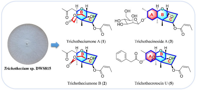
Ten novel trichothecene sesquiterpenoids including two new seco-trichothecenes, trichotheciumones A (1) and B (2), a new trichothecene sesquiterpenoid glycoside, trichothecinoside A (3), and seven new trichothecene sesquiterpenoids, trichothecrotocins T−Z (4−10), together with three new natural products (11−13) and thirteen known compounds (14−26), were isolated from the soil fungus Trichothecium sp. DWS815. The structures and absolute configurations of the new compounds were elucidated by extensive spectroscopic analyses and quantum chemistry ECD calculations. Given the notable anticancer properties of known trichothecenes, the isolated compounds were evaluated for the cytotoxic activities against three cancer cell lines (HCT116, 4T1, MHCC97H) and one normal cell line (GES-1). Cell cycle analysis revealed new compounds 7 and 8 induced G2/M phase arrest in HCT116 cancer cells, which resulted to cell proliferation inhibition activity.Graphical Abstract
Keywords
Trichothecenes Trichothecium sp. Secondary metabolite Cytotoxic activity1 Introduction
Trichothecenes, a family of sesquiterpenoids produced by various fungal genera such as Aspergillus, Cephalosporium, Fusarium, Myrothecium, Stachybotrys, Verticimonosporium and Trichothecium [1, 2], are common contaminants of grains [3]. This family is divided into two main groups, simple and macrocyclic trichothecenes [4]. The simple trichothecenes share a core ring structure and exhibit cytotoxicity by inhibiting eukaryotic protein synthesis and disrupting mitochondrial and ribosomal functions [5–8]. The 12,13-epoxytrichothec-9-ene (EPT) is cited as the cause of this toxicity [9], and structural diversity within this family arises from various substitution patterns on the EPT core. Recent years have witnessed the continuous discovery of trichothecenes from the genus of Trichothecium [10–13].
Building upon the long-standing focus on discovering the structurally novel and biologically active compounds from soil fungi [14–17], our previous chemical investigation of Myrothecium verrucaria PA 57 led to the discovery of novel macrocyclic trichothecenes exhibiting cytotoxic activities [18]. In our persistent endeavor to mine the novel types of trichothecene natural products with higher cytotoxic activities against cancer cell lines and lower cytotoxic activities toward normal cell lines, the fungal strain Trichothecium sp. DWS815 was isolated from a soil sample collected in Daweishan National Forest Park, Hunan Province, China. A comprehensive LC–MS analysis of the fungal fermentation extract revealed a metabolite profile abundant in simple trichothecenes, suggesting the strain's potential to produce novel trichothecene derivatives. Chemical investigation of this strain led to the isolation of ten previously undescribed trichothecene derivatives, trichotheciumones A (1) and B (2), trichothecinoside A (3), and trichothecrotocins T–Z (4–10), together with three new natural products (11–13) and thirteen known compounds (14–26) (Fig. 1). The structures and absolute configurations of all new compounds were established through spectroscopic analysis and quantum chemistry calculations of electronic circular dichroism (ECD). This paper details the purification, structure elucidation, and cytotoxicity of these compounds.
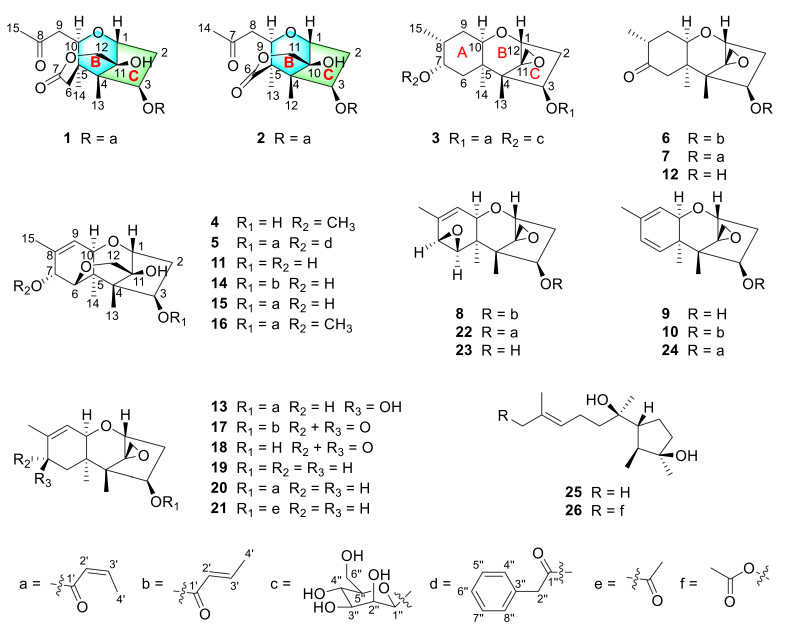
Structures of compounds 1−26
2 Results and discussion
Compound 1 was obtained as a white powder. The molecular formula was determined to be C19H26O7 based on HRESIMS at m/z 367.1758 [M + H]+ (calcd for C19H27O7, 367.1751), corresponding to seven degrees of unsaturation. The 1H NMR spectrum (Table 1) of 1 indicated the presence of four methyl groups at δH 0.98 (s, H3-14), 1.02 (s, H3-13), 2.17 (dd, J = 7.2, 1.8 Hz, H3-4'), and 2.17 (s, H3-15); four methylene groups at δH 2.13/2.53 (ddd, J = 15.8, 4.9, 3.4 Hz, Ha-2; dd, J = 15.8, 8.0 Hz, Hb-2), 2.34/2.89 (d, J = 15.1 Hz, Ha-6; d, J = 15.1 Hz, Hb-6), 2.44/3.10 (dd, J = 16.6, 2.3 Hz, Ha-9; dd, J = 16.6, 9.1 Hz, Hb-9), and 4.21/4.30 (d, J = 13.9 Hz, Ha-12; d, J = 13.9 Hz, Hb-12); five methines including two cis-olefinic methines at δH 5.77 (dq, J = 11.4, 1.8 Hz, H-2') and 6.45 (dq, J = 11.4, 7.2 Hz, H-3') as well as three oxygenated methines at δH 4.01 (d, J = 9.1 Hz, H-10), 4.45 (d, J = 4.8 Hz, H-1), and 5.63 (dd, J = 8.0, 3.4 Hz, H-3). The 13C NMR (Table 4) and DEPT spectra confirmed the presence of 19 carbon resonances assignable to four methyl groups (δC 7.4, 15.6, 20.6, 31.5), four sp3 methylene groups including one downfield oxygenated carbon (δC 36.5, 39.4, 42.0, 66.8), three sp3 oxygenated methine carbons (δC 72.3, 75.3, 81.7), three non-protonated sp3 carbons including one oxygenated carbon (δC 43.1, 53.4, 76.5), two sp2 olefinic carbons (δC 119.3, 147.9), one ketone carbonyl carbon (δC 206.5), and two ester carbonyl carbons (δC 164.8, 173.6).
1H NMR Spectroscopic Data for Compounds 1−4
Key 1H–1H COSY correlations (H-1/H-2/H-3, H-10/H-9, and H-2'/H-3'/H-4') established three independent spin–spin coupling systems (Fig. 2). A crotonyl group was connected to C-3 by HMBC correlations from H-3 and H-2' to C-1' as well as from H-4' to C-2' and C-3'. Further detailed 1D and 2D spectra analysis and comparison with a known co-isolated compound, trichothecene analogue (15) [19], indicated compound 1 bore a resemblance with the trichothecene central skeleton. The B and C rings in 1 were constructed by HMBC correlations from H-1 to C-10 and from H3-13 to C-3, C-5 and C-11. An acetonyl group was attached to C-10 in the B ring based on the HMBC correlations from H-10, H-9 and H3-15 to the carbonyl carbon C-8 (δC 206.5) and from H3-15 to C-9 and 1H–1H COSY correlation between H-10 and H-9. Moreover, HMBC correlations from H2-12 to C-11 and C-7, from H3-14 to C-4, C-10 and C-6, together with a long-range (4JCH) correlation from H3-14 to C-7 confirmed a lactone bridge between C-5 and C-11 across the B ring. The aforementioned planar structure assignment confirmed that compound 1 possessed a 7,8-seco-trichothecene core, representing the first reported isolation of this specific scaffold. NOESY correlations of H-3/H-10/H3-14, as well as H-1/Ha-12 and H3-13/Hb-12, indicated that H-3, H-10, and H3-14 were α-oriented, while H-1, H3-13, and OH-11 resided on the opposite face. This relative configuration of 1 aligned with the biosynthetic origin of trichothecene sesquiterpenoids. The absolute configuration was verified based on the agreement between the experimental and calculated ECD spectra. Finally, the absolute configuration was determined to be 1R,3R,4S,5R,10R,11S (Fig. 4). Thus, 1 was established to be trichotheciumone A, a 7,8-seco-trichothecene.
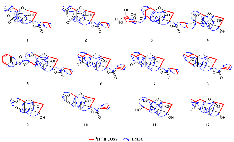
Key HMBC and 1H−1H COSY correlations of compounds 1−12
Compound 2 was obtained as a white powder with the molecular formula of C18H24O7, determined by HRESIMS at m/z 353.1599 [M + H]+ (calcd for C18H25O7, 353.1595) and corresponding to seven degrees of unsaturation. The NMR spectrum for 2 (Tables 1 and 4) showed similarities to those of 1. The key structural difference was the absence of a methylene unit in 2 at the bridged lactone moiety, suggesting it to be a previously undescribed 7,8-seco-7-nor-trichothecene. The B and C rings, along with the crotonyl group and the acetonyl group were assigned by comparison of the key NMR signals (1H–1H COSY, HSQC, and HMBC correlations) with those of 1, as depicted in Fig. 2. The bridged lactone moiety was assigned between C-5 and C-10 across the B ring according to the critical HMBC correlations from H-9, H-11, and H3-13 to C-6 and from H2-11 to C-10 and C-6. Moreover, NOESY correlations of H3-12/H-3/H-9/H3-13, and H3-12/Ha-11, and H-1/Hb-11 supported the relative configuration shown in Fig. 3. The relative configuration of 2 was consistent with the biosynthetic origin of trichothecene sesquiterpenoids. By comparing the experimental and calculated ECD spectra (Fig. 4), the absolute configuration of 2 was defined as 1R,3R,4S,5R,9R,10S. Consequently, 2 was assigned as depicted and named trichotheciumone B, a 7,8-seco-7-nor-trichothecene.
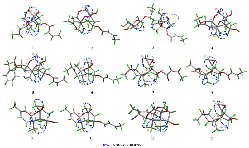
Key NOESY or ROESY (blue arrows) correlations of compounds 1−12
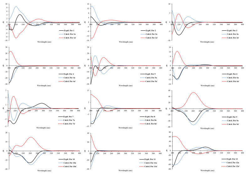
Calculated and experimental ECD spectra of compounds 1−12
Compound 3 was obtained as a white powder, displaying an exact mass of m/z 499.2519 [M + H]+ (calcd for C25H39O10, 499.2538) in HRESIMS, corresponding to a molecular formula of C25H38O10 with eight degrees of unsaturation. Analysis of the NMR data in Tables 1 and 4 indicated that 3 was deemed as a derivative of the trichothecenes. The 13C NMR spectrum displayed six characteristic signals indicative of a glycosyl unit (δC 61.8, 67.5, 70.9, 74.1, 75.6, 102.1). The mannose unit was further evidenced by comparison with the literature data (δC 61.2, 66.6, 71.1, 74.1, 75.9, 100.1) and δH 3.18 (brs), 3.53 (brs), 3.86 (m), 3.87 (brs), 3.97 (brs), and 4.44 (d, J = 0.7 Hz) in CDCl3 [20]. The mannosidic linkage of 1''-O-7 was revealed by a key HMBC correlation from H-1'' to C-7. The β-configuration of the mannose unit was confirmed by the following evidences. Two broad singlets of the anomeric proton H-1'' and H-2'', as well as the critical and obvious NOESY correlations of H-1'' with H-3'' and H-5'', provided compelling evidence for axial situation of anomeric proton H-1'', H-3'', and H-5'' and equatorial situation of H-2''. Based on the NOESY correlations of H3-13/H-3/H-6/H3-14 and analysis in light of biosynthetic origin, the relative configuration of chiral centers C-1, C-3, C-4, C-5, C-10, and C-11 was established. Meanwhile, the overall relative configuration of 3 was determined by interpretation of key NOESY correlations and GIAO 13C NMR calculations with the STS protocol performed for eight isomers (3a–3h) (Table S4). The absolute configuration of 3, including the D-mannose moiety, was determined by ECD calculations using the time-dependent density functional theory (TDDFT) method. As shown in Fig. 4, the calculated ECD curve of 3a (1R,3R,4S,5R,7S,8R,10R,11S-β-D-mannopyranoside) matched well with the experimental ECD curve. Therefore, the absolute configuration of 3 was assigned as 1R,3R,4S,5R,7S,8R,10R,11S-β-D-mannopyranoside, and it was named trichothecinoside A.
Compound 4 was obtained as a white powder, having the molecular formula C16H24O5 based on the protonated ion peak at m/z 297.1700 [M + H]+ (calcd for C16H25O5, 297.1697), corresponding to five degrees of unsaturation. Analysis of 1D data of 4 (Tables 1 and 4) revealed close resemblance to the co-isolated new natural product isocrotocol B (11) [21], indicating a shared trichothecene backbone. A key structural distinction was the presence of an oxygenated methyl group at C-7 in 4. Further analysis of the 2D data confirmed that the remaining structure of 4 were the same as that of 11. The absolute configuration was defined to be 1R,3R,4S,5R,6R,7R,10R,11S by comparison of the experimental ECD spectrum with calculated one (Fig. 4). Therefore, 4 was established as depicted and named trichothecrotocin T.
Compound 5 was obtained as a white powder, exhibiting the molecular formula C27H32O7, based on the protonated molecule peak at m/z 469.2211 [M + H]+ (calcd for C 27H33O7, 469.2221), implying twelve degrees of unsaturation. Analysis of the 1D data (Tables 2 and 4) indicated a structure similar to that of co-isolated compound trichothecrotocin O (16), differing by the replacement of a methoxy group in 16 with a phenylacetoxy moiety at the C-7 position in 5. The proposed structure was supported by 1H–1H COSY correlations for H-4''/H-5''/H-6''/H-7''/H-8'', as well as HMBC correlations from H-7 to C-1'' and from H2-2'' to C-1'', C-3'', C-4'', and C-8''. Key NOESY correlations of H3-13/H-3/H-10/H3-14 revealed the same relative configuration as 16, although the stereochemistry at C-7 remained unassigned. To resolve this, GIAO 13C NMR calculations with the STS protocol were conducted for two diastereomers 5a and 5b (Table S9). The calculated data for 5a matched the experimental data, thereby establishing the relative configuration. Furthermore, the agreement between experimental ECD spectrum and the calculated spectrum for 5 confirmed absolute configuration as 1R,3R,4S,5R,6R,7R,10R,11S (Fig. 4). Therefore, the structure of 5 was validated and assigned the name trichothecrotocin U.
1H NMR Spectroscopic Data for Compounds 5−8
Compounds 6 and 7 were each obtained as a white powder, presenting the same molecular formula C19H26O5, based on their HRESIMS data at m/z 335.1851 [M + H]+ (calcd for C19H27O5, 335.1853) and 335.1863 [M + H]+ (calcd for C19H27O5, 335.1853), respectively, corresponding to seven degrees of unsaturation. Analysis of 1D data for 6 and 7 (Tables 2 and 4) indicated close similarity to those of a new co-isolated natural product dihydrotrichothecolone (12), differing from 12 by the presence of the crotonyl moiety. The connectivity of this moiety was confirmed by the 1H–1H COSY data of H-2'/H-3'/H-4' and HMBC correlations from H-3 and H-2' to C-1' and from H-4' to C-2' and C-3'. The relative configuration of 6 was deemed as being identical to that of 12 through key NOESY correlations as depicted in Fig. 2. The α-orientation of the C-15 was assigned by comparing experimental data with GIAO 13C NMR calculations (STS protocol) performed for two diastereomers 6a and 6b (Table S11). A key distinction between 6 and 7 is the geometry of crotonyl group, which was ascertained as Z in 7, assigned based on the coupling constant (J = 11.4 Hz) between H-2' and H-3'. The relative stereochemistry of 7 was elucidated using the same procedure as for 6. Finally, the absolute configurations of both 6 and 7 were defined to be 1R,3R,4S,5R,8R,10R,11S by comparing their experimental and calculated ECD spectra (Fig. 4). Consequently, compounds 6 and 7 were named trichothecrotocin V and trichothecrotocin W, respectively.
Compound 8 was obtained as a white powder, possessing the molecular formula C19H24O5 based on the proton adduct at m/z 333.1708 [M + H]+ (calcd for C19H25O5, 333.1697) in its HRESIMS, indicating eight degrees of unsaturation. The 1D (Tables 2 and 4) and 2D data disclosed that the structure of 8 was structurally similar to that of a co-isolated known compound crotocin (22), differing only in the E geometry of the crotonyl group. The geometric distinction between 8 and 22 paralleled that observed between 6 and 7, as supported by the large coupling constant (J = 15.6 Hz) between H-2' and H-3' in 8. NOESY correlations of H3-13/H-3/H-10/H3-14/H-6 suggested their co-facial orientation, which facilitated the determination of the relative configuration. The calculated ECD curve of 1R,3R,4S,5R,6R,7S,10R,11S was in good agreement with the experimental data (Fig. 4). Thus, 8 was assigned as depicted and named trichothecrotocin X.
Compounds 9 and 10 were each obtained as a white powder. Compound 9 possessed a molecular formula of C15H20O3 based on the proton adduct in its HRESIMS at m/z 249.1482 [M + H]+ (calcd for C15H21O3, 249.1485), corresponding to six degrees of unsaturation. The molecular formula for 10 was determined to be C19H24O5 by HRESIMS based on the sodium adduct at m/z 339.1563 [M + H]+ (calcd for C19H24O5Na, 339.1567), corresponding to eight degrees of unsaturation. Analysis of 1D data for both compounds 9 and 10 (Tables 3 and 4) exhibited close similarity to those of a co-isolated known compound 7-dehydro-8-dehydroxytrichothecinol B (24). For compound 9, the key structural distinction was the absence of a crotonyl group at C-3. Comprehensive analysis of 2D data confirmed that the remaining planar structure and the relative configuration of 9 were identical to those of 24. The calculated ECD curve for isomer 9a (1R,3R,4S,5R,10R,11S) showed excellent agreement with the experimental data (Fig. 4). For compound 10, the key structural distinction was only in the E geometry of the crotonyl group, supported by HMBC correlations from H-3 to C-1' and from H-4' to C-2' and C-3' as well as the large coupling constant (J = 15.5 Hz) between C-2′ and C-3′, confirming crotonyl group with trans-olefinic double bond. NOESY correlations observed for 10 were similar to those of 9, suggesting an identical relative configuration. Furthermore, the experimental ECD spectrum of 10 (Fig. 4) closely matched that of 9 and was consistent with the calculated spectrum for isomer 10a (1R,3R,4S,5R,10R,11S). Therefore, 9 and 10 were named trichothecrotocin Y and trichothecrotocin Z, respectively.
1H NMR Spectroscopic Data for Compounds 9−13
13C NMR Spectroscopic Data for Compounds 1−13
Compounds 11, 12, and epi-trichothecinol B (13) were isolated from this fungus as new natural products for the first time. The absolute configuration of compound 11 was first determined and the ECD spectra of 13 was provided in Figure S143. The known compounds 14–26 were characterized via analysis of their HRESIMS and 1D data and comparison with literature data. They were identified as trichothecrotocin N (14) [12], trichothecene analogue (15) [19], trichothecrotocin O (16) [12], trichothecrotocin Q (17) [12], trichothecolone (18) [22], trichodermol (19) [23], 8-deoxy-trichothecin (20) [22], trichodermin (21) [24], crotocin (22) [21], crotocol (23) [21], 7-dehydro-8-dehydroxytrichothecinol B (24) [25], cyclonerodiol (25) [26], and cyclonerodiol C (26) [27].
Trichotheciumone A (1) and B (2) possess unusual sesquiterpenoid skeletons based on the trichothecene core. Although structurally distinct, compounds 1 and 2 are likely biosynthesized from the same precursor, trichodiene, a key intermediate in trichothecene biosynthesis [4, 28]. A putative biosynthetic pathway for 1 and 2 is proposed in Scheme 1. The pathway suggests that cleavage of the C-7–C-8 bond in trichothecin, followed by oxidation, would yield the crucial intermediate ⅰ, which belongs to the rare 7,8-seco-trichothecene class. A subsequent nucleophilic attack of the carboxylate anion on the C-12 epoxide would form compound 1, representing the first reported trichothecene-type sesquiterpenoid with an opened A-ring. Alternatively, crotocin (22) could be converted to intermediate 15 via nucleophilic ring-opening of the 6,7-epoxide by a hydroxide ion. Further cleavage of the C-7–C-8 bond in 15, followed by oxidation, would lead to intermediate ⅱ. The oxidative decarboxylation of ⅱ would then yield compound 2, which is the first reported example of a rare A-ring-seco-nor-trichothecene-type sesquiterpenoid.
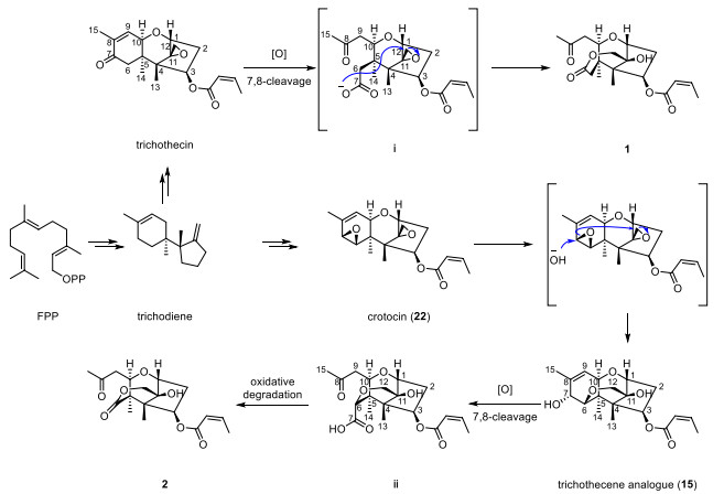
Proposed biogenetic pathway for compounds 1 and 2
The cytotoxic activities of the isolated compounds were evaluated against human colon carcinoma HCT116 cell line, mouse breast cancer 4T1 cell line, and human liver cancer MHCC97H cell line, with one strain of human gastric epithelial GES-1 cells serving as a normal cell lines control, using the CCK-8 method. Seven compounds (7, 8, 10, 13, 17, 18, 20, 24) exhibited significant anti-proliferative activity (Fig. 5A, B). Among them, compound 17 was the most potent compound with IC50 of 0.35 μM, 0.52 μM, 0.57 μM against HCT116, 4T1, and MHCC97H cells respectively (Table 5). Cell cycle assays revealed that new compounds 7 and 8 arrested HCT116 cells at the G2/M phase. Treatment with 0, 1, and 2 μM of compound 7 increased the G2/M population compared with the control (control: 5.25%, 1 μM: 17.22%, 2 μM: 32.42%) and decreased G0/G1 population (control: 64.79%, 1 μM: 52.01%, 2 μM: 24.61%) (Fig. 6A, B). Treatment with 0, 1, and 2 μM of compounds 8 increased the G2/M population compared with the control (control: 4.41%, 1 μM: 7.14%, 2 μM: 12.31%) and decreased G0/G1 population (control: 62.78%, 1 μM: 60.54%, 2 μM: 50.59%) (Fig. 6C, D). However, the test compounds exhibited no observable selectivity in inhibiting proliferation between these three cancer cell lines and GES-1 cell line. Structure–activity relationship (SAR) analysis indicated that the configuration of the C-3 crotonyl group influences toxicity. Specifically, changing the crotonyl group at C-3 from the Z-configuration to the E-configuration results in decreased toxicity, a Z-configuration (as in compounds 7 and 24) conferred greater potency than an E-configuration (as in compounds 6 and 10).
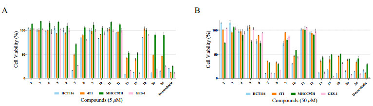
Antiproliferative activity against four cell lines. A, B Cytotoxicity screening of isolated compounds with concentrations 5 μM and 50 μM against four cell lines (HCT116, 4T1, MHCC97H, and GES-1) determined by CCK-8 assay (doxorubicin as positive control)
Cytotoxic activities of compounds 7, 8, 10, 13, 17, 18, 20, and 24a
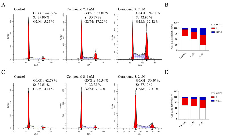
Cell cycle analysis of HCT116 cells after treatment with compounds 7 and 8. A, C Histograms of cell cycle distribution after treatment with 0, 1 and 2 μM of compounds 7 and 8 for 24 h, respectively. B, D Percentage of HCT116 cell in G0/G1, S and G2/M phases after treatment with compounds 7 and 8, respectively. Data are presented as mean ± SD (n = 3)
3 Experimental section
3.1 General experimental procedures
Optical rotations were measured on a Rudolph Research Analytical Autopol IV automatic polarimeter. UV spectra were recorded using a Cary 300 spectrometer (Agilent Technologies, Santa Clara, CA, USA). IR spectra were recorded on a Shimadzu Fourier Transform Infrared Spectrometer using KBr pellets. Circular Dichroism (CD) spectra were obtained on a ChirascanTM-plus Circular Dichroism spectrometer. High-resolution electrospray ionization mass spectrometry (HRESIMS) spectra were acquired with an Agilent 6500 series Q-TOF mass spectrometer (Agilent Technologies, Singapore). NMR spectra were recorded on Bruker Avance Ⅲ HD 600 MHz and 500 MHz spectrometer with tetramethylsilane (TMS) serving as the internal standard. Column chromatography (CC) was performed using macro-porous adsorbent resin D101 (Tianjin Haoju Resin Technology Co., Ltd., Tianjin, China), silica gel (200–300 mesh, Qingdao Marine Chemical, Qingdao, China), and Sephadex LH-20 (GE Healthcare, Uppsala, Sweden). MCI GELTM (Mitsubishi Chemical Corporation, Japan). Semipreparative ZORBAX SB-C18 (Agilent, 5 μm, 9.4 mm × 150 mm), Supersil ODS-B (Elite, 10 μm, 20.0 mm × 250 mm), Supersil ODS2 (Elite, 10 μm, 20.0 mm × 250 mm), Ultimate XB-CN (Welch, 5 μm, 10.0 mm × 250 mm) and preparative SinoPark C18 (Elite, 10 μm, 20.0 mm × 250 mm) were analyzed by an EClassical P3500 prep-HPLC system and a DAD detector (EClassical P3500, Dalian Elite Analytical Instruments Co., Ltd., Dalian, China). The progress of the experiments was monitored by pre-coated silica gel GF254 plates (Qingdao Marine Chemical). Spots were visualized under UV light (254 or 365 nm) or by spraying with 10% H2SO4 in EtOH followed by heating. All other chemicals used in this study were of analytical grade.
3.2 Fungal material
The fungal strain Trichothecium sp. DWS815 was isolated from a forest soil sample collected from Daweishan National Forest Park, Hunan Province. The strain was identified as Trichothecium sp. (GenBank Accession No. ON386019.1) based on their ITS sequences and the phylogenetic analysis (Tables S24 and S25). A voucher specimen of this strain has been deposited at the Xiangya School of Pharmaceutical Sciences, Central South University, Changsha, China.
3.3 Fermentation, extraction and isolation
This fungus was initially cultivated on Sabouraud Dextrose Agar (SDA) medium (containing 20 g/L dextrose, 10 g/L peptone, 1 g/L yeast extract powder, 15 g/L agar and water) for 8 days at 25 ℃. Subsequently, the mycelia on agar plugs were cut and transferred to the sterile rice fermentation medium. The medium was prepared with 100 g of rice and 80 mL of water in 500 mL Erlenmeyer flasks. For large-scale fermentation, a total of 14 kg of rice was distributed into 139 flasks and inoculated with this fungus. The fermentation proceeded at 25 ℃ in the dark for 21 days.
The rice was extracted with ethyl acetate at room temperature 10 times, and the solvent was evaporated under vacuum to yield an extract (170 g). The extract was absorbed onto 500 g macro-porous adsorbent resin D101 and eluted stepwise with water, 80% aqueous MeOH, and 100% MeOH. The 80% aqueous MeOH fraction was collected and condensed to yield 26 g residue. The 80% aqueous MeOH fraction was then fractionated using silica gel CC eluted with a step gradient of Petroleum ether–EtOAc (v/v, 100:0, 20:1, 15:1, 10:1, 5:1, 2:1, 1:1) and EtOAc–MeOH (v/v, 100:0, 10:1, 5:1, 1:1, 0:100) to provide nine fractions (A~I).
The Fr.A was subjected to MCI to give five subfractions (Fr.A1~Fr.A5). Fr.A2 was separated using semipreparative Supersil ODS-B (CH3CN: H2O, 55:45) to afford compound 24 (tR = 20.9 min, 22.5 mg) and Fr.A2-1. Fr.A2-1 was fractionated using Supersil ODS-B (MeOH: H2O, 57:43) to afford compounds 7 (tR = 36.9 min, 3.5 mg) and 21 (tR = 40.1 min, 1.9 mg). Fr.A4 was separated by semipreparative ZORBAX SB-C18 (CH3CN: H2O, 49:51) to obtain compound 20 (tR = 23.9 min, 50.2 mg). Fr.B was subjected to Sephadex LH-20 (MeOH) to give five subfractions (Fr.B1~Fr.B5). Fr.B3 was isolated on the preparative SinoPark C18 (MeOH: H2O, 62:38) to obtain four fractions (Fr.B3-1~Fr.B3-4). Fr.B3-1 was purified using semipreparative Supersil ODS2 (CH3CN: H2O, 28:72) to afford compounds 1 (tR = 21.1 min, 0.3 mg) and 2 (tR = 29.2 min, 0.6 mg). Fr.B3-4 was purified using semipreparative Supersil ODS-B (MeOH: H2O, 53:47) to yield compound 17 (tR = 47.0 min, 24.5 mg). Fr.C was applied to semipreparative ZORBAX SB-C18 with a step gradient of CH3CN-H2O (v/v, from 20:100 to 50:50 in 35 min, flow speed: 3 mL/min) to afford compounds 22 (tR = 19.8 min, 1.6 g), 16 (tR = 17.7 min, 4.9 mg), 8 (tR = 19.4 min, 6.8 mg), and 5 (tR = 32.8 min, 6.58 mg). Fr.D was subjected to Sephadex LH-20 (MeOH) and then was further fractionated using semipreparative Ultimate XB-CN (CH3CN: H2O, 19:81) to give 19 (tR = 14.6 min, 1.1 mg), 18 (tR = 9.5 min, 1.9 mg), 9 (tR = 12.3 min, 1.5 mg), 13 (tR = 28.9 min, 8.4 mg), 26 (tR = 20.0 min, 3.5 mg), 25 (tR = 24.9 min, 9.9 mg), and 12 (tR = 17.0 min, 2.3 mg). Fr.E was subjected to Sephadex LH-20 (MeOH) and then was purified using semipreparative Supersil ODS-B (MeOH: H2O, 26:74) to afford 14 (tR = 15.9 min, 1.4 mg), 15 (tR = 12.2 min, 41.9 mg), 4 (tR = 10.7 min, 3.09 mg), 23 (tR = 23.5 min, 3.3 mg), and 11 (tR = 8.7 min, 1.6 mg). Fr.F was subjected to Sephadex LH-20 (MeOH) and then was purified using semipreparative ZORBAX SB-C18 with a step gradient of CH3CN–H2O (v/v, from 5:95 to 21:79 in 100 min, flow speed: 3 mL/min) to yield 3 (tR = 81.6 min, 1.3 mg). Fr.I was also chromatographed on Sephadex LH-20 (MeOH) and purified using semipreparative Ultimate XB-CN with a step gradient of CH3CN–H2O (v/v, from 25:75 to 45:55 in 40 min, flow speed: 3 mL/min) to give compounds 6 (tR = 29.8 min, 0.5 mg) and 10 (tR = 35.1 min, 1.9 mg).
Trichotheciumone A (1). White powder; [α]D25+ 37.5 (c 0.04, MeOH); UV (MeOH) λmax (log ε): 210 (3.48) nm; IR (KBr) νmax: 3442, 2979, 2950, 2923, 1714, 1647, 1294, 1178, 1115, 1028, 817 cm−1; CD (c 1.19 mM, MeOH): 205 nm (Δε –4.84), 227 nm (Δε + 4.38), 269 nm (Δε –1.91); for 13C NMR (CDCl3, 150 MHz) and 1H NMR (CDCl3, 600 MHz) data, see Tables 1 and 4; HRESIMS (positive) m/z 367.1758 [M + H]+ (calcd for C19H27O7, 367.1751, Δ + 1.9064 ppm).
Trichotheciumone B (2). White powder; [α]D25 + 264.86 (c 0.037, MeOH); UV (MeOH) λmax (log ε): 210 (2.88) nm; IR (KBr) νmax: 3434, 2924, 2855, 2788, 1718, 1578, 1435, 1384, 1180 cm−1; CD (c 1.05 mM, MeOH): 200 nm (Δε + 5.29), 226 nm (Δε –2.83), 270 nm (Δε –0.51); for 13C NMR (CDCl3, 150 MHz) and 1H NMR (CDCl3, 600 MHz) data, see Tables 1 and 4; HRESIMS (positive) m/z 353.1599 [M + H]+ (calcd for C18H25O7, 353.1595, Δ + 1.1326 ppm).
Trichothecinoside A (3). White powder; [α]D25 –2.0 (c 0.05, MeOH); UV (MeOH) λmax (log ε): 200 (3.17) nm; IR (KBr) νmax: 3420, 2962, 2935, 2876, 1715, 1649, 1383, 1291, 1184, 1083, 1027, 959 cm−1; CD (c 1.00 mM, MeOH): 217 nm (Δε –7.13); for 13C NMR (CDCl3, 150 MHz) and 1H NMR (CDCl3, 600 MHz) data, see Tables 1 and 4; HRESIMS (positive) m/z 499.2519 [M + H]+ (calcd for C25H39O10, 499.2538 Δ –3.8057 ppm).
Trichothecrotocin T (4). White powder; [α]D25 –12.5 (c 0.037, MeOH); UV (MeOH) λmax (log ε): 200 (2.82) nm; IR (KBr) νmax: 3459, 3371, 2981, 2932, 2859, 1628, 1457, 1135, 1125, 1098, 1051 cm−1; CD (c 1.35 mM, MeOH): 200 nm (Δε –28.13); for 13C NMR (CDCl3, 150 MHz) and 1H NMR (CDCl3, 600 MHz) data, see Tables 1 and 4; HRESIMS (positive) m/z 297.1700 [M + H]+ (calcd for C16H25O5, 297.1697, Δ + 1.0009 ppm).
Trichothecrotocin U (5). White powder; [α]D25 –10.9 (c 0.064, MeOH); UV (MeOH) λmax (log ε): 200 (3.21) nm; IR (KBr) νmax: 3434, 2978, 2946, 2922, 2859, 1737, 1714, 1647, 1436, 1248, 1179, 1133, 1049 cm−1; CD (c 1.37 mM, MeOH): 200 nm (Δε –30.89), 210 nm (Δε + 0.19), 225 nm (Δε –8.52); for 13C NMR (CDCl3, 150 MHz) and 1H NMR (CDCl3, 600 MHz) data, see Tables 2 and 4; HRESIMS (positive) m/z 469.2211 [M + H]+ (calcd for C 27H33O7, 469.2221, Δ –2.1312 ppm).
Trichothecrotocin V (6). White powder; [α]D25+ 12.5 (c 0.008, MeOH); UV (MeOH) λmax (log ε): 210 (2.94) nm; IR (KBr) νmax: 2971, 2928, 2855, 1717, 1576, 1445, 1384, 1183, 1081, 971 cm−1; CD (c 1.50 mM, MeOH): 208 nm (Δε –11.61), 291 nm (Δε + 0.92); for 13C NMR (CDCl3, 150 MHz) and 1H NMR (CDCl3, 600 MHz) data, see Tables 2 and 4; HRESIMS (positive) m/z 335.1851 [M + H]+ (calcd for C19H27O5, 335.1853, Δ –0.5967 ppm).
Trichothecrotocin W (7). white powder; [α]D25 –6.98 (c 0.043, MeOH); UV (MeOH) λmax (log ε): 210 (3.08) nm; IR (KBr) νmax: 2959, 2928, 1716, 1647, 1466, 1366, 1287, 1183, 1081, 966, 816 cm−1; CD (c 1.29 mM, MeOH): 213 nm (Δε –4.32), 293 nm (Δε + 1.46); for 13C NMR (CDCl3, 150 MHz) and 1H NMR (CDCl3, 600 MHz) data, see Tables 2 and 4; HRESIMS (positive) m/z 335.1863 [M + H]+ (calcd for C19H27O5, 335.1853, Δ + 2.9834 ppm).
Trichothecrotocin X (8). White powder; [α]D25 –16.67 (c 0.024, MeOH); UV (MeOH) λmax (log ε): 208 (3.29) nm; IR (KBr) νmax: 2965, 2925, 2857, 1716, 1602, 1384, 1185, 1083, 969 cm−1; CD (c 0.72 mM, MeOH): 203 nm (Δε –13.10), 230 nm (Δε + 0.50); for 13C NMR (CDCl3, 125 MHz) and 1H NMR (CDCl3, 500 MHz) data, see Tables 2 and 4; HRESIMS (positive) m/z 333.1708 [M + H]+ (calcd for C19H25O5, 333.1697, Δ + 3.3016 ppm).
Trichothecrotocin Y (9). White powder; [α]D25 –51.35 (c 0.037, MeOH); UV (MeOH) λmax (log ε): 200 (2.60) nm; IR (KBr) νmax: 3452, 2965, 2923, 2852, 1664, 1623, 1593, 1447, 1422, 1281, 1185, 1074, 955 cm−1; CD (c 1.49 mM, MeOH): 256 nm (Δε –43.19); for 13C NMR (CDCl3, 125 MHz) and 1H NMR (CDCl3, 500 MHz) data, see Tables 3 and 4; HRESIMS (positive) m/z 249.1482 [M + H]+ (calcd for C15H21O3, 249.1485, Δ –1.2041 ppm).
Trichothecrotocin Z (10). White powder; [α]D25 –63.16 (c 0.019, MeOH); UV (MeOH) λmax (log ε): 210 (3.15) nm; IR (KBr) νmax: 2917, 2852, 1732, 1717, 1606, 1590, 1384, 1189, 1101, 1082, 1064, 1028, 957 cm−1; CD (c 0.60 mM, MeOH): 226 nm (Δε –13.12); for 13C NMR (dimethyl sulfoxide-d6, 150 MHz) and 1H NMR (dimethyl sulfoxide-d6, 600 MHz) data, see Tables 3 and 4; HRESIMS (positive) m/z 339.1563 [M + H]+ (calcd for C19H24O5Na, 339.1567, Δ –1.1793 ppm).
Isocrotocol B (11). White powder; [α]D25 –18.0 (c 0.05, MeOH); UV (MeOH) λmax (log ε): 200 (2.71) nm; IR (KBr) νmax: 3507, 3435, 3332, 2978, 2939, 2876, 1637, 1452, 1321, 1135, 1100, 1055, 973, 898, 817 cm−1; CD (c 1.77 mM, MeOH): 221 nm (Δε + 1.42); for 13C NMR (CDCl3, 150 MHz) and 1H NMR (CDCl3, 600 MHz) data, see Tables 3 and 4; HRESIMS (positive) m/z 283.1542 [M + H]+ (calcd for C15H23O4, 283.1540, Δ + 0.7063 ppm).
Trichothecolone (12). White powder; [α]D25 –5.22 (c 0.23, MeOH); UV (MeOH) λmax (log ε): 200 (2.02) nm; IR (KBr) νmax: 3502, 2982, 2966, 2931, 1715, 1458, 1280, 1081, 957 cm−1; CD (c 8.64 mM, MeOH): 200 nm (Δε –71.20), 292 nm (Δε + 15.78); for 13C NMR (methanol-d4, 150 MHz) and 1H NMR (methanol-d4, 600 MHz) data, see Tables 3 and 4; HRESIMS (positive) m/z 267.1585 [M + H]+ (calcd for C15H23O4, 267.1591 Δ –2.2458 ppm).
Epi-trichothecinol B (13). White powder; [α]D25+ 87.5 (c 0.04, MeOH); UV (MeOH) λmax (log ε): 200 (2.71) nm; IR (KBr) νmax: 3468, 2985, 2962, 2938, 1712, 1643, 1434, 1376, 1313, 1245, 1175, 1078, 1034, 996, 972, 962 cm−1; CD (c 1.77 mM, MeOH): 221 nm (Δε + 1.42); for 13C NMR (CDCl3, 150 MHz) and 1H NMR (CDCl3, 600 MHz) data, see Tables 3 and 4; HRESIMS (positive) m/z 335.1856 [M + H]+ (calcd for C19H27O5, 335.1853, Δ + 0.8950 ppm).
3.4 Quantum chemical calculations
Conformer–rotamer ensemble sampling tool (CREST) was used to search the conformational space of compounds at the GFN0 level of theory in the gas phase [29, 30], followed by optimization at the GFN2-xTB level to avoid the errors caused by poor initial geometry [31], with a 4 kcal/mol energy window to remove high-energy conformers. Optimization and frequency calculation of each conformer were performed at the r2-SCAN-3c level of theory in the conductor-like polarizable continuum model (CPCM) to ensure they were true local minima on the potential energy surface [32, 33]. Under STS protocol [34], DFT GIAO 13C NMR calculation was calculated at the ωB97x-D/6-31G* (IEFPCM, chloroform) level or ωB97x-D/6-31G* (IEFPCM, methanol) level of theory. Time-dependent density-functional theory (TDDFT) ECD calculations were performed at the B3LYP/TZVP (IEFPCM, methanol) level of theory with the consideration of solvent effects. The calculated shielding tensors and ECD curves of conformers were Boltzmann averaged based on Gibbs free energy. In the TDDFT-ECD calculations, 40 excited states were calculated for each conformer. The ECD curves of all conformers were Boltzmann averaged based on Gibbs free energy calculated at the r2SCAN-3c level of theory and generated by Multiwfn [35, 36]. All DFT calculations were performed by Gaussian 16 or ORCA 6.0.0 software packages [37–39]. The DFT optimized geometry data, relative energies, and conformational population of all calculated structures were provided in the supplemental material. The 3D molecular structures were rendered using VMD (1.9.3) [40]. Details are provided in the Supplementary Information.
3.5 Cell lines and culture conditions
The cells of GES-1, HCT116, 4T1 and MHCC97H were cultivated in DMEM medium enriched with 10% fetal bovine serum and 1% penicillin–streptomycin. Cultures were maintained at 37 ℃ in a humidified environment with 5% CO2.
3.6 Cell viability analysis
Cell viability was determined with CCK-8 method [18]. All cell lines were plated into 96-well plates at a concentration of 10,000 cells per well and incubated for an additional 24 h. Then, the cells were treated with concentrations 5 μM and 50 μM of the test compounds and positive control (doxorubicin) for a duration of 48 h. In addition to the experimental groups, blank controls (wells with CCK-8 solution but no cells) and negative controls (wells with vehicle) were included. After treatment, 10 μL of CCK-8 reagent was added to each well and incubated for 1 h. The optical density at 450 nm was then measured on a microplate reader (Feyond-A300, allsheng, Hangzhou, China). The IC50 value represents the half of maximal inhibitory concentration. The cells of HCT116, 4T1, and MHCC97H were treated with a concentration gradient of selected compounds (7, 8, 10, 13, 17, 18, 20, and 24) to obtain IC50 values, using either the range of (0.04 μM–10 μM) or (0.5 μM–50 μM).
3.7 Cell cycle assays
The cell cycle distribution was appraised, using the Propidium Iodide (PI) Flow Cytometry Kit, followed by the flow Cytometry analysis [12]. Briefly, HCT116 cells were planted on 6-well plates and incubated overnight. Next, the cells were treated with 7 and 8 at concentrations of 0, 1 and 2 μM, and incubated for 24 h in triplicate manner. Cells were harvested and washed with cold phosphate buffered saline (PBS), and fixed overnight with ice-cold 70% ethanol. Then, the ethanol was removed, and the cells were rinsed with PBS and stained with the DNA fluorochrome PI at room temperature in the dark for 20 min. CYTEKTM NL-3000 flow cytometer was then used to test samples.
4 Conclusion
In summary, ten previously undescribed trichothecenes, three new natural products and thirteen known compounds were identified by chemical investigation of the EtOAc extract from rice cultures of the fungus Trichothecium sp. DWS815. Notably, trichotheciumones A (1) and B (2) are novel trichothecene skeleton characterized by the cleavage of the A ring found in classical trichothecenes and the formation of a lactone bridge across the B ring. Trichothecinoside A (3), trichothecrotocin V (6), and trichothecrotocin W (7) are unusual natural trichothecene characterized by a hydrogenated EPT moiety. The isolated compounds were evaluated for cytotoxic activity against three human cancer cell lines (HCT116, 4T1, MHCC97H) and one normal cell line (GES-1). Among them, compound 17 was the most potent compound with IC50 of 0.35 μM, 0.52 μM, 0.57 μM against HCT116, 4T1, and MHCC97H cells respectively and new compounds 7 and 8 were found to induce G2/M phase cell cycle arrest in HCT116 colorectal cancer cells, thereby inhibiting cell proliferation. This work not only expands the structural diversity of trichothecene derivatives but also provides a foundation for future studies on structure–activity relationships and mode of action.
Notes
Acknowledgements
The authors thank the Institute for Advanced Study, Central South University for NMR measurement and Center for Medical Research and Innovation, the First Hospital of Hunan University of Chinese Medicine for MS measurement.
Author contributions
Peng-Ju Xu: Writing–original draft, Methodology, Investigation. Ai-Lin Liang: Methodology. Wen-Yu Lu: Methodology. Hong-Ping Long: Methodology. Qing-Hui Xiao: Methodology. Qi-An Chen: Methodology. Meng-Lan Hu: Methodology. Ngoc Nhu Thao Nguyen: Methodology. Shao Liu: Methodology. Jing Li: Writing – review & editing, Investigation, Funding acquisition. Wen-Xuan Wang: Writing–review & editing, Funding acquisition, Conceptualization. All authors read and approved the final manuscript.
Funding
The work was financially supported by National Natural Science Foundation of China (No. 82173713), Natural Science Foundation of Hunan Province (No. 2024JJ8127), Changsha Municipal Natural Science Foundation (No. kq2403041), Hunan Provincial Administration of Traditional Chinese Medicine (No. B2023059), and Fundamental Research Funds for the Central Universities of Central South University (No. 2024ZZTS0990, 2025ZZTS0913).
Data availability
All data generated or analyzed during this study are included within the article and its supplementary information file.
Declarations
Competing interests
The authors declare no conflict of financial interest.
References
-
1.McCormick SP, Stanley AM, Stover NA, Alexander NJ. Trichothecenes: from simple to complex mycotoxins. Toxins 2011;3: 802-14. CrossRef PubMed Google Scholar
-
2.He J, Zhou T, Young JC, Boland GJ, Scott PM. Chemical and biological transformations for detoxification of trichothecene mycotoxins in human and animal food chains: a review. Trends Food Sci Technol 2010;21: 67-76. CrossRef PubMed Google Scholar
-
3.Mahato DK, Pandhi S, Kamle M, Gupta A, Sharma B, Panda BK, et al. Trichothecenes in food and feed: occurrence, impact on human health and their detection and management strategies. Toxicon 2022;208: 62-77. CrossRef PubMed Google Scholar
-
4.Keller NP, Proctor RH, McCormick SP, Kim H-S, Cardoza RE, Stanley AM, et al. Evolution of structural diversity of trichothecenes, a family of toxins produced by plant pathogenic and entomopathogenic fungi. PLoS Pathog 2018;14: e1006946. CrossRef PubMed Google Scholar
-
5.Cundliffe E, Cannon M, Davies JL. Mechanism of inhibition of eukaryotic protein synthesis by trichothecene fungal toxins. Proc Natl Acad Sci U S A 1974;71: 30-4. CrossRef PubMed Google Scholar
-
6.Garreau de Loubresse N, Prokhorova I, Holtkamp W, Rodnina MV, Yusupova G, Yusupov M. Structural basis for the inhibition of the eukaryotic ribosome. Nature 2014;513: 517-22. CrossRef PubMed Google Scholar
-
7.Huang DY, Cui LQ, Dai MH, Wang X, Wu QH, Hussain HI, et al. Mitochondrion: a new molecular target and potential treatment strategies against trichothecenes. Trends Food Sci Technol 2019;88: 33-45. CrossRef PubMed Google Scholar
-
8.Rocha O, Ansari K, Doohan FM. Effects of trichothecene mycotoxins on eukaryotic cells: a review. Food Addit Contam 2005;22: 369-78. CrossRef PubMed Google Scholar
-
9.Wu QH, Dohnal V, Kuca K, Yuan ZH. Trichothecenes: structure-toxic activity relationships. Curr Drug Metab 2013;14: 641-60. CrossRef PubMed Google Scholar
-
10.Yang HX, Ai HL, Feng T, Wang WX, Wu B, Zheng YS, et al. Trichothecrotocins A-C, antiphytopathogenic agents from potato endophytic fungus Trichothecium crotocinigenum. Org Lett 2018;20: 8069-72. CrossRef PubMed Google Scholar
-
11.Yang HX, He J, Zhang FL, Zhang XD, Li ZH, Feng T, et al. Trichothecrotocins D-L, antifungal agents from a potato-associated Trichothecium crotocinigenum. J Nat Prod 2020;83: 2756-63. CrossRef PubMed Google Scholar
-
12.Yang HX, Wu X, Chi MJ, Li ZH, Feng T, Ai HL, et al. Structure and cytotoxicity of trichothecenes produced by the potato-associated fungus Trichothecium crotocinigenum. Bioorg Chem 2021;111: 104874. CrossRef PubMed Google Scholar
-
13.Xing L, Guo YF, Hao KR, Wu YT, Yang AA. Trichothecenes with cytotoxic activity and benzene derivatives with hypolipidemic activity from Trichothecium sp. Nat Prod Res 2024;39: 3181-7. CrossRef PubMed Google Scholar
-
14.Li J, Wang J, Zhou X, Wu XQ, Li Y, Yuan YY, et al. Heterodimeric diketopiperazine alkaloids from Penicillium expansum MA147 and their cytotoxicity. Org Biomol Chem 2024;22: 8869-80. CrossRef PubMed Google Scholar
-
15.Li J, Mo JS, Yuan YY, Liang AL, Lu WY, Xu PJ, et al. Chemical constituents from the soil fungus Penicillium sp. DWS853 and their cytotoxic activity. J Mol Struct 2025;1321: 140025. CrossRef PubMed Google Scholar
-
16.Yuan YY, Li Y, Li J, Lu WY, Liang AL, Xu PJ, et al. Chemical constituents from a soil fungus Xylaria sp. Y01 and their anticancer potential. J Mol Struct 2025;1321: 140274. CrossRef PubMed Google Scholar
-
17.Mo JS, Chen XY, Wang C, Liang AL, Lu WY, Xia XB, et al. A novel fungal alkaloid protects retinal ganglion cells against glutamate-induced excitotoxicity in vitro and in vivo via activation of GABA pathway. Bioorg Chem 2025;166: 109170. CrossRef PubMed Google Scholar
-
18.Mo JS, Tan YF, Ai WJ, Li YY, Yuan YY, Jiang YP, et al. Macrocyclic trichothecenes from Myrothecium verrucaria PA 57 and their cytotoxic activity. Chin J Nat Med 2024;22: 854-63. CrossRef PubMed Google Scholar
-
19.Sy-Cordero AA, Graf TN, Adcock AF, Kroll DJ, Shen Q, Swanson SM, et al. Cyclodepsipeptides, sesquiterpenoids, and other cytotoxic metabolites from the filamentous fungus Trichothecium sp. (MSX 51320). J Nat Prod 2011;74: 2137-42. CrossRef PubMed Google Scholar
-
20.Wangun HV, Dahse HM, Hertweck C. Epicoccamides B-D, glycosylated tetramic acid derivatives from an Epicoccum sp. associated with the tree fungus pholiota squarrosa. J Nat Prod 2007;70: 1800-3. CrossRef PubMed Google Scholar
-
21.J. Gyimesi AM. On the structure of crotocin an antifungal antibiotic. Tetrahedron Lett 1967;8: 1665-73. CrossRef PubMed Google Scholar
-
22.Chinworrungsee M, Wiyakrutta S, Sriubolmas N, Chuailua P, Suksamrarn A. Cytotoxic activities of trichothecenes isolated from an endophytic fungus belonging to order hypocreales. Arch Pharm Res 2008;31: 611-6. CrossRef PubMed Google Scholar
-
23.Jarvis BB, Midiwo JO, Stahly GP, Pavanasasivam G, Mazzola EP. Trichodermadiene: a new trichothecene. Tetrahedron Lett 1980;21: 787-8. CrossRef PubMed Google Scholar
-
24.Godtfredsen WO, Vangedal S, Undheim K, Sjöberg B, Pederson CT, Larsen ER . Trichodermin, a new sesquiterpene antibiotic. Acta Chem Scand 1965. CrossRef PubMed Google Scholar
-
25.Bunyapaiboonsri T, Yoiprommarat S, Lapanun S, Balram U, Chanthaket R, Klaysuban A, et al. Trichothecenes from the fungus Acremonium crotocinigenum BCC 20012. Phytochem Lett 2016;18: 39-43. CrossRef PubMed Google Scholar
-
26.Laurent D, Goasdoue N, Kohler F, Pellegrin F, Platzer N. Characterization of cyclonerodiol isolated from corn infested by fusarium moniliforme sheld.: one‐ and two‐dimensional 1H and 13C NMR study. Magn Reson Chem 2005;28: 662-4. CrossRef PubMed Google Scholar
-
27.Zhou YM, Ju GL, Xiao L, Zhang XF, Du FY. Cyclodepsipeptides and sesquiterpenes from marine-derived fungus Trichothecium roseum and their biological functions. Mar Drugs 2018;16: 519. CrossRef PubMed Google Scholar
-
28.Mostafa AE, El-Hela AA, Mohammad AI, Cutler SJ, Ross SA. New triterpenoidal saponins from Koelreuteria paniculata. Phytochem Lett 2016;17: 213-8. CrossRef PubMed Google Scholar
-
29.Pracht P, Bohle F, Grimme S. Automated exploration of the low-energy chemical space with fast quantum chemical methods. Phys Chem Chem Phys 2020;22: 7169-92. CrossRef PubMed Google Scholar
-
30.Pracht PCE, Ehlert S, Grimme S . A robust non-self-consistent tight-binding quantum chemistry method for large molecules. ChemRxiv 2019. CrossRef PubMed Google Scholar
-
31.Bannwarth C, Ehlert S, Grimme S. GFN2-xTB—an accurate and broadly parametrized self-consistent tight-binding quantum chemical method with multipole electrostatics and density-dependent dispersion contributions. J Chem Theory Comput 2019;15: 1652-71. CrossRef PubMed Google Scholar
-
32.Grimme S, Hansen A, Ehlert S, Mewes JM. r2SCAN-3c: a “Swiss army knife” composite electronic-structure method. J Chem Phys 2021;154: 064103. CrossRef PubMed Google Scholar
-
33.Scalmani G, Frisch MJ. Continuous surface charge polarizable continuum models of solvation. I. General formalism. J Chem Phys 2010;132: 114110. CrossRef PubMed Google Scholar
-
34.Li J, Liu JK, Wang WX. GIAO 13C NMR calculation with sorted training sets improves accuracy and reliability for structural assignation. J Org Chem 2020;85: 11350-8. CrossRef PubMed Google Scholar
-
35.Lu T, Chen F. Multiwfn: a multifunctional wavefunction analyzer. J Comput Chem 2011;33: 580-92. CrossRef PubMed Google Scholar
-
36.Lu T. A comprehensive electron wavefunction analysis toolbox for chemists, Multiwfn. J Chem Phys 2024;161: 082503. CrossRef PubMed Google Scholar
-
37.Frisch MJ, Trucks GW, Schlegel HB, Scuseria GE, Robb MA, Cheeseman JR; Scalmani G, Barone V, Petersson GA, Nakatsuji H, Li X, Caricato M, Marenich AV, Bloino J, Janesko BG, Gomperts R, Mennucci B, Hratchian HP, Ortiz JV, Izmaylov AF, Sonnenberg JL, Williams-Young D, Ding F, Lipparini F, Egidi F, Goings J, Peng B, Petrone A, Henderson T, Ranasinghe D, Zakrzewski VG, Gao J, Rega N, Zheng G, Liang W, Hada M, Ehara M, Toyota K, Fukuda R, Hasegawa J, Ishida M, Nakajima T, Honda Y, Kitao O, Nakai H, Vreven T, Throssell K, Montgomery JA Jr, Peralta JE, Ogliaro F, Bearpark MJ, Heyd JJ, Brothers EN, Kudin KN, Staroverov VN, Keith TA, Kobayashi R, Normand J, Raghavachari K, Rendell AP, Burant JC, Iyengar SS, Tomasi J, Cossi M, Millam JM, Klene M, Adamo C, Cammi R, Ochterski JW, Martin RL, Morokuma K, Farkas O, Foresman JB, Fox DJ. Gaussian 16, Revision C.01. Gaussian, Inc., Wallingford CT. 2016. https://gaussian.com/gaussian16/. PubMed Google Scholar
-
38.Neese F. The ORCA program system. WIREs Comput Mol Sci 2011;2: 73-8. CrossRef PubMed Google Scholar
-
39.Neese F. Software update: The ORCA program system—version 6.0. WIREs Comput Mol Sci 2025;15: e70019. CrossRef PubMed Google Scholar
-
40.Humphrey W, Dalke A, Schulten K. VMD: visual molecular dynamics. J Mol Gr 1996;14: 33-8. CrossRef PubMed Google Scholar
Copyright information
© The Author(s) 2026
Open Access This article is licensed under a Creative Commons Attribution 4.0 International License, which permits use, sharing, adaptation, distribution and reproduction in any medium or format, as long as you give appropriate credit to the original author(s) and the source, provide a link to the Creative Commons licence, and indicate if changes were made. The images or other third party material in this article are included in the article’s Creative Commons licence, unless indicated otherwise in a credit line to the material. If material is not included in the article’s Creative Commons licence and your intended use is not permitted by statutory regulation or exceeds the permitted use, you will need to obtain permission directly from the copyright holder. To view a copy of this licence, visit http://creativecommons.org/licenses/by/4.0/.