
Diversity of oligosaccharides in lipooligosaccharides of Akkermansia muciniphila and its anti-atherosclerotic activity

Abstract
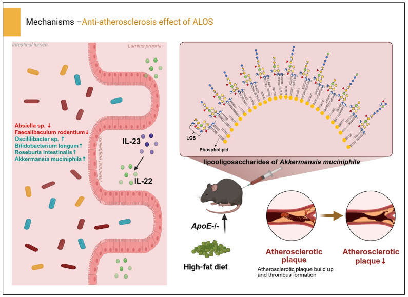
As an important and beneficial gut commensal, Akkermansia muciniphila plays a crucial role in regulating host metabolism and immunity. Lipooligosaccharides from A. muciniphila (ALOS) show anti-obesity effects in high-fat diet-fed mice. Herein, we investigated the chemical characteristics of core oligosaccharides of ALOS and explored its anti-atherosclerotic efficacy. The LC-Q-TOF-MS analysis indicated a high structural diversity of core oligosaccharides in ALOS, comprising fourteen distinct oligosaccharide species with different degrees of phosphorylation. Functionally, administration of ALOS significantly attenuated hyperlipidemia and reduced atherosclerotic plaque burden in high-fat diet-fed ApoE−/− mice. The improvement of these metabolic symptoms was related to the restoration of intestinal barrier integrity. Mechanistically, ALOS upregulated the IL-23/IL-22 immune axis, which in turn promoted intestinal epithelial repair and modulates the microbiota. ALOS intervention reshaped the gut microbiota composition by enriching beneficial genera such as Bifidobacterium longum, Roseburia intestinalis, and Oscillibacter sp., while suppressing potential pathobionts. Our findings highlight the structural diversity and anti-atherosclerotic effect of lipooligosaccharides from A. muciniphila.Graphical Abstract
Keywords
Akkermansia muciniphila Lipooligosaccharide Atherosclerosis IL-22 Gut microbiota1 Introduction
Lipopolysaccharide (LPS), a conserved microbial component, plays a pivotal role in training and shaping mucosal immunity through its interaction with pattern recognition receptors (e.g., TLR-4 and TLR-2), a process critical for gut immune homeostasis. LPS molecules have a common chemical architecture, composed of a lipid A moiety and a saccharide chain[1, 2]. Two groups of LPS have been identified in Gram-negative bacteria, including the smooth-type LPS (S-LPS) possessing a repeating O-polysaccharide domain (O-chain) in the saccharide chain and the rough-type LPS (R-LPS) without O-chain. R-LPS are also termed as lipooligosaccharides (LOS). Gut commensal bacteria-derived LOS from Bacteroides dorei, Bacteroides vulgatus, and Parabacteroides goldsteinii have been chemically characterized and demonstrated to have beneficial effects on the host, enhancing immunological tolerance and alleviating chronic obstructive pulmonary disease [3–5]. The previous work on Segatella copri LPS revealed that a hypo-acylated and hypo-phosphorylated lipid A, coupled with a mannose- and glucose-rich oligosaccharide core, weakly activated the TLR4 receptor and promotes a moderate immune response [6]. Similarly, Cho et al. demonstrated that tetra-acylated monophosphorylated lipid A (4A-MPLA) from Bacteroides fragilis induced a sustained IFN-β response and fosters the expansion of colonic RORγt + Tregs, thereby suppressing inflammation [7]. Hypoacylated derived LPS from Alcaligenes enhanced the IgA production without excessive inflammatory responses via weak TLR4 agonist activity [8].
Akkermansia muciniphila is an important gut mucous layer-colonized bacterium of Verrucobacteria, rich in the gut microbiota of healthy humans. It has been proposed as the representative of the next generation of probiotics for control of obesity[9, 10]. Early studies have demonstrated that oral supplementation of this bacterium improves insulin resistance in both animal and human clinical trials and restores mucosal integrity[11, 12]. Several bioactive components, including the outer pili-like protein Amuc_1100, the branched diacyl phosphatidylethanolamine, tripeptide, and the threonyl-tRNA synthetase, have been reported from A. muciniphila[13–16].
In our early work, a hypoacetylated and mono/bis-phospharylated LOS was identified from A. muciniphila (ALOS) [17]. ALOS is a weak TLR-4 agonist that reduces metabolic disorders in obese mice via activating the TLR-4-IL-23-IL-22 signaling pathway. In this work, we further revealed the structural diversity of oligosaccharide moieties in the ALOS by MS/MS analysis. In addition, we evaluated the anti-atherosclerotic effect of ALOS, which demonstrated protective effects by reducing hyperlipidemia and attenuating atherosclerotic injuries.
2 Results and discussions
2.1 MS/MS analysis of oligosaccharides in the LOS of A. muciniphila
In early reports, oligosaccharide moieties in the ALOS were found to be diverse, with two major oligosaccharides OSG and OSN, determined by NMR and MS analysis [17, 18]. In the current work, to clearly explore the structural diversity of ALOS, the fully deacylated lipopolysaccharides from A. muciniphila were characterized using LC-Q-TOF mass spectrometry. Mass spectra in negative-ion modes and collision-induced dissociation (CID) fragmentation experiments were optimized for each selected parent ion that provided extensive information to elucidate sequences of oligosaccharide chains (OS). As shown in the ion chromatogram, the oligosaccharides showed a high heterogeneity resulting from the presence of fourteen distinct OS species at m/z 869.7, m/z 909.7, m/z 950.3, m/z 990.3, m/z 1104.3, m/z 1144.3, m/z 1185.3, m/z 1225.3, m/z 1224.4, m/z 1266.4, m/z 1306.4, m/z 1347.5, m/z 1387.4 and m/z 1622.0 (Fig. 1A–C). Six of these variants (m/z 869.7, m/z 909.7, m/z 950.3, m/z 990.3, m/z 1224.4, and m/z 1622.0) were also observed in our previous report based on in-source fragmentation HPLC–ESI–MS analysis [14]. Based on the doubly-charged ions detected in the mass range of m/z 850–1650, the reported monosaccharide composition in ALOS, and the structure of OSG and OSN [17, 18], the sugar sequences in OSA-OSN was elucidated. For example, the oligosaccharide OSF was detected at m/z 1144.3572 [M-2H]2− with a retention time of tR = 3.03 min, which was deduced to be the dodecasaccharide composed of three 3-deoxy-D-manno−2-oct-ulopyranosonic acid moieties (Kdo-1, 2, and 3), two fucoses, one heptose, four hexosamines, two hexoses, and two phosphoric acid moieties (Supplementary Table 1). The CID MS/MS spectrum of OSF indicated the presence of daughter ions at m/z 1034.3 (−2), 1789.6, 1569.5, 1349.5, 939.2, 719.1, and 499.0, as shown in Fig. 2A, B. The occurrence of a fragment ion at m/z 1034.3 (−2) was due to the loss of one Kdo moiety from the double-charged parent ion at m/z 1144.3572 (−2). Two paired fragment ions (m/z 1789.6 and 499.0; 1569.5 and 939.2) were formed due to the cleavage of the glycosidic bond between GlcNP and Kdo-1, between Kdo-1 and Kdo-2 from OSF, respectively. The ions at m/z 1569.5 and 719.1 were derived from the daughter ions at m/z 1789.6 and 939.2 due to loss of one Kdo, respectively. By the similar way, the structural sequences of oligosaccharides OSE and OSH-M corresponding to m/z 1104.3 (−2), 1185.3 (−2), 1225.3 (−2), 1266.4 (−2), 1306.4 (−2), 1347.5 (−2), and 1387.4 (−2) were determined (Fig. 2C, Supplementary Fig. 1A-M, and Supplementary Table 1). Of note, the inner core oligosaccharide of ALOS was found to be composed of three Kdo, one fucose, one heptose, and one hexosamine. The structures for oligosaccharides (OSA-OSF and OSH-OSM) need to be further assigned by sophisticated purification and NMR data elucidation. The diverse oligosaccharides reflect the structural diversity of LOS in the commensal A. muciniphila (Scheme 1), which is deduced to play essential roles in the regulation of bacterial colonization and immune regulation.
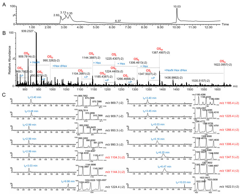
Analysis of core oligosaccharide in the LOS from A. muciniphila by LC-Q/TOF–MS in the negative ion mode. A The total ion chromatogram (TIC) recorded for the core oligosaccharide from A. muciniphila. B Double-charged molecular ion peaks detected in the LC-Q/TOF-MS spectrum. C The extracted ion chromatograms (EICs) of different core oligosaccharide species from A. muciniphila.
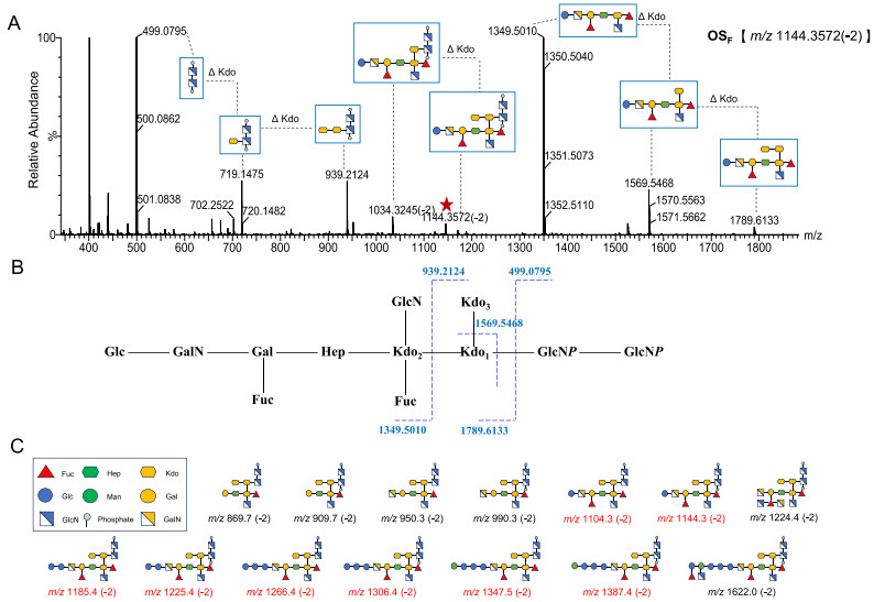
The CID MS/MS spectrum of the oligosaccharide OSF and proposed structures of oligosaccharides detected in A. muciniphila. A CID MS/MS spectrum from the double-charged mother ion at m/z 1144.3. B A proposed fragmentation pattern for OSF. C The proposed structural sequences of the core oligosaccharide from A. muciniphila sketched according to the symbology of monosaccharide residues.“Hex”, hexose; “Hep”, heptose; “Fuc”, fucose; “HexN”, hexosamine; “P”, phosphate; “Kdo”, 3-deoxy-D-manno-2-oct-ulopyranosonic acid, respectively.
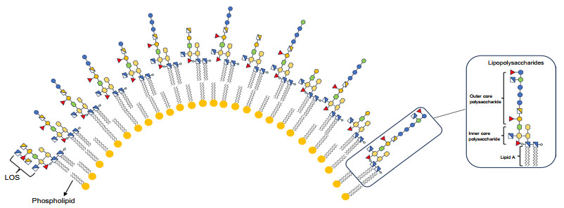
Proposed distribution of lipooligosaccharides on the outer membrane of A. muciniphila
2.2 ALOS inhibits atherosclerosis in ApoE−/− mice
In previous studies, A. muciniphila was reported to attenuate atherosclerotic lesions by reducing metabolic endotoxemia-induced inflammation through restoration of the gut barrier[19]. Interleukin (IL)−23 and its downstream target IL-22 have been reported to limit the development of atherosclerosis by inhibiting the pro-atherosclerotic microbiota[20]. Based on the beneficial effect of A. muciniphila on atherosclerosis and the immunological trait of ALOS, we explored the possible role of ALOS in atherosclerosis. ApoE−/− mice fed with the high-fat diet were intraperitoneally treated with ALOS or vehicle (PBS) for a period of 8 weeks (Fig. 3A). By the end of treatment, plasma lipid levels were significantly improved by ALOS. In comparison with vehicle-treated ApoE−/− mice, reductions in the levels of total cholesterol (TC; 55% and 29%), low-density lipoprotein cholesterol (LDL-C; 44% and 38.9%), total triglycerides (T-TG; 40% and 32%) were achieved by ALOS at doses of 0.2 and 0.5 mg kg−1, respectively (Fig. 3B). The levels of very-low-density lipoprotein (VLDL) and oxidized LDL (ox-LDL) were also significantly reduced by ALOS in a concentration-dependent manner (Fig. 3C–D). The high-fat diet induced formation of atherosclerotic lesions, as demonstrated by hematoxylin and eosin staining, Oil Red O staining, and Masson staining of aortic root regions (Fig. 3E). Compared to Mod, treatment with ALOS substantially reduced the aortic root lesion (Fig. 3E). After 12 weeks of high-fat diet, the accumulation of lipid in the whole aorta was shown in Fig. 3F. Treatment with ALOS substantially reduced the lesion area, necrotic core burden, and the content of collagen (Fig. 3E–G). In addition, ALOS also reduced the lipid accumulation in the liver (Fig. 3H). Based on these results, ALOS showed promising anti-atherosclerosis efficacy.
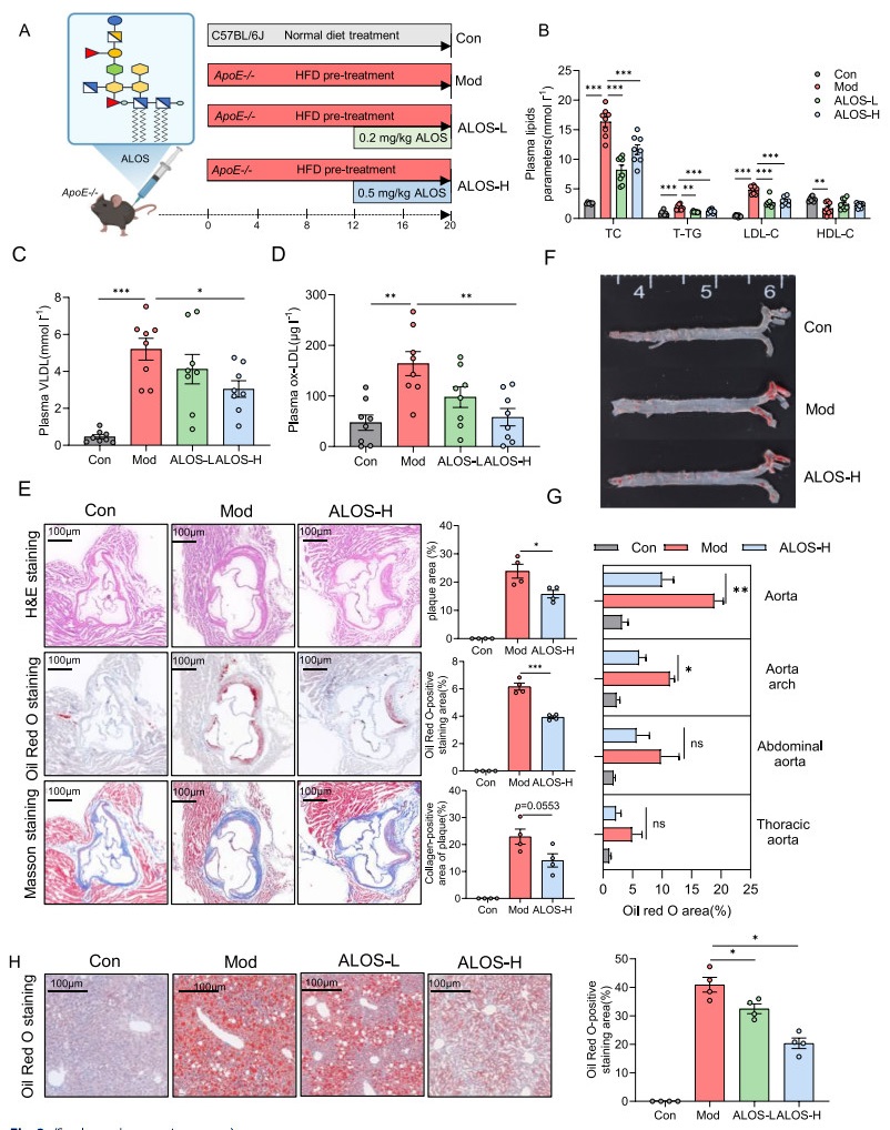
ALOS attenuates atherosclerosis in ApoE−/− mice. A Experimental design showing groups and durations (n = 8). ALOS: lipooligosaccharides derived from A.muciniphila. ALOS-L: 0.2 mg/kg ALOS treated- ApoE−/−. ALOS-H: 0.5 mg/kg ALOS treated- ApoE−/−. B Plasma lipid parameters (TC, T-TG, LDL-C, and HDL-C). C Plasma very low-density lipoprotein (VLDL). D Oxidized low-density lipoprotein (ox-LDL). E Representative H&E staining, Oil Red O staining, and Masson staining of cross-sections of aortic roots and quantitative data of plaque area, ecrotic core area, and collagen-positive area (scale bar, 100 μm, n = 4). F Lipid content of the whole aorta visualized by staining with Oil Red O. G The quantitative data of Oil Red O-positive staining area; n = 4 mice for each group. H Representative Oil Red O staining of liver (scale bar, 100 μm, n = 4). *P < 0.05; **P < 0.01. One-way ANOVA (with Tukey's HSD) test in B–H.
2.3 ALOS ameliorates gut barrier function and promotes IL-22 production
Endotoxemia caused by the increased intestinal epithelial permeability due to long-term high-fat diet is recognized as one of the pathogenesis mechanisms underlying the development of atherosclerosis[21, 22]. Compared with atherosclerosis model mice, ALOS-treated mice showed significantly lower plasma LPS levels and high-sensitivity C-reactive protein (hs-CRP) (Fig. 4A–B). Furthermore, ALOS decreased the plasma levels of proatherogenic cytokines, such as IL-6, TNF-α, and increased the levels of anti-inflammatory IL-10 (Fig. 4C–D, and F), while the level of IL-1β remained unaltered (Fig. 4E). The intestinal epithelium serves as the first defense barrier of the gastrointestinal tract and its compromise directly induces inflammation. AB-PAS staining revealed significant reductions of intestinal tissue in goblet cell count in the atherosclerosis model group (Fig. 4G), while ALOS supplementation mitigated the damage to epithelial integrity. In our earlier study on metabolic syndrome, it has been confirmed that ALOS has a weak TLR4 agonist and can elicit the IL-23/IL-22 axis, which is essential for maintaining the integrity of the intestinal barrier and modulation of gut microbiota[17]. We hypothesize that IL-23/IL-22 might be one of the mechanism by which ALOS suppress atherosclerosis. In ApoE−/− mice, the plasma level of IL-22 showed a marked decrease compared to that of normal mice. Compared to atherosclerosis model mice, a high dose of ALOS treatment significantly increased the plasma levels of IL-23 and IL-22 (Fig. 4H–I). The production of intestinal IL-22 was also reduced in the mice with atherosclerosis, while ALOS treatment significantly increased the protein levels of IL-22, as shown by immunofluorescent staining and WB (Fig. 4J–K). These results suggest that the repair of ALOS on the intestinal barrier in atherosclerotic mice is mechanistically related to the activation of the IL-23/IL-22 pathway.
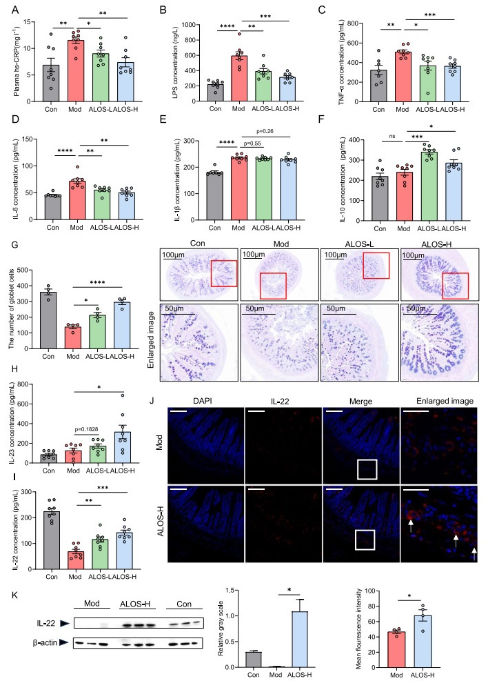
ALOS ameliorates gut barrier function and promotes IL-22 production. A C-reactive protein (hs-CRP). B LPS. C Tumour necrosis factor-alpha (TNF-α). D Interleukin-6 (IL-6). E Interleukin 1β (IL-1β). F Interleukin 10 (IL-10). G Representative AB-PAS staining of ileum pathological sections (scale bar, 100 μm, n = 4). H Plasma IL-23. I Plasma IL-22. J Representative immunofluorescent staining of IL-22 in the ileum. n = 4 mice for each group. Scale bar, 20 μm. K Western blotting of the IL-22 and β-actin levels in the ileum tissue of ApoE−/− mice. *P < 0.05; **P < 0.01. One-way ANOVA (with Tukey's HSD) test in A–I. Unpaired t-test in J–K.
2.4 ALOS reshapes the structure of the intestinal microbiota
Given the role of intestinal IL-22 in regulating antimicrobial peptides production and shaping gut microbiota composition[23–26], we next examined the influence of ALOS on the composition of intestinal microbiota in ApoE−/− mice by the high-throughput sequencing of 16S rRNA in the cecal content. A total of 1256267 sequences were obtained after denoising with DADA2, averaging 89,733 sequences in each sample. Treatment with ALOS did not affect the α-diversity of the bacterial community, as indicated by the Chao1 index and Dominance index, suggesting that ALOS did not change the richness and diversity of the gut microbiome (Fig. 5A–B). Principal coordinates analysis (PCoA) analysis revealed a distinct clustering of gut microbiota composition between the two groups (Fig. 5C), and the UPGMA clustering analysis also showed a clear separation between the model and ALOS groups (Fig. 5D). To further investigate specific alterations of bacterial communities, we compared the relative abundance of the major microbial taxa at the phylum, genus, and species levels. Firmicutes, Bacteroidetes, and Verrucomicrobia were the dominant bacterial phyla in both groups (Fig. 5E). The Firmicutes to Bacteroidetes (F/B) ratio was significantly lower in the ALOS-treated groups (3.18) compared to the mod groups (9.53), corresponding to a 66.63% decrease (Fig. 5E). Meanwhile, ALOS mainly up-regulated the abundance of the genus of Akkermansia, Roseburia, Oscillibacter, and Butyricimonas and decreased the abundance of pathogenic bacteria belonging to Helicobacter and Mucispirillum (Fig. 5F). The linear discriminant analysis effect size (LEfSe) identified 15 operational taxonomic units (OTUs) altered by ALOS, with a significant increase in Bifidobacterium, Robinsoniella, and a decrease in Absiella (Fig. 5G). As to the abundance of species, ALOS treatment decreased the relative abundance of Limosilactobacillus reuteri, and Faecalibaculum rodentium, while enriching the abundance of Bifidobacterium longum, Roseburia intestinalis, Phocaeicola vulgatus, Phocaeicola faecalis, and Akkermansia muciniphila (Fig. 5H–N). These shifts conferred metabolically benefits. F. rodentium, which was decreased by ALOS, contributes to the susceptibility of metabolic disorders by suppressing segmented filamentous bacteria and reducing intestinal Th17 cells [27]. Conversely, B. longum enriched by ALOS exhibited beneficial effects against metabolic disorders by regulating the mRNA expression of renin-angiotensin system factors and ameliorating lipid and glucose levels [28]. R. intestinalis, a promising next-generation probiotic, reinforced barrier function and suppressed pro-inflammatory signaling via butyrate production[29].
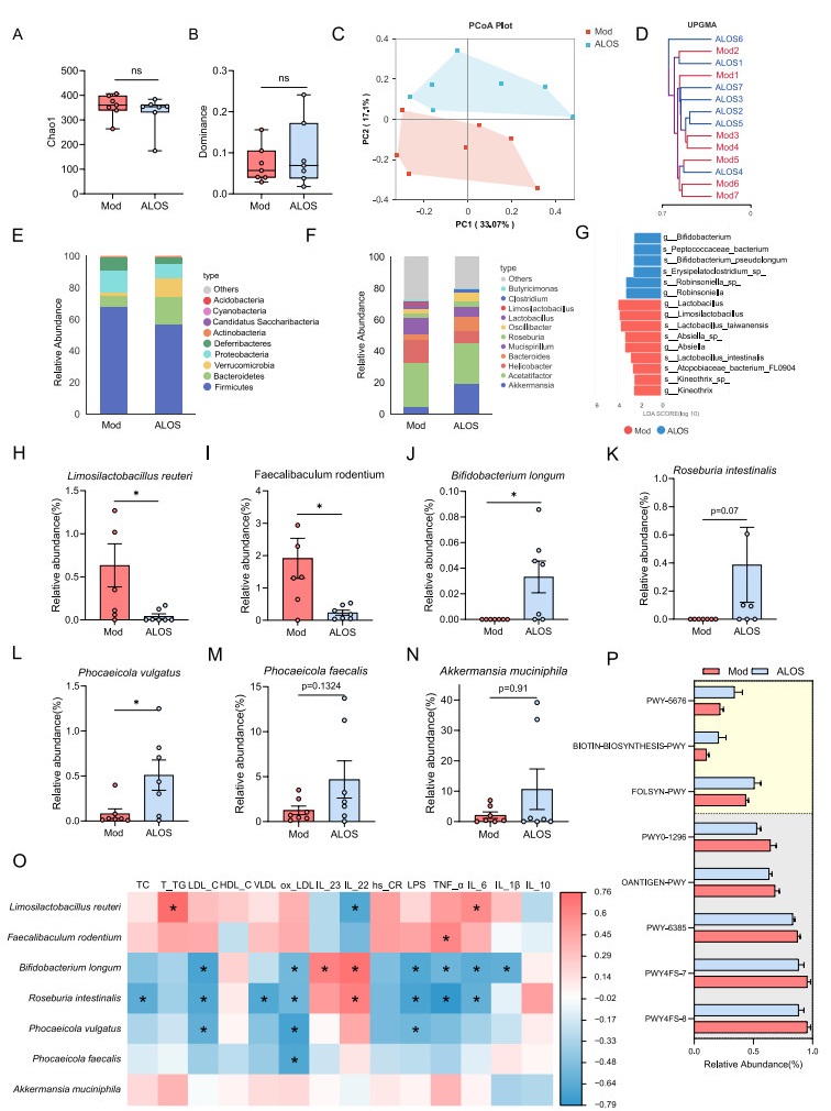
ALOS reshapes the structure of the intestinal microbiota. A The Chao 1 index. B The dominance index. C Principal coordinate analysis. D UniFrac distance-based unweighted pair-group method with arithmetic (UPGMA) means analysis. The shorter the branch length between samples, the higher the similarity between the two samples. E Relative abundance of gut microbiota at the phylum level. F Relative abundance of gut microbiota at the generic level. G LEfSe analysis of microbiota. H–N The relative abundance of differentially abundant flora identified at different taxonomic levels. O Spearman’s correlation analyses with serum biochemical parameters and IL‐22 levels. P Predict microbial function on the METACYC database. PWY-5676 (acetyl-CoA fermentation to butanoate Ⅱ), BIOTIN-BIOSYNTHESIS-PWY (biotin biosynthesis Ⅰ), FOLSYN-PWY (superpathway of tetrahydrofolate biosynthesis and salvage), PWY0-1296 (purine ribonucleosides degradation), OANTIGEN-PWY (O-antigen building blocks biosynthesis (E. coli)), PWY-6385 (peptidoglycan biosynthesis Ⅲ (mycobacteria)), PWY4FS-7 (phosphatidylglycerol biosynthesis Ⅰ (plastidic)), PWY4FS-8 (phosphatidylglycerol biosynthesis Ⅱ (non-plastidic)). *P < 0.05; **P < 0.01. Unpaired t-test in A–B, H–J, L–O. Mann–Whitney U test in K, P.
To further investigate the correlation of the key bacteria induced by ALOS and the atherosclerosis-associated symptoms, a Spearman correlation analysis was conducted. As a result, L. reuteri, Absiella sp., and F. rodentium, which were decreased by ALOS, were positively correlated with the parameters of atherosclerosis, while the ALOS-enriched bacteria, such as Oscilllibacter sp., B. longum, R. intestinalis, P. vulgatus, and P. faecalis, were negatively correlated with the symptoms of atherosclerosis (Fig. 5O). To predict the functional alteration in the gut microbiota after treatment, the PICRUSt2 analysis was employed. The results revealed that ALOS upregulated the metabolic pathways of PWY-5676 (acetyl-CoA fermentation to butanoate Ⅱ), BIOTIN-BIOSYNTHESIS-PWY (biotin biosynthesis Ⅰ) and FOLSYN-PWY (superpathway of tetrahydrofolate biosynthesis and salvage), which could benefit for ameliorating atherosclerosis [30–32], while downregulated the pathways, such as PWY0-1296 (purine ribonucleosides degradation), OANTIGEN-PWY (O-antigen building blocks biosynthesis (E. coli)), PWY-6385 (peptidoglycan biosynthesis Ⅲ (mycobacteria)), PWY4FS-7(phosphatidylglycerol biosynthesis Ⅰ (plastidic)), PWY4FS-8(phosphatidylglycerol biosynthesis Ⅱ (non-plastidic)) (Fig. 5P). Collectively, these data suggest that ALOS treatment partially regulated the structure and functions of gut microbiota in ApoE−/− mice. In conclusion, ALOS treatment ameliorates the dysbiosis of gut microbiota in mice with atherosclerosis, thereby contributing to the alleviation of atherosclerotic injuries.
3 Discussions
In this study, the distribution and diversity of oligosaccharide chains in the lipooligosaccharids of A. muciniphila was proposed. MS/MS analysis of oligosaccharides in the ALOS identified fourteen mono/bis-phosphorylated oligosaccharide species. Based on our prior and current findings demonstrating that ALOS mediated activation of the IL-23/IL-22 in both obesity and ApoE−/− mice models [17, 18], we propose that the unique structural features and high diversity of ALOS may coordinately contribute to its ability to elicit a balanced immune activation, favoring the IL-23/IL-22 pathway over pro-inflammatory cytokine cascades. Consistent with their unique chemical structures and activation of IL-23/IL-22 immune response, the lipooligosaccharides derived from A. muciniphila are associated with attenuated hyperlipidemia, reduced plaque burden, improved intestinal barrier markers, and a reshaped gut microbiota in ApoE−/− mice.
As a relatively weak TLR4 agonist, ALOS exhibits favorable safety profiles, as demonstrated in our previous studies through acute endotoxin toxicity models, comparing to the conventional LPS from Escherichia coli [17]. LPS molecules are easily degraded in the acidic gastric environment, limiting oral bioavailability. Consequently, intraperitoneal administration was used in the current study. The well-established crosstalk between immunity and the gut microbiota[33] suggests that systemic ALOS-induced activation of the IL-23/IL-22 axis may promote the expansion of metabolically beneficial gut commensals, such as Bifidobacterium longum, Roseburia intestinalis, Phocaeicola vulgatus, Phocaeicola faecalis, and A. muciniphila.
Despite these promising findings, several limitations of the current study should be acknowledged. The decomposition of LPS under acidic condition hinders the application of oral delivery, a preferable route for gut microbiota modulation and relatively safe for therapeutic applications. In the following study, coating ALOS with suitable material in formulation to avoid acidic condition in stomach is necessary for establishing an oral delivery system for ALOS. In early studies, we and other researchers have chemically identified two main oligosaccharides in LOS of A. muciniphila through detailed analysis of MS and NMR data[17, 18]. However, duo to its high heterogeneity, separation of other oligosaccharides is unsuccessful after great efforts. In this work, we turned to LC–MS/MS analyses to show oligosaccharide diversity in ALOS. The obtained MS/MS data together with previously reported structural information for two main oligosaccharides (OSG and OSN) revealed structural diversity in oligosaccharides of ALOS, but the exact linking sites between sugar units and complete NMR assignment remain to be confirmed. To achieve purification of these unique oligosaccharides(OSA-OSF and OSH-OSM), new sophisticated separation approaches are needed to be developed. Moreover, further investigations are required to establish the safety, immune activation thresholds, and the translational dosingof ALOS.
In conclusion, our study revealed the structural characteristics and diversity of oligosaccharides in the lipooligosaccharides of A. muciniphila, demonstrated the anti-atherosclerotic activity and gut microbiota modulation of ALOS in mice. These findings advance our understanding of microbiome-host crosstalk in cardiovascular disease and suggest the therapeutic potential of gut bacterial glycolipids.
4 Experimental sections
4.1 Isolation and characterization of oligosaccharides from LOS of A. muciniphila
A. muciniphila HW07 strain was cultured anaerobically at 37 ℃ in modified brain–heart infusion (BHI) broth. The extraction and purification of LOS from A. muciniphila (ALOS) were conducted according to our early study [17]. The fully deacylated oligosaccharides linked to the lipid A sugar skeleton were subsequently obtained from the purified ALOS by reduction and alkaline hydrolysis procedure. Briefly, the purified ALOS (1 mg) was treated with hydrazine solution and stirred (37 ℃, 1.5 h). Subsequently, the resulting reduced product was then heated with 4 M KOH (120 ℃, 16 h). After cooling, the mixture was allowed to precipitate. The aqueous phase containing the deacylated oligosaccharide was collected, neutralized with 2 M HCl, and dialyzed (molecular mass cut-off: 500 Da) to remove salts and small molecules. The Q-TOF MS and MS/MS analysis of the deacylated oligosaccharides was acquired on a Water Xevo G3 Q-TOF mass spectrometry in the negative ion mode. For CID-MS/MS analysis, the sample was dissolved in water and analyzed using a Shim-pack GIST C18-AQ column (250 × 4.6 mm; 5 μm; SHIMADZU, Japan) at a flow rate of 0.3 mL/min. The mass spectrometer was set to scan over an m/z range of 60–3000. A gradient elution beginning with Solvent A (0.01% formic acid) for 3 min, then with an increase of Solvent B (acetonitrile) to 100% (v/v) over 10 min, followed by 100% B for 5 min and a decrease to 100% A in 8 min, was applied.
4.2 Mice
C57BL/6 J and ApoE−/− mice (6–8 weeks, male) were obtained from the Experimental Animal Center, Chinese Academy of Medical Sciences. All animals were normally housed under specific pathogen-free (SPF) conditions. The chow diet (13.5% calories from fat) and a high-fat diet (HFD, 60% kcal fat, D12492, New Brunswick) were used for feeding. Experiments were conducted according to approval by the Institute of Microbiology, Chinese Academy of Sciences (IMCAS).
4.3 The effects of ALOS on high-fat diet-induced ApoE−/− mice
C57BL/6 J fed with a normal diet (ND), ApoE−/− mice fed with HFD at 8 weeks of age, diet pretreatment for 12 weeks, and then divided into three groups. Intraperitoneal injection of ALOS 0.2 mg/kg (ALOS-L) or 0.5 mg/kg (ALOS-H) once every two days for 8 weeks.
4.4 Biochemical analyses
The interleukin-6 (IL-6) (SEKM-0007; Solarbio), tumor necrosis factor-alpha (TNF-α) (RK00027; Abclonal), interleukin-1β (IL-1β) (MM-0040M1; MEIMIAN), interleukin-10 (IL-10) (1418033–25-6; Solarbio), interleukin-22 (IL-22) (E-MSEL-M0040; Elabscience BiotechnologyCo., Ltd.), interleukin-23 (IL-23) (RK00017; Abclonal), oxidized LDL (ox-LDL), very low density lipoprotein cholesterol (VLDL) (ml037709-2; Mlbio), and lipopolysaccharide (LPS) (WLCSJZF202535; AMOY LUNCHANGSHUO) levels in the plasma and ileum were determined in commercial ELISA kits. High-sensitivity C-reactive protein (Hs-CRP) was determined by commercial ELISA kits (EnBo, Beijing, China). The total cholesterol (T-CHO), triglycerides (TG), low-density lipoprotein cholesterol (LDL-C), and free fatty acid (FFA) levels in plasma were determined by commercially available kits (Nanjing Jiancheng Bioengineering Institute, Jiangsu, China).
4.5 Histological analysis
The aortic roots, liver, adipose tissues, and intestinal tissues were isolated and fixed in 4% paraformaldehyde solution. The aortic roots were stained with H&E, Oil Red O, or Masson’s; the livers were stained with Oil Red O; the intestinal tissues were stained with AB-PAS. The lesion area, the number of goblet cells, the relative area of Oil Red O, and Collagen volume fraction (CVF) were measured using ImageJ software.
4.6 DNA extraction and 16S rRNA sequencing analysis
According to the manufacturer’s instructions, fecal DNA was extracted using the QIAamp Fast DNA stool Mini Kit (Qiagen, Cat# 51604). Just as in previous study[34], Primers for the 16S rRNA variable region 3–4 (V3-V4) were 338 F (5′-ACTCCTACGGGAGGCAGCA-3′) and 806R (5′-GGACTACHVGGGTWTCTAAT-3′). DNA library was performed on an Illumina NovaSeq sequencing platform.
4.7 Immunofluorescence staining analysis
The fresh intestinal tissues were isolated and immediately fixed in 4% paraformaldehyde solution for 24 h and blocked with 5% bovine serum albumin for 30 min. Then, the 3–5 μm thick slides were incubated at 4 ℃ overnight with primary antibodies: anti-IL-22 rabbit pAb (R30198; Zenbio), followed by the secondary antibody for 50 min at room temperature with goat anti-rabbit IgG-Cy3 (Servicebio, GB21303). The ImageJ software was used to quantify the images.
4.8 Western blotting
After the ileal tissue of mice was homogenized in the cold RIPA lysis buffer at 50 mg/L, the protein concentration of the supernatant was determined by the BCA method. The protein was isolated by sodium dodecyl sulfate polyacrylamide gel electrophoresis (SDS-PAGE) (36276ES10; Yeasen) and transferred to polyvinylidene fluoride (PVDF, Servicebio; G6044-0.45) membrane. First, the membranes were closed with 5% non-fat milk at room temperature for 1 h. Secondly, the membranes were incubated overnight at 4 ℃ with different primary antibodies: anti-IL-22 (R30198; Zenbio) and β-actin (700068; Zenbio). Finally, HRP-linked goat anti-rabbit IgG (511203; Zenbio) secondary antibody was incubated for 1 h. Protein was visualized using ECL Plus Western Blot assay (36208ES7636208ES76; Yeasen). β-actin was used to normalize the expression of IL-22.
4.9 Statistical analysis
Correlations between species and factors were analyzed using Spearman's correlation coefficient. Spearman's correlation analysis calculates Spearman's ρ, ρ = 1 indicates a completely positive linear correlation, ρ = −1 indicates a complete negative linear correlation, and ρ = 0 indicates no linear correlation. The normality of the distribution was tested by the Kolmogorov–Smirnov test. Comparisons between the two groups are represented by Welch's t‐test, unpaired t‐test, and Mann–Whitney U test. Welch's t‐test required normality in both groups but did not require equal variances. The unpaired t‐test required both normality and equal variances. The Mann–Whitney U test required neither normality nor equal variances. One‐way ANOVA test (post‐event verification method: Tukey's HSD) was used for multiple comparisons. The data in the figure were expressed as mean ± SEM. GraphPad Prism 9.0 and ImageJ were used to analyze all the data and showed as: ns p > 0.05, *p < 0.05, **p < 0.01, ***p < 0.001, ****p < 0.0001.
Notes
Acknowledgements
This study was financially supported by the National Natural Science Foundation of China (92578117) and the National Key Research and Development Program of China (2025YFA1309200). The authors express their gratitude to Dr. Xiaofei Jia from Waters Technologies(Beijing) Co., Ltd. for her contributions in MS analysis.
Author contributions
Yuting Zhang: conceptualization, Validation, Formal analysis, Investigation, Writing-original draft, Visualization; Wang Dong: conceptualization, Validation, Formal analysis, Investigation, Writing-original draft, Visualization; Jinghan Lin: methodology; Jingzu Sun: formal analysis; Ruopeng Yin: validation; Xun Lv: supervision, Conceptualization, Funding acquisition; Wenzhao Wang: validation; Tao Wang: writing-review and editing; Hongwei Liu: supervision, Conceptualization, Writing-review and editing, Project administration, Funding acquisition. All authors read and approved the manuscript.
Funding
National Natural Science Foundation of China,92578117,Hongwei Liu,Key Research and Development Program of China,2025YFA1309200,Hongwei Liu
Data availability
Raw 16S rDNA data had been deposited on NCBI at https://submit.ncbi.nlm.nih.gov/subs/bioproject/SUB15789623/overview with BioProject submission: SUB15789623. All other data supporting the findings of the study are available within the article and its supplementary information files, or from the corresponding authors upon reasonable request.
Declarations
Ethics statement and consent to participate
The ethics application (IMCAS2024126) was approved by the Institute of Microbiology, Chinese Academy of Sciences of Research Ethics Committee.
Competing interests
The authors declare no competing financial interest.
References
-
1.Garcia-Vello P, Di Lorenzo F, Zucchetta D, Zamyatina A, De Castro C, Molinaro A. Lipopolysaccharide lipid A: a promising molecule for new immunity-based therapies and antibiotics. Pharmacol Ther 2022;230: 107970. CrossRef PubMed Google Scholar
-
2.Coats SR, Do CT, Karimi-Naser LM, Braham PH, Darveau RP. Antagonistic lipopolysaccharides block E.coli lipopolysaccharide function at human TLR4 via interaction with the human MD-2 lipopolysaccharide binding site. Cell Microbiol 2007;9: 1191-202. CrossRef PubMed Google Scholar
-
3.Vatanen T, Kostic AD, d’Hennezel E, Siljander H, Franzosa EA, Yassour M, et al. Variation in microbiome LPS immunogenicity contributes to autoimmunity in humans. Cell 2016;165: 842-53. CrossRef PubMed Google Scholar
-
4.Di Lorenzo F, Pither MD, Martufi M, Scarinci I, Guzmán-Caldentey J, Łakomiec E, et al. Pairing Bacteroides vulgatus LPS structure with its immunomodulatory effects on human cellular models. ACS Cent Sci 2020;6: 1602-16. CrossRef PubMed Google Scholar
-
5.Lai H-C, Lin T-L, Chen T-W, Kuo Y-L, Chang C-J, Wu T-R, et al. Gut microbiota modulates COPD pathogenesis: role of anti-inflammatory Parabacteroides goldsteinii lipopolysaccharide. Gut 2022;71: 309-21. CrossRef PubMed Google Scholar
-
6.De Simone Carone L, Barra G, Cirella R, Ziaco M, Mercogliano M, Olmeo F, et al . Decoding a gut commensal signal: structural and immunological profiling of Segatella Copri Lipopolysaccharide. Angew Chem Int Ed 2025. CrossRef PubMed Google Scholar
-
7.Cho H-S, Yoo J-S, Song X, Goh B, Diallo A, Lee J, et al. Structure of gut microbial glycolipid modulates host inflammatory response. Cell 2025;188: 5295-5312.e5218. CrossRef PubMed Google Scholar
-
8.Shibata N, Kunisawa J, Hosomi K, Fujimoto Y, Mizote K, Kitayama N, et al. Lymphoid tissue-resident Alcaligenes LPS induces IgA production without excessive inflammatory responses via weak TLR4 agonist activity. Mucosal Immunol 2018;11: 693-702. CrossRef PubMed Google Scholar
-
9.Reunanen J, Kainulainen V, Huuskonen L, Ottman N, Belzer C, Huhtinen H, et al. Akkermansia muciniphila adheres to enterocytes and strengthens the integrity of the epithelial cell layer. Appl Environ Microbiol 2015;81: 3655-62. CrossRef PubMed Google Scholar
-
10.Depommier C, Everard A, Druart C, Plovier H, Van Hul M, Vieira-Silva S, et al. Supplementation with Akkermansia muciniphila in overweight and obese human volunteers: a proof-of-concept exploratory study. Nat Med 2019;25: 1096-103. CrossRef PubMed Google Scholar
-
11.Everard A, Belzer C, Geurts L, Ouwerkerk JP, Druart C, Bindels LB, et al. Cross-talk between Akkermansia muciniphila and intestinal epithelium controls diet-induced obesity. Proc Natl Acad Sci USA 2013;110: 9066-71. CrossRef PubMed Google Scholar
-
12.Cani PD, Depommier C, Derrien M, Everard A, de Vos WM. Akkermansia muciniphila: paradigm for next-generation beneficial microorganisms. Nat Rev Gastroenterol Hepatol 2022;19: 625-37. CrossRef PubMed Google Scholar
-
13.Bae M, Cassilly CD, Liu X, Park S-M, Tusi BK, Chen X, et al. Akkermansia muciniphila phospholipid induces homeostatic immune responses. Nature 2022;608: 168-73. CrossRef PubMed Google Scholar
-
14.Sanz Y, Ottman N, Reunanen J, Meijerink M, Pietilä TE, Kainulainen V, et al . Pili-like proteins of Akkermansia muciniphila modulate host immune responses and gut barrier function. PLoS ONE 2017. CrossRef PubMed Google Scholar
-
15.Xie S, Li J, Lyu F, Xiong Q, Gu P, Chen Y, et al. Novel tripeptide RKH derived from Akkermansia muciniphila protects against lethal sepsis. Gut 2024;73: 78-91. CrossRef PubMed Google Scholar
-
16.Kim S-M, Park S, Hwang S-H, Lee E-Y, Kim J-H, Lee GS, et al. Secreted Akkermansia muciniphila threonyl-tRNA synthetase functions to monitor and modulate immune homeostasis. Cell Host Microbe 2023;31: 1021-1037.e1010. CrossRef PubMed Google Scholar
-
17.Sun L, Zhang Y, Dong W, Sun J, Wang T, Shao F, et al . Akkermansia muciniphila‐derived hypoacylated rough‐type lipopolysaccharides alleviate diet‐induced obesity via activation of TLR4−IL‐23−IL‐22 immune axis. iMeta 2025. CrossRef PubMed Google Scholar
-
18.Garcia-Vello P, Tytgat HLP, Elzinga J, Van Hul M, Plovier H, Tiemblo-Martin M, et al. The lipooligosaccharide of the gut symbiont Akkermansia muciniphila exhibits a remarkable structure and TLR signaling capacity. Nat Commun 2024;15: 8411. CrossRef PubMed Google Scholar
-
19.Li J, Lin S, Vanhoutte PM, Woo CW, Xu A. Akkermansia muciniphila protects against atherosclerosis by preventing metabolic endotoxemia-induced inflammation in Apoe−/− mice. Circulation 2016;133: 2434-46. CrossRef PubMed Google Scholar
-
20.Fatkhullina AR, Peshkova IO, Dzutsev A, Aghayev T, McCulloch JA, Thovarai V, et al. An interleukin-23-interleukin-22 axis regulates intestinal microbial homeostasis to protect from diet-induced atherosclerosis. Immunity 2018;49: 943-957.e949. CrossRef PubMed Google Scholar
-
21.Nie H-Z-R, Zhou Y-W, Yu X-H, Yin C-G, Li L-F, Hao H-Q, et al. Intestinal epithelial Krüppel-like factor 4 alleviates endotoxemia and atherosclerosis through improving NF-κB/miR-34a-mediated intestinal permeability. Acta Pharmacol Sin 2024;45: 1189-200. CrossRef PubMed Google Scholar
-
22.Parantainen J, Barreto G, Strandberg TE, Mars N, Nurmi K, Eklund KK . Increased intestinal mucosal permeability and metabolic endotoxemia predict the risk of cardiovascular mortality. Atherosclerosis 2025. CrossRef PubMed Google Scholar
-
23.Mar JS, Ota N, Pokorzynski ND, Peng Y, Jaochico A, Sangaraju D, et al. IL-22 alters gut microbiota composition and function to increase aryl hydrocarbon receptor activity in mice and humans. Microbiome 2023;11: 47. CrossRef PubMed Google Scholar
-
24.Wang X, Ota N, Manzanillo P, Kates L, Zavala-Solorio J, Eidenschenk C, et al. Interleukin-22 alleviates metabolic disorders and restores mucosal immunity in diabetes. Nature 2014;514: 237-41. CrossRef PubMed Google Scholar
-
25.Yin R, Wang T, Sun J, Dai H, Zhang Y, Liu N, et al. Postbiotics from Lactobacillus johnsonii activates gut innate immunity to mitigate alcohol-associated liver disease. Adv Sci 2024;12: e2405781. CrossRef PubMed Google Scholar
-
26.Yin R, Wang T, Dai H, Han J, Sun J, Liu N, et al. Immunogenic molecules associated with gut bacterial cell walls: chemical structures, immune-modulating functions, and mechanisms. Protein Cell 2023;14: 776-85. CrossRef PubMed Google Scholar
-
27.Kawano Y, Edwards M, Huang Y, Bilate AM, Araujo LP, Tanoue T, et al. Microbiota imbalance induced by dietary sugar disrupts immune-mediated protection from metabolic syndrome. Cell 2022;185: 3501-3519.e3520. CrossRef PubMed Google Scholar
-
28.Machado AS, Oliveira JR, Lelis DF, de Paula AMB, Guimaraes ALS, Andrade JMO, et al. Oral probiotic Bifidobacterium longum supplementation improves metabolic parameters and alters the expression of the Renin-Angiotensin System in obese mice liver. Biol Res Nurs 2021;23: 100-8. CrossRef PubMed Google Scholar
-
29.Nie K, Ma K, Luo W, Shen Z, Yang Z, Xiao M, et al . Roseburia intestinalis: a beneficial gut organism from the discoveries in genus and species. Front Cell Infect Microbiol 2021. CrossRef PubMed Google Scholar
-
30.Dicks LMT . Butyrate produced by gut microbiota regulates atherosclerosis: a narrative review of the latest findings. Int J Mol Sci 2025. CrossRef PubMed Google Scholar
-
31.Kockx MM, Muhring J, Bortier H, De Meyer GR, Jacob W. Biotin- or digoxigenin-conjugated nucleotides bind to matrix vesicles in atherosclerotic plaques. Am J Pathol 1996;148: 1771-7. PubMed Google Scholar
-
32.Qiao SS, Liu C, Sun L, Wang T, Dai HQ, Wang K, et al. Gut Parabacteroides merdae protects against cardiovascular damage by enhancing branched-chain amino acid catabolism. Nat Metab 2022;4: 1271-86. CrossRef PubMed Google Scholar
-
33.Zheng D, Liwinski T, Elinav E. Interaction between microbiota and immunity in health and disease. Cell Res 2020;30: 492-506. CrossRef PubMed Google Scholar
-
34.Wang T, Han J, Dai H, Sun J, Ren J, Wang W, et al . Polysaccharides from Lyophyllum decastes reduce obesity by altering gut microbiota and increasing energy expenditure. Carbohydr Polym 2022. CrossRef PubMed Google Scholar
Copyright information
© The Author(s) 2026
Open Access This article is licensed under a Creative Commons Attribution 4.0 International License, which permits use, sharing, adaptation, distribution and reproduction in any medium or format, as long as you give appropriate credit to the original author(s) and the source, provide a link to the Creative Commons licence, and indicate if changes were made. The images or other third party material in this article are included in the article’s Creative Commons licence, unless indicated otherwise in a credit line to the material. If material is not included in the article’s Creative Commons licence and your intended use is not permitted by statutory regulation or exceeds the permitted use, you will need to obtain permission directly from the copyright holder. To view a copy of this licence, visit http://creativecommons.org/licenses/by/4.0/.