Limonene and its metabolite perillyl alcohol inhibit Chlamydia trachomatis growth by altering host isoprenoid metabolism

Abstract
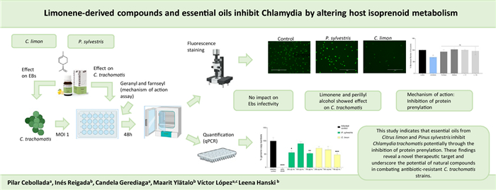
The obligate intracellular bacterium Chlamydia trachomatis is the most common bacterial sexually transmitted infection globally, with approximately 131 million new cases each year. It contributes to widespread reproductive health issues, including infertility and chronic pelvic pain. The unique cell morphology and biphasic life cycle pose challenges to their effective eradication. Guided by the identification of essential oils (EOs) capable of suppressing C. trachomatis intracellular growth, this study evaluated the antichlamydial properties of the monoterpene limonene and its metabolites perillyl alcohol and perilic acid. The antichlamydial activity was assessed through qPCR-based quantification of bacterial genome copy numbers and immunofluorescence staining of chlamydial inclusions to monitor bacterial growth and infectious progeny production. Elementary body membrane integrity was assessed with viability PCR. Citrus limon essential oil exhibited dose-dependent inhibition of C. trachomatis growth, decreasing infectious progeny by over 90%. Pinus sylvestris EO displayed consistent but non-dose-dependent effects. Limonene, a major constituent of the EOs, exhibited significant suppression of chlamydial growth and progeny production, particularly for its R-enantiomer. While viability-PCR data indicated that the EOs and limonene affected EB membrane permeability, EB infectivity was not affected by the treatments. Perillyl alcohol suppressed chlamydial growth at a lower concentration (100 µg mL−1) than the parent compound limonene. The antichlamydial activity of both limonene and perillyl alcohol was suppressed when infected cultures were supplemented with farnesyl or geranylgeranyl, indicating that the antichlamydial mechanism of the two monoterpenes involves competitive inhibition of protein prenylation. Hence, targeting host protein prenylation may be an effective antichlamydial strategy.Graphical Abstract
Keywords
Intracellular bacteria Protein prenylation Membrane structural integrity Perillyl alcohol Monoterpen1 Introduction
Chlamydia trachomatis is an obligate intracellular pathogen, most notably for causing sexually transmitted infections (STIs) and ocular infections. This bacterium exhibits multiple serovars, among which A to C are responsible for trachoma, a form of infectious blindness, while D to K represent the most common bacterial STIs worldwide and L1 to L3 usually lead to lymphatic system infections. C. trachomatis infections may cause cervicitis, fibrotic endometritis, and pelvic inflammatory disease in women. Vertical transmission during delivery can result in neonatal conjunctivitis [1, 2]. Most infected women and half of infected men are asymptomatic, which complicates the management as the non-treated infection may progress, leading to a chronification with severe consequences such as pelvic inflammatory disease, ectopic pregnancy, and obstructive infertility [3]. People unaware of their infection may also risk their sexual partners' health, therefore contributing to the pathogen dissemination. In fact, in a context where the incidence of STIs is increasing, it is worth to highlight that C. trachomatis infections represent the most prevalent bacterial STI globally, with 131 million new cases annually [4].
C. trachomatis exhibits a distinctive lifecycle, in which the bacteria can exist primarily in two different forms: the infectious elementary body (EB) and the reproductive reticulate body (RB). EBs, the extracellular forms, are responsible for the infection of the host cells. EBs internalize into the host eukaryotic cell inside endocytic vacuoles which fuse and mature to form an inclusion where the EBs transform into RBs, the replicative form of C. trachomatis [5]. RBs replicate by binary fission utilizing the host cell nutrients, filling the cytoplasm and dislocating the nucleus. Besides, the bacteria exploit host vesicular trafficking by recruiting and modulating Rab GTPases, to facilitate their intracellular survival and replication [6]. RBs convert back into EBs, which are then released into the extracellular space either by host cell lysis or by extrusion of the intact inclusion, allowing them to infect new host cells [7].
Under stress conditions such as antibiotic or interferon-γ exposure, nutrient deprivation, or coinfection, C. trachomatis can enter a persistent state marked by enlarged, non-infectious aberrant RBs, resulting in more complex management of the infection [8, 9]. Apart from the ability to enter persistent state, C. trachomatis also presents a challenge for treatment due to antibiotic resistance, a problem common among many other bacteria. C. trachomatis infections are usually treated with inhibitors of the protein synthesis, namely tetracyclines (doxycycline) and macrolides (azithromycin). Despite its ability to induce chlamydial persistence, in some cases amoxicillin is also considered as a therapeutic option [10]. Reported failure rates range from 5 to 23%, often linked to resistance driven by prolonged exposure to subinhibitory drug concentrations [11]. Besides the two lipid membranes of the gram-negative bacterium itself, the host-derived inclusion membrane and host cell plasma membrane constitute additional permeability barriers which limit the local antibiotic concentrations reaching the replicating chlamydial RBs. These challenges underscore the need for alternative antimicrobials and novel treatment strategies.
Essential oils (EOs) are plant-derived volatile products consisting of a mixture of terpenes, sesquiterpenes and aromatic compounds, typically obtained through steam or hydro distillation or in the case of citrics, by mechanical pressure. The antimicrobial properties of EOs and individual EO-derived terpenes have been extensively studied, indicating that most of these lipophilic compounds exert direct bactericidal effects through disruption of microbial membranes and subsequent loss of membrane polarization. To date, data on the impact of EOs or EO-derived terpenes on intracellular bacteria have remained very scarce, presumably due to often limited selectivity in targeting prokaryotic versus eukaryotic membranes and hence cytotoxicity on host cells. In the case of Chlamydia, chances for selective chlamydiocidal activity are further compromised due to presence of host-derived lipids in both bacterial plasma membrane and outer membrane. Two previous studies have addressed the effects of EOs and EO-derived terpens on EB infectivity [12–14] and one study has suggested an inhibitory effect of a Mentha EO on RB replication stage [14].
The current study was established with the hypothesis that identifying non-toxic EOs capable of suppressing C. trachomatis growth during the intracellular stages of its life cycle can guide the identification of antichlamydial terpenes with mechanisms of action distinct from direct disruption of chlamydial membranes. Following this reasoning, we identified limonene as a previously unknown C. trachomatis growth inhibitor based on its dominant proportion in the composition of two commercially available antichlamydial EOs, Citrus limon and Pinus sylvestris. Limonene was found to suppress C. trachomatis intracellular growth and progeny production but not affect EB infectivity. Drawing on the known ability of limonene and its metabolite perillyl alcohol to suppress protein prenylation in eukaryotic cells, we hypothesized a role of altered host protein prenylation in the observed antichlamydial effect and conducted supplementation experiments with the prenylation substrates farnesyl and geranylgeranyl to experimentally test this concept.
2 Materials and methods
2.1 Chemicals
The EOs selected for this work were obtained from Pranarom (Ghislenghien, Belgium). The main characteristics of the EOs are listed in Table 1.
Summary of the key characteristics of the EOs studied in this work, including their name, batch, plant part used, country of origin, and primary compounds
S-(−)-limonene and R-(+)-limonene (Sigma-Aldrich, St. Louis, MO, US) (Fig. 1) were used to compare its activity with the EOs. For cell culture, Dulbecco's Modified Eagle's Medium (DMEM; Cytiva, Marlborough, Massachusetts, United States), gentamicin (Sigma-Aldrich, St. Louis, MO, US), and heat-inactivated fetal bovine serum (FBS; Invitrogen, Thermo Scientific, Waltham, MA, US) were used. Azithromycin (AZM; Cayman Chemical, Ann Arbor, MI, US) served as a positive control in antichlamydial assays. For bacterial genome quantification, the GeneJET Genomic DNA Purification Kit (Thermo Fisher Scientific, Massachusetts, USA), primers, and Master Mix (Applied Biosystems, Thermo Scientific, Waltham, MA, US) were used. For inclusion staining, Chlamydia LPS Monoclonal Antibody (B410F), Goat anti-Mouse IgG (H + L) Highly Cross-Adsorbed Secondary Antibody (Invitrogen, Thermo Scientific, Waltham, MA, US), Alexa FluorTM Plus 488 (Invitrogen, Thermo Scientific, Waltham, MA, US), Evans Blue, Propidium monoazide (PMA) and cycloheximide (Sigma-Aldrich, St. Louis, MO, USA) were used. The antichlamydial activity of perillyl alcohol (Sigma-Aldrich, St. Louis, MO, US) and perillic acid (Santa Cruz Biotechnology), metabolites of limonene, was also evaluated. The chemical structure of limonene and its metabolites herein tested is presented in Fig. 1. For mechanism of action studies, farnesyl pyrophosphate ammonium salt and geranylgeranyl pyrophosphate ammonium salt (Merck Life Sciences) were obtained.
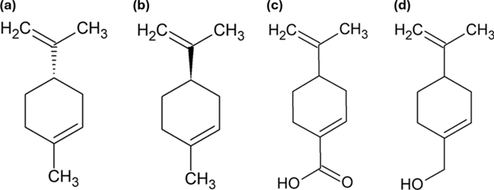
Chemical structures of a (S)-(−)-limonene, b (R)-(+)-limonene, c perillic acid, and d perillyl alcohol. Structures were generated using ChemSketch 2.0
2.2 Chemical characterization
The chemical composition of both EOs was determined using gas chromatography coupled with mass spectrometry (GC–MS). Analyses were performed on an Agilent Technologies GC 6890 system interfaced with a 5973-mass selective detector and fitted with an HP-5MS-IU capillary column (30 m × 0.25 mm, 0.25 µm film thickness; Agilent Technologies). Samples were introduced in split mode at a ratio of 100:1, with an injection volume of 0.2 µL.
The oven temperature program began at 60 ℃ and was ramped to 240 ℃ at a rate of 3 ℃ per minute. Helium served as the carrier gas at a constant flow rate of 1.0 mL/min. Mass spectrometric detection was conducted in electron impact (EI) mode at 70 eV, scanning over an m/z range of 41–250. Compound identification was achieved by comparing both retention times and mass spectra with reference data from the NIST library and previously published literature [15].
Additionally, linear retention indices (LRIs) were calculated using a homologous series of n-alkanes (C8–C30) run under identical conditions. These experimental indices were cross-referenced with literature values to support the provisional identification of analytes. Where possible, compound assignments were further corroborated by matching both fragmentation patterns and LRIs with those reported in the Wiley275 library and by Adams (2009).
2.3 Bacterial strains
The chlamydial species utilized was an isolate of C. trachomatis, Serovar K (ATCC VR-887). It was propagated in HeLa-229 (Homo sapiens, human epithelial cells; HeLa-229 ATCC (CCL-2.1); LOT: 59501359) cells and stored in 0.2 M sucrose, 0.02 M sodium phosphate (pH 7.4) and 5 mM glutamic acid (SPG-buffer) at − 80 ℃ until used.
2.4 Mammalian cells maintenance and cytotoxicity assessment
HeLa-229 cells were cultured in DMEM, supplemented with 20 µg mL−1 gentamicin and 10% FBS. The cells were maintained at 37 ℃ with 5% CO2 in a humidified incubator (HeracellTM 240i, Thermo Scientific, Waltham, MA, US). The viability of HeLa cells upon exposure to EOs, limonene, perllyl alcohol and perillic acid was determined by seeding HeLa-229 cells into 96-well plates (6 × 10⁴ cells/well) and incubating them for 24 h before treatment with various substance concentrations. After 48 h, cells were washed with PBS, stained with 20 µM resazurin, and incubated for 2 h. Fluorescence (λex = 560 nm, λem = 590 nm) was recorded using a Varioskan LUX microplate reader (Thermo Scientific, Waltham, MA, US).
2.5 Determination of activity against C. trachomatis
2.5.1 Infection procedure
HeLa-229 cells were plated in 24-well plates (4 × 10⁵ cells/well) on glass coverslips. After a 24 h incubation, the cells were infected with C. trachomatis, Serovar K (ATCC VR-887) at a multiplicity of infection (MOI) of one in infection media (DMEM + Cycloheximide 1 µg mL−1) and the plate was centrifugated for 1 h at 550 g and further incubated for another hour at 37 ℃.
2.5.2 Activity against C. trachomatis
To study the effect of the EOs, limonene, metabolites and AZM 40 nM (positive control) on C. trachomatis, the infection procedure described above was followed. After the 1 h incubation at 37 ℃, the inoculum was replaced with fresh infection media with different concentrations of EO. The plate was then further incubated for 48 h.
2.5.3 Infectious progeny assay
To assess the impact on chlamydial infectious progeny production, the procedure described in 2.5.2 was followed. Following incubation, the infected cells were detached from the wells and frozen at −80 ℃ to enable the lysis. The cell lysate was then used to infect fresh HeLa-229 cells in a 24-well plate as in 2.5.1.
2.5.4 Direct impact on EBs
C. trachomatis (4 × 10⁵ Inclusion Forming Units) was pretreated with different concentrations of EOs or limonene in infection media and incubated for 1 h at 4 ℃. After this pretreatment, the EOs and AZM were removed in a 1 h centrifugation (4 ℃ and 21 000 g). Subsequently, the EBs were resuspended in fresh medium and inoculated into the HeLa-229 cells, as described in 2.5.1.
2.5.5 DNA extraction and quantitative PCR measurement
Following incubation, the cells were scraped and centrifuged at 2000 × g for 5 min, resuspended and stored at − 80 ℃. Genomic DNA was extracted using the GeneJET Genomic DNA Purification Kit. Quantification was performed using a StepOnePlusTM Real-Time PCR System with StepOneTM Software v2.3 (Thermo Fisher Scientific, Waltham, MA, USA). Each qPCR reaction contained 20 ng of template DNA, Fast SYBRTM Green Master Mix, and 0.4 mM of each primer (Forward: GGCGTATTTGGGCATCCGAGTAACG; Reverse: TCAAATCCAGCGGGTATTAACCGCT), in 96-well MicroAmp optical plates (Thermo Fisher Scientific). Thermal cycling was set to 95 ℃ for 20 s, followed by 40 cycles of 95 ℃ for 3 s and 60 ℃ for 30 s. A melting curve analysis followed, consisting of 95 ℃ for 15 s, 60 ℃ for 1 min, and 95 ℃ for 15 s. Each run included non-template and negative controls. Standard curves for genome equivalent (GE) quantification were generated by serial dilution of purified C. trachomatis DNA with a known titer.
2.5.6 Inclusion staining
At 48 h post-infection, cells grown on coverslips were washed with PBS, fixed in methanol for 10 min, and blocked with 0.5% BSA for 15 min. They were then incubated with 75 µL of Chlamydia LPS Monoclonal Antibody (B410F) diluted 1:100 for 1 h in a humid chamber. After washing, Goat anti-Mouse IgG (H + L), Alexa FluorTM Plus 488 diluted 1:2000, was applied. Following a second wash, cells were stained with 100 µL of Evans Blue for 20 min, rinsed three times with Milli-Q water, and then analyzed under a microscope.
2.6 Mechanism of action studies
2.6.1 EBs viability PCR
To complement the EB infection-based assays and evaluate whether the EOs and limonene affected its membrane, the viability-PCR (V-PCR) approach described by Janssen et al. was adapted [16]. Briefly, EBs suspensions were incubated for 1 h at 4 ℃ in the presence EOs (750 µg mL−1), or R(+)-limonene (525 µg mL−1). Untreated EBs were included as viability controls. Following incubation, samples were centrifuged at 21,000 × g for 1 h at 4 ℃ and resuspended in 500 µL SPG buffer. Two additional EB aliquots were heat-killed (95 ℃, 10 min) to serve as non-viable controls. PMA was added to all preparations to a final concentration of 50 µM, and samples were incubated for 10 min in the dark at 4 ℃, followed by light activation for 15 min at 465 nm. After a final centrifugation step (21,000 × g, 1 h, 4 ℃), DNA was extracted and subjected to qPCR as described in Sect. 2.5.5. A marked reduction in amplification signal was interpreted as loss of EB viability due to compromised membrane integrity. An infected control non-treated and a heat-treated control were prepared to confirm the assay's reliability.
2.6.2 Inhibition of protein prenylation
To determine whether the antichlamydial effect of limonene and perillyl alcohol is mediated through the inhibition of protein prenylation, cell cultures were supplemented with farnesyl pyrophosphate and geranylgeranyl pyrophosphate (substrates of prenyltransferase enzymes) to assess whether the additional substrate could counteract limonene's antichlamydial activity. To do so, infection with C. trachomatis was performed as explained in Sect. 2.5.1. After that, cells were treated with 10 µM of farnesyl pyrophosphate or geranylgeranyl pyrophosphate alone or in combination with R-(+) limonene (525 µg mL−1) or perillyl alcohol (100 µg mL−1).
2.7 Statistical analysis
Statistical analyses were performed using GraphPad Prism 10, with outliers identified via Grubbs' test. One-way ANOVA followed by Dunnett's or Bonferroni's post hoc tests was used to assess differences between sample concentrations and the control (p-values reported). For bacterial inclusion quantification, images were processed using a custom Python script employing libraries such as skimage, numpy, matplotlib, and pandas. The script converted images to grayscale, applied Gaussian filtering, thresholding, and morphological operations, and labelled regions to detect and count fluorescent signals, compiling the results into a DataFrame for statistical analysis performed as previously described. GraphPad Prism 10 was also used to create the Figures.
3 Results
3.1 The impact of C. limon and P. sylvestris EOs on C. trachomatis growth and progeny production
In our search for antichlamydial EOs showing no cytotoxicity on the HeLa-229 epithelial cell line serving as the C. trachomatis infection host, C. limon and P. sylvestris EOs stood out (Supplementary Information). The impact of EOs on C. trachomatis growth in HeLa-229 cells was studied by administering the EOs after C. trachomatis EB internalization (starting the treatment at 2 h post-infection). At the end of a 48-h infection, both bacterial genome copy numbers and bacterial inclusion counts were quantified. C. limon reduced the genome copy numbers in a dose-dependent manner, showing a 27.7% and 33.6% reduction at 250 and 500 µg mL−1 respectively (Fig. 2a). A statistically significant reduction in C. trachomatis genome copy numbers was reached at the highest concentration (52.6% reduction; p < 0.001). C. limon EO also reduced the number of chlamydial inclusions (Fig. 2b) at 500 (40.6% reduction; p < 0.001) and 750 µg mL−1 (76.1% reduction; p < 0.001). Figure 2 also shows that P. sylvestris also displayed activity at some of the concentrations but did not show a concentration-dependent activity within the tested conditions.
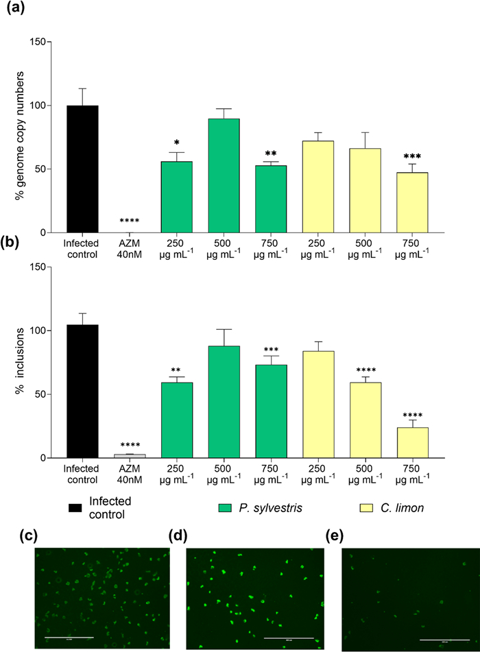
Impact of EOs on C. trachomatis growth. a Percentage of genome copy numbers and b percentage of inclusions of C. trachomatis remaining after treatment with C. limon and P. sylvestris EOs. Data were normalized to the untreated control. Different concentrations of EOs ranging from 250 to 750 µg mL−1 were studied. Mean and SEM values are displayed, n = 4. Representative images for each condition are presented in panels c, d, and e, corresponding to the untreated control, treatment with P. sylvestris EO (750 µg mL−1) and treatment with C. limon EO (750 µg mL−1) respectively. Significant differences were determined by ANOVA (statistical significance: *p < 0.05; **p < 0.01; ***p < 0.001; ****p < 0.0001)
The effects of both EOs on preventing the generation of infectious Chlamydia progeny was determined by infecting fresh HeLa-229 cells with lysates collected from EO-treated infections. C. limon EO reduced the number of inclusions in the infectious progeny assay a 16.9%, 41.0% and 92.7% at concentrations of 250 µg mL−1, 500 µg mL−1 (p < 0.01) and 750 µg mL−1 (p < 0.0001) respectively (Fig. 3a). P. sylvestris proved to be active (p < 0.05, p < 0.001 and p < 0.0001) at the same concentrations reducing the number of inclusions by 32.5%, 51.3% and 66.4% respectively (Fig. 3b).
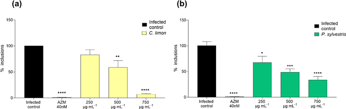
Impact of C. limon and P. sylvestris EOs on C. trachomatis infectious progeny production. The figure shows the percentage of bacterial inclusion of C. trachomatis in HeLa-229 cells infected with the infectious progeny obtained from a first infection in another set of HeLa-229 cells treated with C. limon (a) and P. sylvestris (b) at different concentrations (250–750 µg mL−1), as well as azithromycin (AZM) used as a positive control and untreated controls. Data were normalized to the untreated control. Mean and SEM values are displayed, n = 4. Significant differences were determined by the ANOVA test (statistical significance, when compared to the untreated control: *p < 0.05; **p < 0.01. ***p < 0.001. ****p < 0.0001)
3.2 Chemical composition of the EOs
The chemical composition of both EOs was determined using gas chromatography coupled with mass spectrometry GC–MS. 29 compounds were identified in C. limon EO, with limonene representing the 64.13% of the EO (Table 2). Other compounds found in the EO were gamma-terpinene (11.022%) and beta-Pinene (10.767%). For P. sylvestris, a total of 46 compounds were identified (Table 3). The three major constituents were alpha-pinene (32.06%), beta -pinene (19.63%), and limonene + beta -phellandrene (11.52%). Chemical structures of the main compounds found in both EO are presented in the Supplementary Information.
Chemical characterization of C. limon EO
Chemical characterization of P. sylvestris EO
3.3 Impact of limonene on C. trachomatis growth
Given its remarkable proportion in the composition of both studied EOs, the impact of commercially obtained pure limonene was studied under the same conditions against C. trachomatis. Limonene concentrations were adjusted to correspond to its quantities present in C. limon EO at concentrations exhibiting antichlamydial activity. The impact of both enantiomers on C. trachomatis growth was studied as regard to genome copy numbers (Fig. 4a) and to bacterial inclusion counts (Fig. 4b). In both cases, R-Limonene showed a higher reduction than the one displayed by S-Limonene. In fact, the highest R-limonene concentration 525 µg mL−1 significantly reduced the genome copy numbers of C. trachomatis (64.5% reduction; p < 0.05) (Fig. 4a). Both S-Limonene and R-Limonene dose-dependently reduced the number of chlamydial inclusions (Fig. 4b). The inhibitory effect reached statistical significance for S-limonene at the highest concentration 525 µg mL−1 (24.64% reduction; p < 0.0001), while R-limonene showed reduction for both 350 µg mL−1 (16.2% reduction; p < 0.05) and 525 µg mL−1 (34.9% reduction, p < 0.0001).
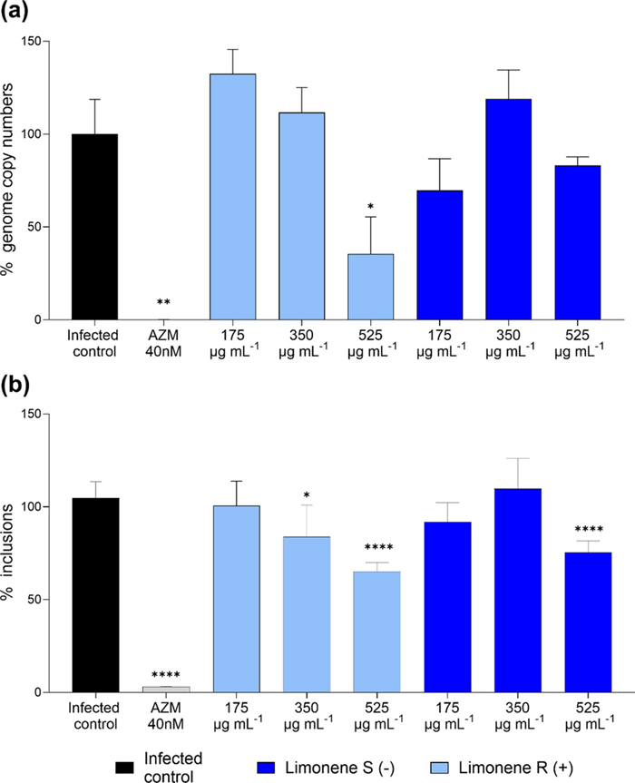
Impact of limonene on C. trachomatis growth. a Remaining genome copy numbers and b remaining inclusion counts of C. trachomatis after treatment with the R and S enantiomers of limonene. Data were normalized to the untreated control. Different concentrations of limonene ranging from 175–525 µg mL−1 were studied. In the graphs mean and SEM values are displayed, n = 4. Significant differences were determined by ANOVA (statistical significance: *p < 0.05; **p < 0.01; ***p < 0.001; ****p < 0.0001)
The impact of limonene on the C. trachomatis infectious progeny production was also studied for both S-limonene and R-limonene (Fig. 5). For both enantiomers, the effect was dose-dependent and reached statistical significance (p < 0.05) at the highest concentration (525 µg mL−1), reducing the number of chlamydial inclusions by 47.9% for R-limonene and 51.8% for S-limonene.
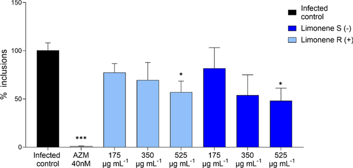
Impact of limonene on the infectious progeny. The figure shows the percentage of remaining bacterial inclusion counts of C. trachomatis in HeLa-229 cells infected with the infectious progeny obtained from a first infection in another set of HeLa-229 cells treated with limonene (175–525 µg mL−1), azithromycin (AZM) and untreated controls. Data were normalized to the untreated control, with mean values and SEM being displayed (n = 4). Significant differences were determined by ANOVA (statistical significance: *p < 0.05; **p < 0.01. ***p < 0.001. ****p < 0.0001)
3.4 Impact of limonene and the limonene-containing EOs on C. trachomatis EBs
To determine whether limonene affected the C. trachomatis EB membrane, viability-PCR was performed. In this assay, EB suspensions are exposed to the studied substances for 1 h and subsequently exposed to propidium monoazide (PMA). EBs with compromised membrane integrity allow PMA to enter the cell and intercalate with the DNA, thereby preventing DNA amplification in the subsequent PCR reaction. As shown in Fig. 6, when V-PCR is performed, significant differences were found between the infectious control and the samples. Both EOs at 750 µg mL⁻1 significantly reduced the genome copy numbers (p < 0.001). Also, the pure limonene affected EB membranes (p < 0.05), although the effect was less pronounced than the one exhibited by the EOs.
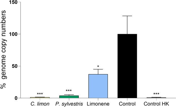
Viability-PCR analysis of C. trachomatis EBs after treatment with essential oils and limonene. Percentage of C. trachomatis genome copy numbers obtained by Viability-PCR after EB treatment with C. limon and P. sylvestris EOs (750 µg mL⁻1) or R (+)-limonene (525 µg mL⁻1). A heat killed (HK) EBs were used as a positive control. Data were normalized to the untreated control, with mean values and SEM being displayed (n = 4). Significant differences were determined by ANOVA (statistical significance: *p < 0.05; **p < 0.01. ***p < 0.001. ****p < 0.0001)
Since the viability-PCR data indicated that limonene and limonene-containing EOs may affect C. trachomatis EB membrane permeability (Fig. 6), we wanted to determine whether this effect translates to altered EB infectivity. To this end, EB suspensions were exposed to limonene or the EOs for 1 h before inoculating them to HeLa-229 monolayers. In Fig. 7, the impact of both EOs (Fig. 7a) and limonene enantiomers (Fig. 7b) on the C. trachomatis EB infectivity is depicted. The results showed that neither the pure limonene enantiomers nor the limonene-containing EOs have significant effect decreasing the EB infectivity. Only the positive control azithromycin showed significant differences (p < 0.0001).
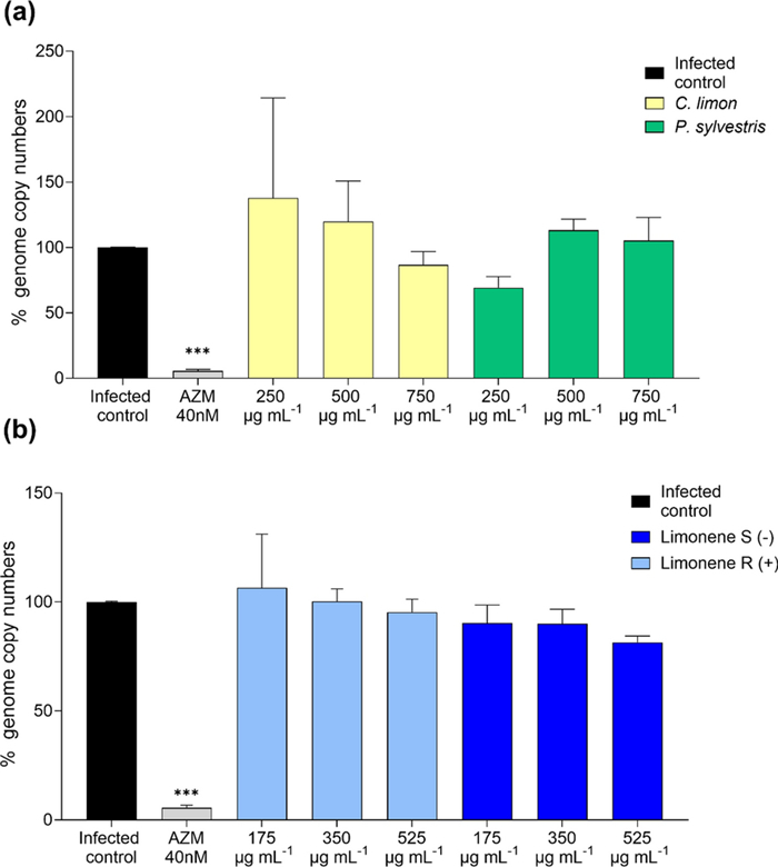
Impact of the C. limon and P. sylvestris EOs on C. trachomatis EB infectivity. Genome copy numbers of C. trachomatis were obtained from HeLa-229 cells infected with EBs pre-treated with EOs (a) or limonene (b) at different concentrations. Azithromycin (AZM) was used as a positive control. Data were normalized to untreated control. Mean and SEM values are displayed, n = 4. Statistical analysis with ANOVA (statistical significance: *p < 0.05; **p < 0.01. ***p < 0.001. ****p < 0.0001)
3.5 Involvement of host protein prenylation in the antichlamydial activity
As the above-described data indicated that direct targeting of chlamydial membranes does not contribute to the observed antichlamydial effect, we tested an alternative hypothesis on the underlying mechanism, namely the involvement of host cell altered protein prenylation. To this end, two interconnected approaches were taken. First, the impact of two limonene metabolites, perillyl alcohol and perillic acid were evaluated for their ability to reduce the number of chlamydial inclusions. While no significant differences between the infected control and the perillic acid were found (data not shown), perillyl alcohol (100 µg mL−1) reduced the inclusion counts by 66.2% (p < 0.0001; Fig. 8).
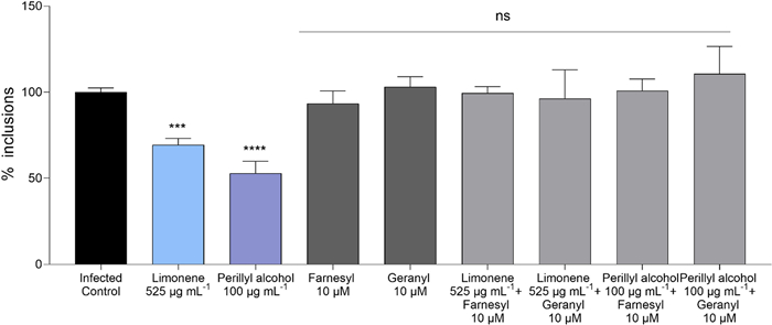
Impact of Farnesyl pyrophosphate and Geranylgeranyl pyrophosphate supplementation on the antichlamydial activity of limonene and perillyl alcohol. The figure shows the percentage of remaining inclusions of C. trachomatis after the treatment with R-limonene (525 µg mL−1), perillyl alcohol (100 µg mL−1), farnesyl (10 µM), geranylgeranyl (10 µM), limonene (525 µg mL−1) + farnesyl (10 µM), limonene (525 µg mL−1) + geranylgeranyl (10 µM), perillyl alcohol (100 µg mL−1) + farnesyl (10 µM) and perillyl alcohol (100 µg mL−1) + geranylgeranyl (10 µM). Data were normalized to untreated control. Mean and SEM values are displayed (n ≥ 4). Significant differences between groups were determined using ANOVA followed by Dunnett's post hoc correction for multiple comparisons (statistical significance, when compared to the infected control: statistical significance: *p < 0.05; **p < 0.01. ***p < 0.001. ****p < 0.0001, ns: non-significant). No significant differences between the infected control and the perillic acid were found (not represented)
Second, the impact of supplementing the cultures with external prenyltransferase substrates was used to assess the involvement of possible competitive inhibition of this cellular process. The chlamydial growth inhibition experiment with both limonene and perillyl alcohol were repeated under the same conditions as in previous experiments, adding farnesyl pyrophosphate and geranylgeranyl pyrophosphate to the cultures. The results shown in Fig. 8 demonstrate that both limonene and perillyl alcohol consistently reduce the number of C. trachomatis inclusions. The addition of farnesyl pyrophosphate or geranylgeranyl pyrophosphate alone did not affect inclusion counts (Fig. 8). However, when perillyl alcohol and limonene-treated infections were supplemented with either of the lipids, the inhibitory effect of both limonene and its metabolite were almost entirely lost, showing no significant differences with the infected control.
4 Discussion
Although terpenes and EOs have been widely studied for their bioactive properties, their activity against intracellular bacteria remains mostly unexplored. While C. trachomatis is an obligate intracellular pathogen, several non-obligate bacteria can also adopt an intracellular lifestyle as a virulence strategy, gaining access to nutrients, evading immune responses, and establishing reservoirs that contribute to persistence and reinfection [17]. This highlights the relevance of exploring therapeutic approaches targeting intracellular bacterial forms. The results of the current study indicate that limonene, a monoterpene found in various EOs, inhibits C. trachomatis intracellular growth and progeny production but does not significantly reduce C. trachomatis EB infectivity in vitro. Furthermore, perillyl alcohol, the main metabolite of limonene, was found to be more potent C. trachomatis growth inhibitor than its parent compound, and the antichlamydial effect of both limonene and perillyl alcohol was found to be abolished by supplementing the infections with protein prenyltransferase substrates.
The identification of limonene as an antichlamydial compound was guided by the observation that two limonene-rich EOs, namely lemon and pine, showed antichlamydial activity when administered to epithelial cells after C. trachomatis EB internalization. Lemon essential oil, widely used in cosmetics, hygiene products, foods and aromatherapy, has been extensively studied for its bioactive properties and found to exhibit diverse properties including antimicrobial activity [18, 19]. Pine trees, widely found in Europe, Asia, North America and North Africa, yield EOs which have been reported to carry various bioactivities, including antiviral and antibacterial effects [20]. While we found P. sylvestris EO to reproducibly reduce C. trachomatis growth, its impact on both bacterial genome copy numbers and chlamydial inclusion counts did not show clear concentration-dependence and was lower than that displayed by C. limon EO.
For both studied EOs, the efficacy seemed to be lower for the genome copy numbers compared to the reduction of inclusion counts under the same conditions. This may reflect the fact that qPCR is able to detect DNA also from non-viable C. trachomatis, indicating that the presence of genetic material might not necessarily reflect the intracellular bacterial viability [16].
A recent study reported that P. sylvestris and C. limon EOs, as well as their major constituent limonene are active against drug-resistant Neisseria gonorrhoeae [21]. N. gonorrhoeae a is a sexually transmitted pathogen with complications like those of C. trachomatis, such as pelvic inflammatory disease. These two bacteria not only produce similar symptoms, but 40–46% of individuals infected with N. gonorrhoeae are concurrently infected with C. trachomatis [22]. Thus, the present findings may have translational relevance for STI management, as the EOs could act against both pathogens without harming human cervical epithelial cells (see Supplementary material).
Our comprehensive chemical characterization of both lemon and pine EOs demonstrated findings consistent with previous reports, involving a mixture of a diverse set of terpenoids and other volatile compounds. Limonene, a monocyclic monoterpene was found in major quantities in both EOs, being a major compound for C. limon EO and one of the main components in P. sylvestris. Hence, the antichlamydial activity of limonene was confirmed through C. trachomatis genome copy and inclusion count quantification as well as infectious progeny production assays. Both enantiomers, S-limonene and R-limonene, were studied using concentrations adjusted to correspond those in the applied lemon EO to evaluate whether the compound contributed to the activity against C. trachomatis. Notably, R-limonene (also known as D-limonene), reported in the literature as the enantiomer found in C. limon EO [19], exhibited greater antichlamydial activity in our study (Figs. 4 and 5). Even though limonene makes up roughly two thirds of the studied C. limon EO, the EO proved to have higher activity against C. trachomatis (Fig. 2) than its isolated major constituent (Fig. 4). Although falling beyond the scope of the current study, the various additional compounds found in the EO presumably have potentiating effects which may contribute to the overall activity of the EO. Similarly, while the differences in the antichlamydial activity between lemon and pine EOs could be attributed to the different amount of limonene found, the role of other compounds present in the EO and potential synergies between them cannot be overlooked.
Besides the two EOs included in this study, limonene is found widely in plants such as sweet orange, bitter orange, neroli, mandarin or petitgrain [19]. Limonene is widely used for its citric fragrance as an additive and has been linked to several biological effects, including antioxidant, anti-inflammatory, and cardioprotective activities [23].
As for the possible antibacterial mechanisms of action, many previous studies on limonene against planktonic bacteria have demonstrated its membrane disruption potential. At least in Escherichia coli, limonene seems to damage the cell membrane producing alterations in membrane permeability and disrupting the membrane integrity [24]. To assess whether limonene or the EOs could affect C. trachomatis EB membrane integrity, viability-PCR assay was performed. According to these data, the EBs treated with EOs and limonene indeed had experienced damage in the EB membrane, indicated by the altered PCR amplification after PMA exposure (Fig. 6). However, despite the effect on the EB membrane permeability, the EB infectivity was not compromised by treatment with the EOs or limonene, as the EB pretreatment did not reduce C. trachomatis genome copy numbers after the infection on HeLa-229 cells (Fig. 7). The chlamydial EB form, whose morphology and composition have become increasingly well characterized, is encased by the chlamydial outer membrane complex (COMC). This structure is heavily reinforced by disulfide-cross-linked proteins, including the major Outer Membrane Protein (MOMP), OmcA and OmcB, which confer exceptional rigidity and restrict membrane permeability. Together with surface lipopolysaccharide and additional outer-membrane components, the COMC protects EBs from osmotic and mechanical stress during their extracellular phase [5, 25, 26]. When the EBs are internalized, there is a reorganization of this structure since RBs present reduced disulfide bonds in their COMC proteins, resulting in a less rigid structure essential for their replication and adequate for the stable environment of the inclusion [5, 25]. Hence, the maintained EB infectivity despite lipid membrane alterations may result from unique chlamydial cell wall characteristics and the changes that occur after internalization. In fact, protein components of the EB envelope play a direct role in the internalization process. Several well-characterized surface-exposed and secreted proteins contribute to host cell entry. For instance, OmcB acts as an adhesin that mediates the initial attachment to host cell receptors, while preformed type Ⅲ secretion system effectors are delivered into the host cell upon contact, triggering actin rearrangements required for uptake. Following internalization, inclusion membrane proteins further remodel the host endomembrane system to promote intracellular survival. Therefore, although limonene and limonene-containing EOs seem to damage the EB lipid membrane, internalization may still occur due to these protein factors [27, 28]. In fact, a recent study on monoclonal antibodies targeting MOMP epitope exposed on the C. trachomatis EB surface suggested that despite effectively coating the outer membrane, the antibodies failed to reduce EB infectivity, as they were rapidly lost from the bacterial surface after EB internalization. This illustrates that the membrane remodeling and other changes that EBs undergo after internalization can modify or diminish the functional effects of surface-associated alterations. Regardless of the underlying molecular level interactions, our results indicate that viability-PCR alone may not serve as a reliable method for assessing the infectivity of chlamydial EBs.
Since our data indicated that the impact of the EOs and limonene on EB membrane permeability did not have a significant role in their antichlamydial activity, other mechanisms of action were explored. Anticancer properties of limonene have been widely described, being competitive inhibition of protein prenylation one of the known mechanisms of action [29]. Protein prenylation is a crucial post-translational modification that regulates the localization and function of some membrane-associated proteins in eukaryotes, most notably small GTPases including members of the Ras, Rho, and Rab families. This process involves the covalent attachment of isoprenoid groups, such as farnesyl (C15) or geranylgeranyl (C20) moieties, to conserved cysteine residues at the C-terminus of these proteins [30]. C. trachomatis exploits host vesicular trafficking pathways through the recruitment and manipulation of small GTPases, particularly Rab proteins, to facilitate its intracellular survival and replication [6]. However, the inhibition of protein prenylation has never been studied as an antichlamydial strategy. Nevertheless, one earlier study on a closely related species, Chlamydia pneumoniae, reported this pathogen to increase small GTPase prenylation in its host cells [31]. In the same study, geranylgeranyl supplementation was found to counteract statin-mediated inhibition of this process. Inspired by these findings, we investigated the involvement of limonene's competitive inhibition of eukaryotic protein prenylation in its antichlamydial activity. Both farnesyl and geranylgeranyl supplementation of infected cell cultures were found to reverse the inhibitory effect of limonene (Fig. 8). These data suggest that one potential mechanism of action of limonene against C. trachomatis is related to the inhibition of protein prenylation through competitive inhibition, since the antichlamydial activity of limonene is lost when the prenyltransferase substrates farnesyl and geranylgeranyl are added to the culture.
In previous literature, limonene has been shown to inhibit protein prenylation, specifically targeting farnesyl transferase (FTase) and geranylgeranyl transferase (GGTase) activity of small GTPases [29, 32]. However, its inhibitory effect is relatively weak, with an IC50 exceeding 40 mM (5449.6 µg mL−1) when assayed on enzymes isolated from rat brain cytosol. Given the high IC50, it is evident that relatively high limonene concentrations are required to achieve meaningful effects on cellular protein prenylation patterns. While the concentrations applied in our current work to achieve chlamydial inhibition were relatively high, they are still lower than the above-mentioned value for the inhibition of isolated FTase and GGTase. Another earlier study reported that limonene inhibits isoprenylation in Plasmodium falciparum, a process essential for the function of its Ras- and Rap-like proteins, with an IC50 value of 1.22 mM (166.2 µg mL−1), [33, 34]. The discrepancies in IC₅₀ values between the previously mentioned study, which used enzymes extracted from rat brain cytosol, and the results obtained in cell-based models are particularly noteworthy. Since previous reports have shown that limonene metabolites exhibit greater potency as prenylation inhibitors than limonene itself [32, 35], these differences may reflect some degree of modification or metabolic transformation occurring within the cellular environment. One previous study reported that, although metabolic transformations of limonene have been observed in vivo, no such metabolism was detected in cell culture [36]. However, this result was obtained on a 3 h incubation in a mouse embryonic fibroblast cell line, leaving open the possibility that cell types more competent in xenobiotic metabolism or longer incubation times, such as the 48-h used in our experiment, could have different outcomes.
To elucidate whether limonene metabolites displayed antichlamydial activity, the impact of perillic acid and perillyl alcohol was evaluated (Fig. 8). Perillyl alcohol is the major metabolite of limonene that naturally occurs in the essential oils of several plants, including mint, lavender, lemongrass, and sage. In human plasma, one of the main metabolites derived from both limonene and perillyl alcohol is perillic acid [29]. According to our data, perillyl alcohol reduced the number of C. trachomatis inclusions at concentrations significantly lower than those tested for limonene. Besides, as shown in Fig. 8, the antichlamydial activity of perillyl alcohol was also impaired by the addition of farnesyl and geranylgeranyl. These results indicate that perillyl alcohol displays higher antichlamydial potency than limonene and is also related to the inhibition of protein prenylation. On the other hand, perillic acid did not show antichlamydial activity (data not shown). Duelund et al. [37] demonstrated that perillic acid shows virtually no partitioning into the lipid bilayer, indicating that it does not significantly interact with the hydrophobic core of biological membranes [37]. Therefore, it is possible that this compound is not internalized to eukaryotic cells and consequently lacks antichlamydial activity, despite previous reports suggesting its inhibitory activity on protein prenylation [32, 38].
The herein suggested host–pathogen interface-targeting approach (disrupting host isoprenoid-dependent processes) that limonene and its metabolite perillyl alcohol possess, represents a previously undescribed pharmacological approach for tackling chlamydial infections. Our findings suggest that future studies on protein prenyltransferase inhibitors or other small molecules targeting cellular isoprenoid synthesis may pave the wave for drug repurposing to treat intracellular bacterial infections. These results hence emphasize the relevance of natural products in the identification of novel targets and mechanisms of action for the treatment of infectious diseases.
Notes
Acknowledgements
We acknowledge Gobierno de Aragón (Subvenciones para la contratación de personal investigador predoctoral en formación—Convocatoria 2023-2027) for providing the research grant and Programa de Becas Ibercaja-CAI. Authors also thank Government of Aragon for financial support for the Phyto-Pharm group (ref. B44_23R) and Universidad San Jorge for funding Internal Project 2425013 through the Call for Internal Research Projects (Academic Year 2024–2025).
Author contributions
Conceptualization: I.R., M.Y., V.L., and L.H.; methodology: I.R., M.Y., and L.H.; formal analysis: P.C.; investigation: P.C., M.Y., I.R., and C.G.; resources: L.H.; data curation: P.C.; M.Y., and I.R.; writing—original draft preparation: P.C.; writing—review and editing: I.R., L.H., M.Y., and V.L.; supervision: I.R., L.H., M.Y., and V.L.; project administration: L.H.; funding acquisition: L.H. and V.L. All authors have read and agreed to the published version of the manuscript.
Funding
Open Access funding provided by University of Helsinki (including Helsinki University Central Hospital). The work has been financially supported by the Research Council of Finland grants #333291 and #358425 to LH. This research was partially funded by Cátedra Pranarom-Universidad San Jorge. The funders had no role in study design, results, analysis, and conclusions.
Data availability
All data are available from the authors upon request.
Declarations
Competing interests
The authors declare no competing interests.
References
-
1.Grygiel-Górniak B, Folga BA. Chlamydia trachomatis—an emerging old entity? Microorganisms. 2023;11: 1283. CrossRef PubMed Google Scholar
-
2.Sharma V, Khan MM. Current progress and future perspective of Chlamydia trachomatis infection: a rising threat to women health. Curr Microbiol. 2025;82: 42. CrossRef PubMed Google Scholar
-
3.Passos LG, Terraciano P, Wolf N, De Oliveira FDS, De Almeida I, Passos EP. The correlation between Chlamydia trachomatis and female infertility: a systematic review. RBGO Gynecol Obstet. 2022;44: 614. CrossRef PubMed Google Scholar
-
4.World Health Organization. New report flags major increase in sexually transmitted infections, amidst challenges in HIV and hepatitis.. Geneva: World Health Organization, 2024. PubMed Google Scholar
-
5.Yang S, Zeng J, Yu J, Sun R, Tuo Y, Bai H. Insights into Chlamydia development and host cells response. Microorganisms. 2024;12: 1302. CrossRef PubMed Google Scholar
-
6.Faris R, Merling M, Andersen SE, Dooley CA, Hackstadt T, Weber MM. Chlamydia trachomatis CT229 subverts Rab GTPase-dependent CCV trafficking pathways to promote chlamydial infection. Cell Rep. 2019;26: 3380-3390. e5. CrossRef PubMed Google Scholar
-
7.Gitsels A, Sanders N, Vanrompay D. Chlamydial infection from outside to inside. Front Microbiol. 2019;10: 2329. CrossRef PubMed Google Scholar
-
8.Wang M, Qiu X, Xu Y, et al. Impacts of penicillin G on Chlamydia trachomatis serovar D and host HeLa cells in genital infections. Arch Microbiol. 2025;207: 164. CrossRef PubMed Google Scholar
-
9.Kozusnik T, Adams SE, Greub G. Aberrant bodies: an alternative metabolic homeostasis allowing survivability? Microorganisms. 2024;12: 495. CrossRef PubMed Google Scholar
-
10.World Health Organization. Guidelines for the treatment of Chlamydia trachomatis. Geneva: World Health Organization, 2016. PubMed Google Scholar
-
11.Mestrovic T, Ljubin-Sternak S. Molecular mechanisms of Chlamydia trachomatis resistance to antimicrobial drugs. Front Biosci (Landmark Ed). 2018;23: 656-70. CrossRef PubMed Google Scholar
-
12.Mosolygó T, Mouwakeh A, Ali MH, Kincses A, Mohácsi-Farkas C, Kiskó G, et al. Bioactive compounds of Nigella sativa essential oil as antibacterial agents against Chlamydia trachomatis. Microorganisms. 2019;7: 370. CrossRef PubMed Google Scholar
-
13.Hanski LL, Kapp K, Tiirola TM, Orav A, Vuorela HJ, Püssa T, et al. Mint flavorings from candies inhibit the infectivity of Chlamydia pneumoniae. Nat Prod Commun. 2016;11(11): 1725-8. PubMed Google Scholar
-
14.Sessa R, Di Pietro M, De Santis F, Filardo S, Ragno R, Angiolella L. Effects of Mentha suaveolens essential oil on Chlamydia trachomatis. BioMed Res Int. 2015;2015: 508071. CrossRef PubMed Google Scholar
-
15.Adams RP. Identification of essential oil components by gas chromatography/mass spectrometry. 4th ed. Carol Stream: Allured Publishing Corporation, 2009. PubMed Google Scholar
-
16.Janssen KJH, Hoebe CJPA, Dukers-Muijrers NHTM, Eppings L, Lucchesi M, Wolffs PFG. Viability-PCR shows that NAAT detects a high proportion of DNA from non-viable Chlamydia trachomatis. PLoS ONE. 2016;11: e0165920. CrossRef PubMed Google Scholar
-
17.Lewis AJ, Richards AC, Mulvey MA. Invasion of host cells and tissues by uropathogenic bacteria. Microbiol Spectr. 2016. CrossRef PubMed Google Scholar
-
18.Agarwal P, Sebghatollahi Z, Kamal M, Dhyani A, Shrivastava A, Singh KK, et al. Citrus essential oils in aromatherapy: therapeutic effects and mechanisms. Antioxidants. 2022;11: 2374. CrossRef PubMed Google Scholar
-
19.Dosoky NS, Setzer WN. Biological activities and safety of Citrus spp. essential oils. Int J Mol Sci. 2018;19: 1966. CrossRef PubMed Google Scholar
-
20.Dziedziński M, Kobus-Cisowska J, Stachowiak B. Pinus species as prospective reserves of bioactive compounds with potential use in functional food—current state of knowledge. Plants. 2021;10: 1306. CrossRef PubMed Google Scholar
-
21.Jurado P, Uruén C, Martínez S, Lain E, Sánchez S, Rezusta A, et al. Essential oils of Pinus sylvestris, Citrus limon and Origanum vulgare exhibit high bactericidal and anti-biofilm activities against Neisseria gonorrhoeae and Streptococcus suis. Biomed Pharmacother. 2023;168: 115703. CrossRef PubMed Google Scholar
-
22.Creighton S, Tenant-Flowers M, Taylor CB, Miller R, Low N. Co-infection with gonorrhoea and chlamydia: how much is there and what does it mean? Int J STD AIDS. 2003;14: 109-13. CrossRef PubMed Google Scholar
-
23.Anandakumar P, Kamaraj S, Vanitha MK. D-limonene: a multifunctional compound with potent therapeutic effects. J Food Biochem. 2021;45: e13566. CrossRef PubMed Google Scholar
-
24.Gupta A, Jeyakumar E, Lawrence R. Strategic approach of multifaceted antibacterial mechanism of limonene traced in Escherichia coli. Sci Rep. 2021;11: 13816. CrossRef PubMed Google Scholar
-
25.Cossé MM, Hayward RD, Subtil A. One face of Chlamydia trachomatis: the infectious elementary body. Curr Top Microbiol Immunol. 2018;412: 35-58. CrossRef PubMed Google Scholar
-
26.Degn LLT, Bech D, Christiansen G, Birkelund S. Lack of neutralization of Chlamydia trachomatis infection by high avidity monoclonal antibodies to surface-exposed major outer membrane protein variable domain Ⅳ. Mol Immunol. 2023;163: 163-73. CrossRef PubMed Google Scholar
-
27.Gitsels A, Van Lent S, Sanders N, Vanrompay D. Chlamydia: what is on the outside does matter. Crit Rev Microbiol. 2020;46: 100-19. CrossRef PubMed Google Scholar
-
28.Strange N, Luu L, Ong V, Wee BA, Phillips MJA, McCaughey L, et al. HtrA, fatty acids, and membrane protein interplay in Chlamydia trachomatis to impact stress response and trigger early cellular exit. J Bacteriol. 2024;206: e00371-23. CrossRef PubMed Google Scholar
-
29.Mukhtar YM, Adu-Frimpong M, Xu X, Yu J. Biochemical significance of limonene and its metabolites: future prospects for designing and developing highly potent anticancer drugs. Biosci Rep. 2018;38: BSR20181253. CrossRef PubMed Google Scholar
-
30.Jung D, Bachmann HS. Regulation of protein prenylation. Biomed Pharmacother. 2023;164: 114915. CrossRef PubMed Google Scholar
-
31.Dechend R, Gieffers J, Dietz R, Joerres A, Rupp J, Luft FC, et al. Hydroxymethylglutaryl coenzyme A reductase inhibition reduces Chlamydia pneumoniae-induced cell interaction and activation. Circulation. 2003;108: 261-5. CrossRef PubMed Google Scholar
-
32.Hardcastle IR, Rowlands MG, Moreno Barber A, Grimshaw RM, Mohan MK, Nutley BP, et al. Inhibition of protein prenylation by metabolites of limonene. Biochem Pharmacol. 1999;57: 801-9. CrossRef PubMed Google Scholar
-
33.Maier AG, Matuschewski K, Zhang M, Rug M. Plasmodium falciparum. Trends Parasitol. 2019;35: 481-2. CrossRef PubMed Google Scholar
-
34.Moura IC, Wunderlich G, Uhrig ML, Couto AS, Peres VJ, Katzin AM, et al. Limonene arrests parasite development and inhibits isoprenylation of proteins in Plasmodium falciparum. Antimicrob Agents Chemother. 2001;45: 2553-8. CrossRef PubMed Google Scholar
-
35.Crowell PL, Ren Z, Lin S, Vedejs E, Gould MN. Structure-activity relationships among monoterpene inhibitors of protein isoprenylation and cell proliferation. Biochem Pharmacol. 1994;47: 1405-15. CrossRef PubMed Google Scholar
-
36.Crowell PL, Chang R, Ren Z, Elson CE, Gould MN. Selective inhibition of isoprenylation of 21–26-kDa proteins by the anticarcinogen d-limonene and its metabolites. J Biol Chem. 1991;266: 17679-85. CrossRef PubMed Google Scholar
-
37.Duelund L, Amiot A, Fillon A, Mouritsen OG. Influence of the active compounds of Perilla frutescens leaves on lipid membranes. J Nat Prod. 2012;75: 160-6. CrossRef PubMed Google Scholar
-
38.Rolim TS, Sampaio ALF, Mazzei JL, Moreira DL, Siani AC. Synthesis, bioproduction and bioactivity of perillic acid: a review. Molecules. 2025;30: 528. CrossRef PubMed Google Scholar
Copyright information
© The Author(s) 2026
Open Access This article is licensed under a Creative Commons Attribution 4.0 International License, which permits use, sharing, adaptation, distribution and reproduction in any medium or format, as long as you give appropriate credit to the original author(s) and the source, provide a link to the Creative Commons licence, and indicate if changes were made. The images or other third party material in this article are included in the article’s Creative Commons licence, unless indicated otherwise in a credit line to the material. If material is not included in the article’s Creative Commons licence and your intended use is not permitted by statutory regulation or exceeds the permitted use, you will need to obtain permission directly from the copyright holder. To view a copy of this licence, visit http://creativecommons.org/licenses/by/4.0/.