
Pyridone alkaloids from an Antarctic endolichenic Tolypocladium sp.

Abstract
Three new pyridone alkaloids, tolypyrone A (1), tolypyrosides A (2) and B (3), and a new tetramic acid 2′-epitolypoalbin (4), along with ten known compounds, were obtained from Tolypocladium sp. CPCC 401485 isolated from an unidentified Antarctic lichen specimen. Structural elucidation of new compounds was achieved by analyses of HRESIMS and NMR spectroscopic data combined with X-ray crystallography, advanced Marfey's method, and ECD calculation. Tolypyrone A (1) was the first 4-pyridone alkaloid with an unusual octahydro-1H-isochromeno[3,4-b]pyridin-4-one triheterocyclic ring system. Compounds 1, 4 and 6 exhibited antimicrobial activities with the minimum inhibition concentration of 16−64 μg/mL.Graphical Abstract

Keywords
Pyridone alkaloids Tetramic acid Antarctic microbes Endolichenic fungus Tolypocladium sp. Antimicrobial activities1 Introduction
Extremophile organisms represent a valuable source of bioactive compounds for medicinal application [1]. The continent of Antarctica is widely recognized by the scientific community as the most extreme region on Earth, distinguished by its unparalleled meteorological conditions. Microorganisms surviving in Antarctica are anticipated to have evolved a variety of strategies to maintain activity and metabolic function against the unfavorable environmental conditions, such as very low temperature, intense UV-radiation, elevated salinity, and nutritional deficiency [2]. Among the strategies Antarctic microorganisms used to interact with the environment, specialized metabolites are particularly important for their survival under the harsh conditions [3]. The genomes of Antarctic microbes have been shaped by environmental pressures and long-term isolation, indicating that they may biosynthesize unique metabolites with novel structures and specific bioactivity. A growing number of diverse natural products exhibiting various bioactivities have been identified from Antarctic microbes [1, 4–6].
2-Pyridone alkaloids are a family of compounds featuring a 2-pyridone core, most of which are produced by filamentous fungi [7–9]. They show a variety of bioactivities, such as antibacterial (leporin A) [10], antifungal (ilicicolin H) [11], antimalarial (torrubiellone A and cordypyridone A) [12, 13], and antitumor (TMC-69 and maximiscin) [14] activities. This family of compounds exhibit diverse structures resulted from modification of the pyridone core, including 4-hydroxy-3-acyl/alkyl/alkoxy, 4-hydroxy-5-alkyl/aryl, 4-hydroxy-6-alkyl, and 4-oxy-3-alkoxy/alkyl [7, 8]. Due to their diverse bioactivities, 2-pyridones have attracted much attention as fascinating lead compounds for drug development and agrochemical industries [7, 8].
During our ongoing efforts to discover novel antibiotics from fungi [15–17], the cultures of Tolypocladium sp. CPCC 401485 obtained from an unidentified lichen, were found to show significant antimicrobial activities. Bioassay-guided isolation and chemical characterization yielded three new pyridones (1−3) and a new tetramic acid (4), as well as ten related analogues (5−14) (Fig. 1). Compound 1 represented the first case of 4-pyridone natural products with a novel octahydro-1H-isochromeno[3,4-b]pyridin-4-one triheterocyclic ring skeleton. The obtained compounds were assessed for antimicrobial activities against bacterial and fungal pathogens and cytotoxicities against cancerous cells.
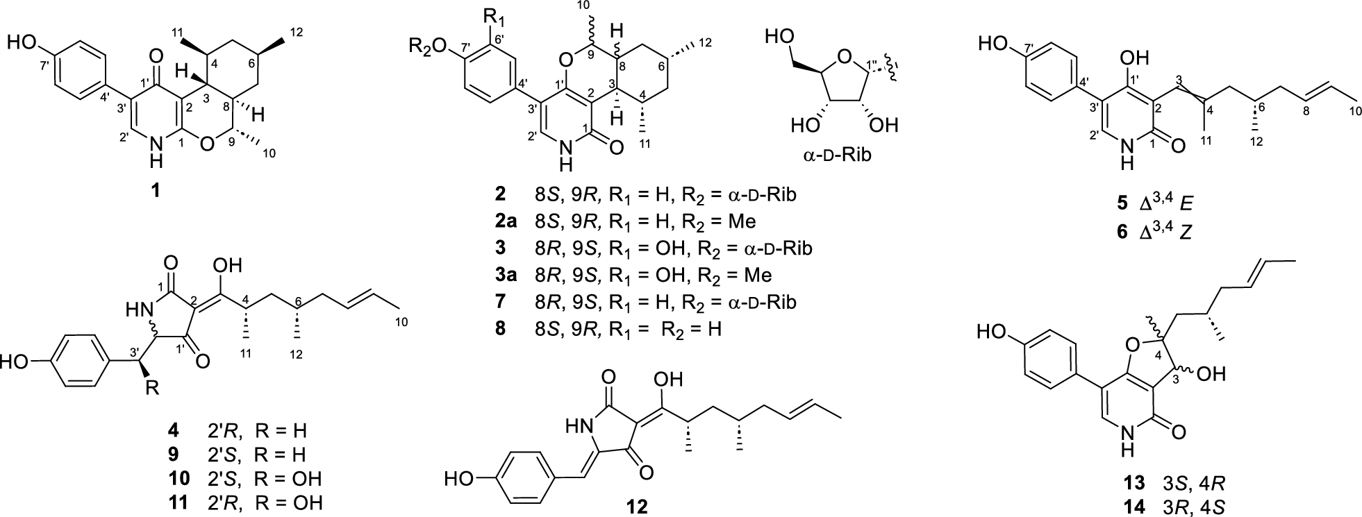
Chemical structures of compounds 1–14
2 Results and discussion
Compound 1 was isolated and characterized as colorless, needle-like crystals. Analysis of HRESIMS data allowed to assign the molecular formula as C21H25NO3. Its IR spectrum exhibited an intense absorption peak at 1693 cm−1 attributed to an unsaturated carbonyl group. The 1H NMR spectrum of 1 revealed an A2B2 pattern of a para-disubstituted aromatic ring (δH 7.33 and 6.70, d, each 2H) and an isolated olefinic methine at δH 7.19 (s) (Table 1). Two exchangeable protons signals, observed in DMSO-d6, were assigned to one phenolic hydroxy proton (δH 9.35) and one amine proton (δH 11.36). The 13C NMR and HSQC spectra indicated one carbonyl at δC 178.1, additional five quaternary sp2 carbons, five sp2 methines, two sp3 methylenes, five aliphatic methines, and three methyls. These data indicated that 1 was an analogue of the pyridone alkaloid tolypyridone A [18].
1H (600 MHz) and 13C NMR (150 MHz) Data for Compounds 1−3a
The COSY correlations revealed three spin systems (Fig. 2A), including one aromatic ring; one tetra-substituted cyclohexane ring involving the aliphatic protons from C-3 to C-9 with three methyl substituents at C-4, C-6, and C-9; and one enamine fragment containing H-2′ and NH. In the HMBC experiment, H-3 showed correlations with C-1 (δC 155.2), C-2 (δC 107.3), C-8, and C-9 while H-9 was correlated to C-1. These data revealed the presence of a dihydropyran ring fused with the cyclohexane ring to form an isochromene moiety. Further, the dihydropyran ring was deduced to be fused with a 4-pyridone ring according to the cross-peaks of H-3/C-1′ (δC 178.1) and H-2′/C-1′, C-3′, and C-1. A 7′-hydroxybenzene moiety was assigned at C-3′ of the pyridone moiety based on the cross-peaks of OH with C-6′, C-7′, and C-8′ as well as H2-5′, 9′ with C-3′, completing the establishment of the whole structure. The relative configuration was elucidated with help of the coupling constants of relevant protons and ROESY experiment (Fig. 3). The large value for 3JH-3,4 and 3JH-3,8 (10.2 Hz) indicated their trans 1,3-diaxial relationships. In addition, H-3 showed ROESY cross-peaks with H-9 and H3-11, revealing that they have the same orientation on the isochromene skeleton. On the contrary, the protons H-4, H-6, and H-8 were assigned on the opposite side of the cyclohexane moiety according to cross-peaks of H-8/H-4 and H-6.
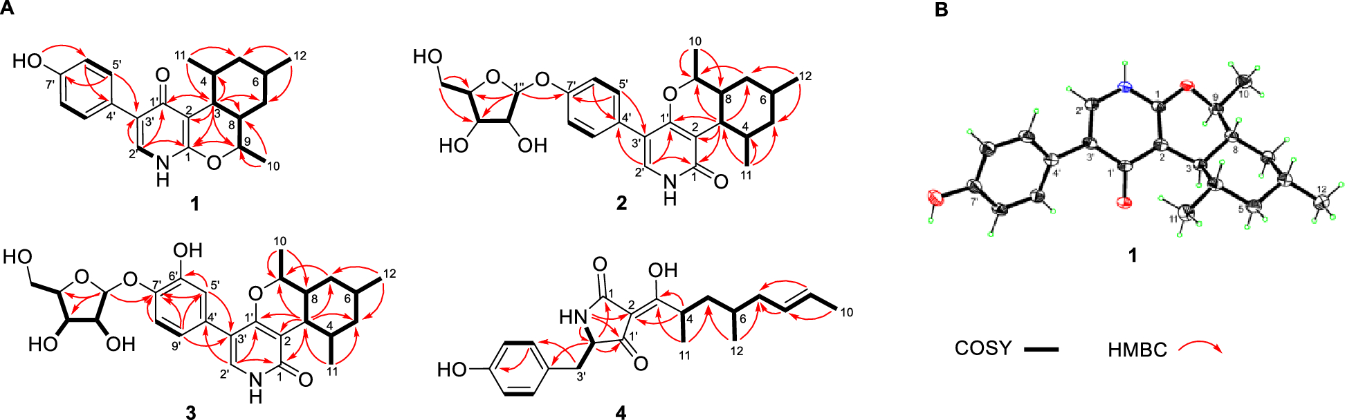
A The key 2D correlations of compounds 1−4. B X-ray crystallographic structure of 1
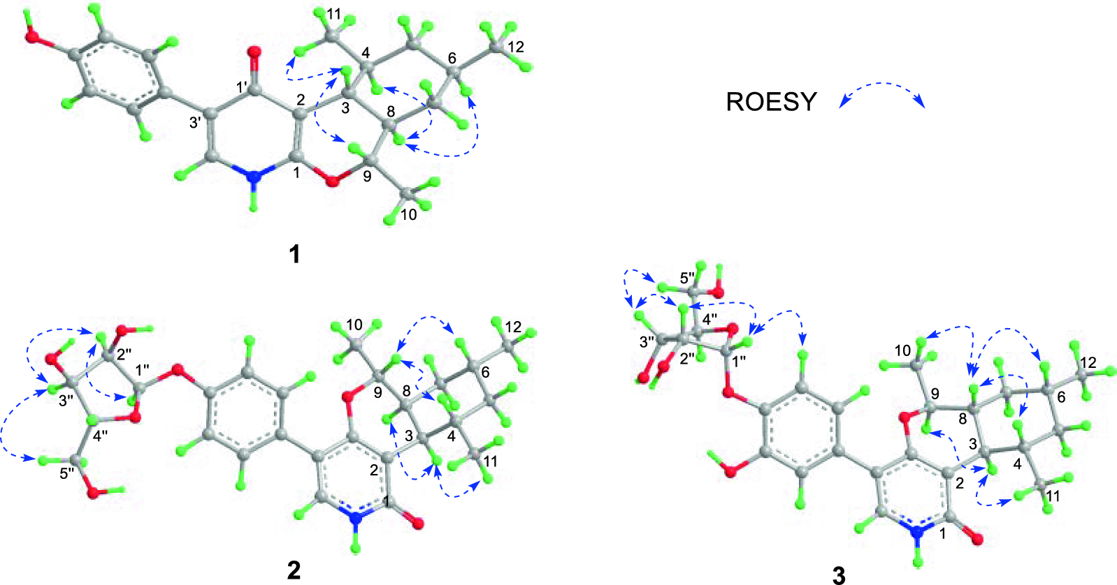
The ROESY correlations (blue dashed line) of 1−3
Very recently, Shim group reported the identification of tolypyridinol A [19], wherein the core structure was determined as 4-hydroxypyridine. Compared to those of tolypyridinol A, the NMR data of 1 recorded in the same solvent CD3OD (Table S2) showed significant differences for H-2′, C-1′, and C-2′, indicative of a distinct core structure for 1. Furthermore, the COSY cross-peaks between NH (δH 11.36) and H-2′ (δH 7.19) recorded in DMSO-d6 for 1 unequivocally precluded the possibility of a pyridine ring in 1. The structure was finally consolidated by X-ray crystallographic analysis (Fig. 2B), which indicated the absolute configuration as 3R, 4S, 6R, 8R, 9S by a small value of the refined Flack parameter − 0.06 (9). This was further verified by the ECD calculation (Fig. 4) [20]. Thus, 1 was elucidated as a novel 4-pyridone alkaloid and named tolypyrone A.
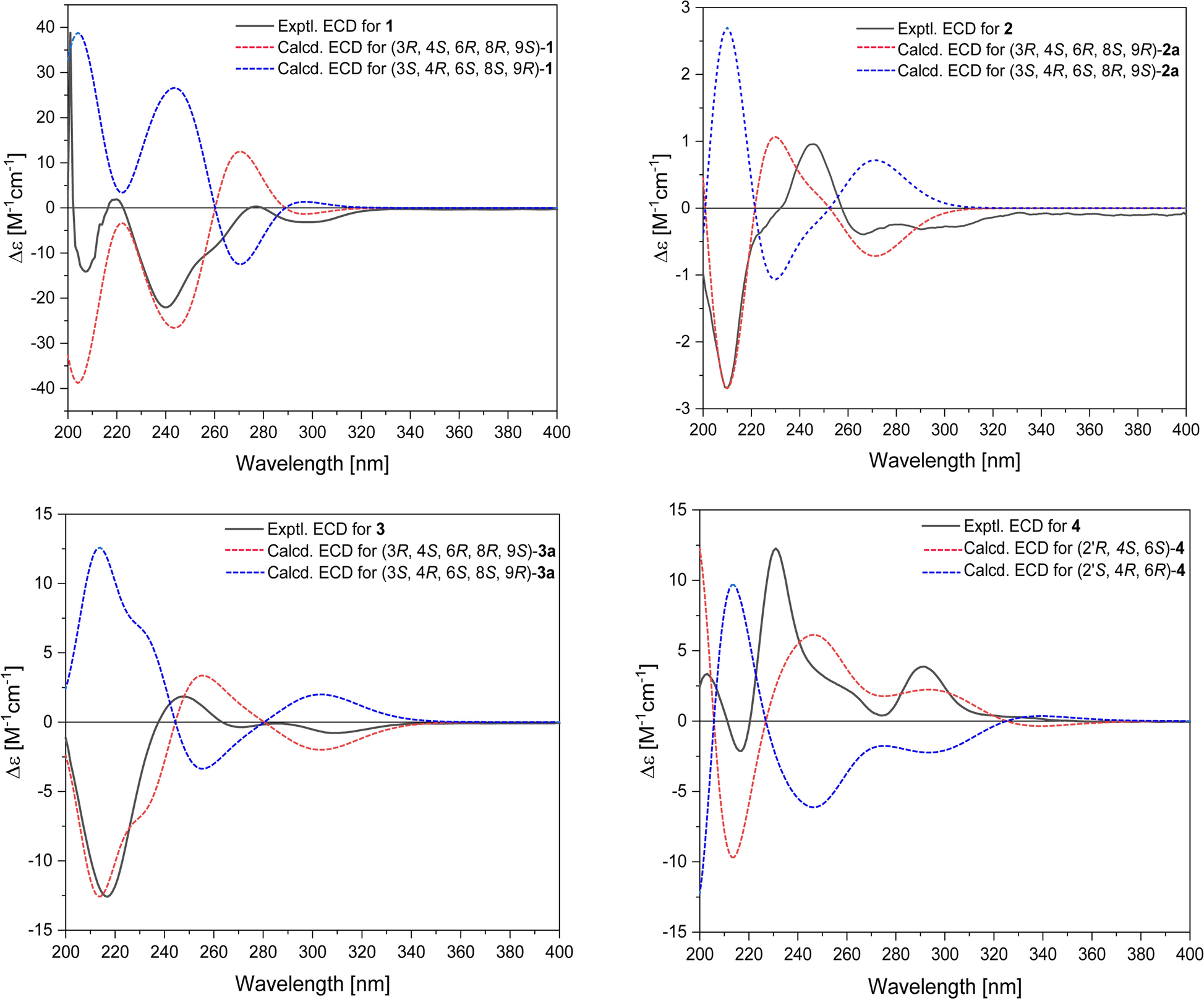
Comparison of the experimental and calculated ECD spectra for compounds 1−4
Compound 2 was obtained as a white noncrystalline powder and analyzed for C26H33NO7 by HRESIMS and NMR data. The 1H and 13C NMR spectra of 2 displayed signals closely similar to those of the co-isolated trichodin B (7) [21], including a ribofuranosyl moiety, a para-disubstituted aromatic ring, and a substituted cyclohexane ring with three methyls. However, the resonance for H-3 appeared as a double doublet at δH 2.60 (J = 10.8, 4.2 Hz in CD3OD) in 2 while it showed a triplet at δH 2.26 (J = 10.1 Hz) in 7. A combination of 2D NMR data led to assignment of the identical planar structure of 2 with that of 7. The large coupling JH-8, 9 (12.0 Hz) and JH-3,4 (10.8 Hz) revealed their 1,3-diaxial positions whereas the small value of JH-3,8 (4.2 Hz) suggested a cis fusion between the dihydropyran and cyclohexane rings. ROESY correlations of H-3/H-8 and H3-11 revealed that they were cis oriented on the cyclohexane moiety. On the contrary, H-9 exhibited ROESY cross-peaks with H-4 and H-6, suggesting that they positioned on the other side of the ring skeleton. Therefore, the relative configuration of the aglycone moiety in 2 was determined as 3R*, 4S*, 6R*, 8S*, 9R*, consistent with that of tolypyridone L [22] (8, also named as tolypyridone I by Jung et al. [23]). The resonance for C-1′′ (δC 102.4 in CD3OD; δC 100.4 in DMSO-d6) and the relatively large value for JH-1′′,2′′ (4.2 Hz) were suggestive of an α- anomeric configuration for the ribofuranosyl [24–26], which was consistent with the cross-peaks between H-1′′/H-2′′, H-2′′/H-3′′, and H-3′′/H-5′′ observed in the ROESY experiment. Of note, the ribofuranosyl in trichodin B (7) was previously assigned to be of β- anomeric configuration [21]. However, the typical values of JH-1′′,2′′ (4.2 Hz) and δC-1′′ (102.4 ppm in CD3OD) reported for 7 unambiguously indicated that the ribofuranosyl in trichodin B (7) should be of α-configuration according to those of the synthetic O-aryl and O-alky ribofuranosides (α-: JH-1′′,2′′ 4.0 Hz, δC-1′′ 102 ± 1.0; β-: JH-1′′,2′′ 0 Hz, δC-1′′ 107 ± 1.0) [24, 25].
Compound 2 displayed an ECD curve similar to tolypyridone L [22, 23], indicating that the aglycone of 2 had the same absolute configuration as tolypyridone L. This deduction was further verified by ECD calculation, wherein the calculated spectrum for the truncated model compound 3R, 4S, 6R, 8S, 9R-2a matched well with the experimental for 2, leading to assignment of the absolute stereochemistry for 2. The ribosyl residue was assigned as D-configuration by using the naphthimidazole derivatization method followed by chiral LC–MS analysis (Figure S3) [27]. Thus, 2 was elucidated as the 7′-O-α-D-ribofuranosyl-tolypyridone L and given the trivial name tolypyroside A.
Compound 3 possessed the molecular formula C26H33NO8 from HRESIMS, containing one oxygen atom more than 2. In contrast to 2, the NMR data revealed an ABX system of a trisubstituted aromatic ring and an exchangeable proton at δH 8.61 corresponding to a phenol group in 3. The heteronuclear correlations from H-5′ to C-3′, C-6′ (δC 146.6), and C-7′ (δC 143.3), and H-9′ to C-3′, C-5′, and C-7′ revealed the presence of a 6′,7′-dioxygenated aromatic ring in 3 in place of the 7′-oxygenated aromatic ring in 2. Specifically, C-7′ was deduced to be attached with an O-ribofuranosyl residue by the cross-peaks from H-1′′ to C-7′ whereas the chemical shift (δC 146.6) of C-6′ suggested that it was substituted by a hydroxy group. The large coupling constant of 3JH-3/8 and 3JH-3/4 (both 10.2 Hz) revealed their trans relationships. Analysis of ROESY data revealed that the protons H-4, H-6, and H-8 and the methyl group at C-9 positioned on the same orientation of the ring skeleton whereas H-3, H-9, and the methyl group at C-4 were on the opposite orientation. The ribofuranosyl moiety in 3 was assigned as α-D-configuration according to the resonances for H-1′′ and C-1′′, ROESY data, and chiral HPLC analysis of the hydrolysate. The aglycone was determined to have the 3R, 4S, 6R, 8R, 9S configuration by ECD calculation (Fig. 4).
Compound 4 was analyzed for the molecular formula C21H27NO4 by HRESIMS and NMR data. The NMR spectra of 4 measured in CDCl3 (Tables 2 and S4) were almost identical with those of the co-isolated tolypoalbin (9) [28], showing the resonances characteristic for a tetramic acid (δC 175.5, 100.6, 193.7, 194.1). However, the optical rotation ([α]D25 + 156.2 in MeOH) and the ECD pattern (Figure S9) of 4 were totally opposite to those of 9 ([α]D25 − 180.8 in MeOH). These data indicated that 4 was a stereoisomer of 9. Of note, the NMR spectra of 4 (Figures S42-43) recorded in CDCl3, like those of 3-acyltetramic acids, showed two set of resonances corresponding to two equilibrating Δ2,3 geometric isomers (4:1) [27, 29]. The resonances of C-1 (δC 175.5) and C-3 (δC 193.7) suggested the major isomer with a Δ2,3 Z geometry. The large value of 3JH-8,9 (15.0 Hz) suggested an E configuration for Δ8,9. To elucidate the absolute configuration of the pyrrolidine-2,4-dione ring, 4 was firstly subjected to alcoholysis with 1 M HCl/EtOH and then alkaline hydrolysis with 0.5 M KOH to avoid the epimerization of C-2′ [28]. The resultant hydrolysate was then applied to Marfey's analysis [30]. Comparative LC–MS analysis of the derivatives of 4 with those of L-tyrosine (Tyr) and D-Tyr standards unambiguously determined the R configuration for C-2′ in 4 (Figure S2). Since previous studies revealed that the tetramic acids and 4-hydroxy pyridones share a common PKS-NRPS origin [31–33], compound 4 was deduced to possess the same configuration 4S, 6S as in 9. This was further confirmed by ECD calculation for 4. Therefore, 4 was characterized as a 2′-epimer of 9 and named 2′-epitolypoalbin.
1H (600 MHz) and 13C NMR (150 MHz) Data for Compounds 4−6a
Compounds 5 and 6 were determined as a pair of geometric isomers with two double bonds Δ3,4 and Δ8,9 in the side chain. The geometry of the double bond Δ3,4 was determined as E in 5 and Z in 6 by ROESY data (Figure S6). The E geometry of Δ8,9 was indicated by comparing resonances of the allylic carbons C-7 (δC 39.5 and 39.2) and C-10 (δC 17.9 and 17.9) with those reported for the 2-pyridone and tetramic acid derivatives [29, 33]. The S configuration was assigned for C-6, like those in tolypyridone C and 1−3, according to the proposed biosynthesis of 4-hydroxy-2-pyridones [31–33].
During the preparation of this manuscript, Shim group reported two compounds tolypyridones K and L [19], whose structures were assigned to be identical with those of 5 and 6, respectively. However, NMR data for several 1H and 13C atoms reported for tolypyridones K and L were inconsistent with those of 5 and 6 (Table S6). Elaborative examination of NMR spectra supplemented in the Shim's paper indicated that these data for tolypyridones K and L had been incorrectly assigned. Of note, the names tolypyridones K and L had been designated for other two compounds discovered earlier by Wang group [22]. In addition, a search of 2-pyridones named beginning with the term "tolypyridone" isolated from the genus Tolypocladium reported in the literatures showed that each of tolypyridones I, J [22, 23] K, and L [19, 22] represented two compounds with different structures whereas tolypyridones I [23] and L [22] had the identical structures (Table S1 and Figure S1). To avoid the homonymous issues, compounds 5 and 6 were re-named as tolypyridones K2 and L2 in this study, respectively, wherein the suffix number 2 indicated a compound discovered later.
Compounds 7−14 were identified as trichodin B (7) [21], tolypyridone L (8) [22], tolypoalbin (9) [28], F-14329 (10) [29], chaunolidines A (11) and C (12) [29], tolypyridones E (13) and G (14) [33], respectively.
In in vitro antimicrobial assay for the obtained compounds, 1 showed inhibitory activities against Bacillus subtilis, Staphylococcus epidermidis, S. aureus, and Candida albicans with a minimum inhibitory concentration (MIC) at 16 μg/mL (Table 3). In contrast, the 2-pyridones 2, 3, 7, and 8 were inactive (MIC > 128 μg/mL), indicating that the fusing pattern of the isochromene and pyridone rings are crucial to antimicrobial effect. Compound 6 exhibited weak activities towards C. albicans and other Gram-positive bacteria (MIC: 32−64 μg/mL). Interestingly, the Δ3,4 geometric isomer 5 was completely inactive (MIC > 128 μg/mL). All the tetramic acid analogues 4 and 9−12 showed modest antimicrobial activities against the tested microorganisms except for Gram negative bacteria. Among the isolated tetramic acids, compound 12 was the most potent with the MIC value down to 2 μg/mL, suggesting that the double bond Δ2′,3′ could increase the antimicrobial activity. All the compounds displayed no inhibitory activities toward P. aeruginosa and E. coli. In the cytotoxic assay, only compound 6 exhibited weak inhibitory effects against the proliferation of HCT116 and H460 cell lines with IC50 values of 11.8 and 14.9 μM, respectively.
Antimicrobial Activities of Compounds 1−14 (MIC, μg/mL)
Although hundreds of 2-pyridone natural products have been characterized from fungi [7, 9], to the best of our knowledge, 4-pyridone alkaloids have never been discovered in nature. The biosynthesis of fungus-derived 2-pyridone alkaloids has been well investigated [31–33]. The pyridone core was proved to be generated from the acyl tetramic acid precursor by oxidative ring expansion catalyzed by a P450 oxidase [31]. Compound 1 might be derived from tolypyridone C via the reduction and subsequent dehydration of the 3-keto group and the following intramolecular hetero Diels–Alder cyclization (Figure S5) [33]. Although the versatile pericyclase LepI was discovered to catalyze the hetero D-A reaction in the biosynthesis of leporin C [34], a fungal 2-pyridone congener, we did not find its homologous enzyme in the genome of strain CPCC 401485 by genomic analysis, suggesting that the formation of 1 might be achieved by a nonenzymatic process [33].
3 Experimental section
3.1 General experimental procedures
The instrumentations were described in the Supplementary Information.
3.2 Biological material
The fungus CPCC401485 was isolated from an unidentified lichen specimen collected at Fildes Peninsula (62° 12′ 04" S, 58° 57′ 44" W), Antarctica. Its ITS gene sequence (577 nucleotides, GenBank no. PX482356) showed a similarity of 96.62% with Tolypocladium ovalisporum CBS 700.92 (GenBank no. NR_155019.1). In combination with morphological features, the strain was identified as belonging to the genus Tolypocladium.
3.3 Fermentation and isolation
The fungus was grown on PDA Petri dishes at 18 ℃ for five days and then transferred into Erlenmeyer flasks (500 mL) filled with 100 mL of the liquid medium (potato starch 3.0 g/L, glucose 20 g/L). After cultivation at 18 ℃ with shaking at 200 rpm for 3 days, the primary cultures were transferred into 45 flasks of 500 mL capacity each containing the production medium composed of 0.3 g of peptone, 100 g of rice, and 100 mL of water. Still cultures were maintained at 18 ℃ for 30 days. Extraction of the cultures with EtOAc/MeOH (9:1, v/v, 3 × 13.5 L) furnished a crude extract. The crude extract (93 g) was fractioned by C18 RP-MPLC (59 × 7.2 cm, 1500 g), successively eluting with 10%, 30%, 50%, 70%, 90% MeOH in water and 100% MeOH, and separated into nine fractions (F1-F9). Fraction F6 (2.9 g, eluted from MPLC by 70% MeOH) was submitted to gel chromatography on a Sephadex LH-20 column, using CH2Cl2/MeOH (1: 1, v/v) as the mobile phase, to obtain eight mixtures (F6-1-F6-8). Purification of the mixture F6-5 (247 mg) by RP-HPLC on a Capcell PAK C18 column (MGII 5 μm, 10 × 250 mm) with 37% MeCN/H2O containing 0.1% formic acid (FA) led to isolation of 1 (3.4 mg) and 5 (1.2 mg). The other fraction F5 (5.1 g) eluted by 70% MeOH was re-chromatographed by silica gel with a gradient mixture of CH2Cl2/MeOH from 100: 1 to 0: 1 to give five mixtures (F5-1-F5-5). The mixtures F5-2 (544 mg) and F5-4 (440 mg) were decolored by Sephadex LH-20 chromatography (CH2Cl2/MeOH 1: 1, v/v) and further purified on the same RP-HPLC column mentioned above. This led to isolation of 2 (2.7 mg) and 3 (5.1 mg) from F5-4 eluting by 33% MeCN/H2O and 6 (6.0 mg) from F5-2 by 35% MeCN/H2O. Fraction F8 (7.1 g, 90% MeOH-eluting) was firstly decolored by Sephadex LH-20 chromatography (CH2Cl2/MeOH 1: 1) and then fractioned on a silica gel column with a gradient mixture of CH2Cl2/MeOH (100: 1 ~ 0: 1), resulting in the separation of 13 mixtures (F8-5–1-F8-5–13). Finally, compound 4 (12.9 mg) was purified from the mixture F8-5–9 (362 mg) by semi-preparative RP-HPLC (Cosmosil MS-Ⅱ C18, 10 × 250 mm) with 47% MeCN/H2O containing 0.1% FA.
Tolypyrone A (1): colorless needle crystal; mp 305.8−306.5 ℃; [α]D25 − 254.3 (c 0.28, MeOH); UV (MeOH) λmax (log ε) 241 (4.62), 279 (4.22) nm; ECD (c 2.95 × 10⁻⁴ M, MeOH) λmax (Δε) 207 (−14.11), 240 (− 22.05), 298 (− 3.16) nm; IR νmax 3280, 2952, 2926, 1693, 1614, 1454, 1377, 1210, and 1140 cm−1; 1H NMR (DMSO-d6 and CD3OD, 600 MHz), Tables 1 and S2 in the Supplementary Information; 13C NMR (DMSO-d6 and CD3OD, 150 MHz), Tables 1 and S2; HRESIMS m/z 340.1925 [M + H]+ (calcd 340.1913 for C21H26NO3).
Tolypyroside A (2): white, amorphous powder; [α]D25 + 10.0 (c 0.12, MeOH); UV (MeOH) λmax (log ε) 210 (4.03), 247 (3.86) nm; ECD (c 1.06 × 10⁻³ M, MeOH) λmax (Δε) 210 (− 2.70), 246 (+ 0.96), 266 (− 0.39) nm; IR νmax 3299, 2946, 2925, 1641, 1511, 1437, 1237, and 1046 cm−1; 1H NMR (CD3OD and DMSO-d6, 600 MHz), Tables 1 and S3; 13C NMR (CD3OD and DMSO-d6, 150 MHz), Tables 1 and S3; HRESIMS m/z 472.2319 [M + H]+ (calcd 472.2335 for C26H34NO7).
Tolypyroside B (3): white, amorphous powder; [α]D25 − 19.2 (c 0.25, MeOH); UV (MeOH) λmax (log ε) 214 (4.29), 250 (3.98), 287 (3.75) nm; ECD (c 1.03 × 10⁻³ M, MeOH) λmax (Δε) 217 (− 12.59), 248 (+ 1.85), 308 (− 0.78) nm; IR νmax 3272, 2949, 2923, 1641, 1616, 1511, 1440, 1237, and 1048 cm−1; 1H NMR (DMSO-d6, 600 MHz), Table 1; 13C NMR (DMSO-d6, 150 MHz), Table 1; HRESIMS m/z 488.2291 [M + H]+ (calcd 488.2284 for C26H34NO8).
2′-Epitolypoalbin (4): white, amorphous powder; [α]D25 + 156.2 (c 0.85, MeOH); UV (MeOH) λmax (log ε) 223 (4.12), 280 (4.16) nm; ECD (c 5.61 × 10⁻⁴ M, MeOH) λmax (Δε) 217 (− 2.13), 231 (+ 12.27), 291 (+ 3.86) nm; IR νmax 3293, 2965, 2923, 1657, 1608, 1516, 1451, 1336, 1243 and 968 cm−1; 1H NMR (CDCl3 and DMSO-d6, 600 MHz), Tables 2, S4 and S5; 13C NMR (CDCl3 and DMSO-d6, 150 MHz), Tables 2, S4 and S5; HRESIMS m/z 358.2009 [M + H]+ (calcd 358.2018 for C21H28NO4).
Tolypyridone K2 (5): white, amorphous powder; [α]D20 − 6.0 (c 0.80, MeOH); UV (MeOH) λmax (log ε) 210 (4.01), 252 (3.76) nm; ECD (c 5.90 × 10⁻⁴ M, MeOH) λmax (Δε) 200 (− 0.56), 263 (− 0.21) nm; IR νmax 3242, 2954, 2926, 1611, 1516, 1444, 1378, 1212 cm−1; 1H NMR (DMSO-d6 and CD3OD, 600 MHz), Tables 2 and S6; 13C NMR (DMSO-d6 and CD3OD, 150 MHz), see Tables 2 and S6; HRESIMS m/z 340.1925 [M + H]+ (calcd 340.1913 for C21H26NO3).
Tolypyridone L2 (6): white, amorphous powder; [α]D20 + 9.5 (c 0.80, MeOH); UV (MeOH) λmax (log ε) 210 (4.72), 254 (4.49) nm; ECD (c 5.90 × 10⁻⁴ M, MeOH) λmax (Δε) 215 (− 0.94), 228 (+ 0.54), 266 (− 0.20) nm; IR νmax 3212, 2956, 1641, 1614, 1516, 1448, 1270, 1223, and 836 cm−1; 1H NMR (DMSO-d6 and CD3OD, 600 MHz), Tables 2 and S6; 13C NMR (DMSO-d6 and CD3OD, 150 MHz), see Tables 2 and S6; HRESIMS m/z 340.1917 [M + H]+ (calcd 340.1913 for C21H26NO3).
3.4 Crystallographic analysis of 1
The crystals of 1 were yielded by re-crystallization in MeOH. A single crystal (0.25 × 0.03 × 0.02) was selected for crystallographic analysis on a Rigaku XtaLAB Synergy diffractometer with Cu Kα radiation (λ = 1.54184 Å). Crystal data of 1: colorless needle crystal, C21H25NO3, M = 339.42, orthorhombic, space group P212121 (no. 19); a = 8.6674(14) Å, b = 10.2984(16) Å, c = 20.3542(3) Å, α = 90°, β = 90°, γ = 90°, V = 1816.82(5) Å3, Z = 4, Dx = 1.241 g/cm3, F (000) = 728.0, μ (Cu Kα) = 0.658 mm−1. 17, 480 measured reflections (8.688° ≤ 2θ ≤ 154.328°), 3726 independent unique reflections (Rint = 0.0507, Rsigma = 0.0325), R1 = 0.0361, wR2 = 0.0934, S = 1.055, Flack parameter = − 0.06(9). The crystallographic data is accessible through the Cambridge Crystallographic Data Centre (CCDC 2499034, https://www.ccdc.cam.ac.uk).
3.5 Advanced Marfey's analysis
A sample of 4 or 9 (1 mg) was dissolved in 1 M HCl/EtOH (500 μL) and refluxed at 110 ℃ for 15 min. After evaporation to dryness, the residue was re-dissolved in 0.5 M KOH (500 μL) and stirred at rt for 2 h. The resultant hydrolysate was applied to advanced Marfey's analysis using a protocol described previously [15]. The details were given in the Supplementary Information.
3.6 Determination of absolute configuration of the ribose moiety in 2 and 3.
Compound 2 or 3 (0.5 mg) was treated with 2 M HCl (1 mL) and heated at 110 ℃ for 4 h. The hydrolyzed products were concentrated in vacuo to dryness and then re-suspended in acetic acid (2 mL) and added 2,3-naphthalenediamine (2 mg) and molecular iodine (0.5 mg). The mixtures was agitated at rt for 6 h to afford the fluorescent naphthimidazole derivative [27]. The derivatives of L- and D-ribose authentic standards were similarly prepared. The derivatized materials were applied to LC–MS analysis on a chiral column (Capcell CD-ph 5 μm, 10 × 250 mm; 18% MeCN with 0.1% FA, 2 mL/min; column temperature: 35 ℃). The standard L- and D-ribose-naphthimidazole derivatives gave retention times at 10.91 and 17.13 min, respectively, while the derivatives of 2 and 3 were eluted at 17.19 and 17.13 min (Figure S3). Thus, the ribose moiety in 2 and 3 was determined to be of D-configuration.
3.7 ECD calculation
See the Supplementary Information.
3.8 Antimicrobial assay
Antimicrobial assay was performed against Staphylococcus aureus R6101, S. aureus ATCC 29213, S. epidermidis ATCC 12228, Bacillus subtilis ATCC 6633, Escherichia coli ATCC 25922, Pseudomonas aeruginosa ATCC 27853, Edwardsiella ictaluri ATCC 33202, E. tarda QDIO-2, Candida albicans ATCC 10231, Mucor racemosus CICC 3112, and Magnaporthe oryzae 131 according to the procedure described previously [35].
3.9 Cytotoxicity assay
Cytotoxicity assay was conducted in accordance with the protocol described previously [36].
Notes
Acknowledgements
The HPCC of Peking Union Medical College is acknowledged for computational facilities.
Author contributions
Shasha Li carried out the experiment of isolation, structural elucidation, ECD calculations and wrote the original draft; Ting Yu screened the antimicrobial activities; Yue Shang and Shuzhen Chen designed and performed the cytotoxicity assay. Jianju Feng, Tao Zhang and Liyan Yu conducted the sample collection and strain isolation. Maoluo Gan was responsible for conceptualization, supervision, writing and revising original draft. All authors read and approved the final manuscript.
Funding
This work was financially supported by CAMS Innovation Fund for Medical Sciences (CIFMS, 2021-I2M-1-055 and 2024-I2M-TS-016), National Natural Science Foundation of China (Grant no. 82273830), the National Key R&D Program of China (Grant no. 2024YFC2309604), and the National Microbial Resources Center (No. NMRC-2025-3).
Data availability
The data generated in this study were included in the article and its supplementary materials.
Declarations
Competing interests
The authors declare no competing financial interest.
References
-
1.Giordano D. Bioactive molecules from extreme environments. Mar Drugs. 2020;18: 640. CrossRef PubMed Google Scholar
-
2.De Pascale D, De Santi C, Fu J, Landfald B. The microbial diversity of Polar environments is a fertile ground for bioprospecting. Mar Genomics. 2012;8: 15-22. CrossRef PubMed Google Scholar
-
3.Medeiros W, Kralova S, Oliveira V, Ziemert N, Sehna L. Antarctic bacterial natural products: from genomic insights to drug discovery. Nat Prod Rep. 2025;42: 774-87. CrossRef PubMed Google Scholar
-
4.Liu JT, Lu XL, Liu XY, Gao Y, Hu B, Jia BH, et al. Bioactive natural products from the antarctic and arctic organisms. Mini Rev Med Chem. 2013;13: 617-26. CrossRef PubMed Google Scholar
-
5.Núñez-Pons L, Shilling A, Verde C, Baker BJ, Giordano D. Marine terpenoids from Polar latitudes and their potential applications in biotechnology. Mar Drugs. 2020;18: 401. CrossRef PubMed Google Scholar
-
6.Bratchkova A, Ivanova V. Bioactive metabolites produced by microorganisms collected in Antarctica and the Arctic. Biotechnol Biotechnol Equip. 2011;25: 1-7. CrossRef PubMed Google Scholar
-
7.Jessen HJ, Gademann K. 4-hydroxy-2-pyridone alkaloids: structures and synthetic approaches. Nat Prod Rep. 2010;27: 1168-85. CrossRef PubMed Google Scholar
-
8.Sangwan S, Yadav N, Kumar R, Chauhan S, Dhanda V, Walia P, et al. A score years' update in the synthesis and biological evaluation of medicinally important 2-pyridones. Eur J Med Chem. 2022;232: 114199. CrossRef PubMed Google Scholar
-
9.Chen XL, Zhao XY, Cong MJ, Hu YW, Zhang CS, Liu YH, et al. Fungal 4-hydroxy-2-pyridone alkaloids: structures, synthesis, biosynthesis, and bioactivities (2010-2024). Eur J Med Chem. 2025;297: 117953. CrossRef PubMed Google Scholar
-
10.TePaske MR, Gloer JB, Wicklow DT, Dowd PF. Leporin A: an antiinsectan N-alkoxypyridone from the sclerotia of Aspergillus leporis. Tetrahedron Lett. 1991;32: 5687-90. CrossRef PubMed Google Scholar
-
11.Hayakawa S, Minato H, Katagiri K. The ilicicolins, antibiotics from Cylindrocladium ilicicola. J Antibiot. 1971;24: 653-4. CrossRef PubMed Google Scholar
-
12.Isaka M, Chinthanom P, Supothina S, Tobwor P, Hywel-Jones NL. Pyridone and tetramic acid alkaloids from the spider pathogenic fungus Torrubiella sp. BCC 2165. J Nat Prod. 2010;73: 2057-60. CrossRef PubMed Google Scholar
-
13.Isaka M, Tanticharoen M, Kongsaeree P, Thebtaranonth Y. Structures of cordypyridones A-D, antimalarial N-hydroxy- and N-methoxy-2-pyridones from the insect pathogenic fungus Cordyceps nipponica. J Org Chem. 2001;66: 4803-8. CrossRef PubMed Google Scholar
-
14.Hirano N, Kohno J, Tsunoda S, Nishio M, Kishi N, Okuda T, et al. TMC-69, a new antitumor antibiotic with Cdc25A inhibitory activity, produced by Chrysosporium sp. TC1068. Taxonomy, fermentation and biological activities. J Antibiot. 2001;54: 421-7. CrossRef PubMed Google Scholar
-
15.Li SS, Li YN, Yu T, Wei XW, Zhang YY, Chen B, et al. Discovery of potent antiosteoporotic cyclic depsipeptides with an unusual nitrile hydroxy acid from Microascus croci. Bioorg Chem. 2025;155: 108133. CrossRef PubMed Google Scholar
-
16.Hao XM, Li SS, Li JR, Wang GY, Li J, Peng ZG, et al. Acremosides A-G, sugar alcohol-conjugated acyclic sesquiterpenes from a sponge-derived Acremonium species. J Nat Prod. 2024;87: 1059-66. CrossRef PubMed Google Scholar
-
17.Li Q, Li SS, Li SW, Hao XM, Wang AQ, Si SY, et al. Antimicrobial and anti-inflammatory cyclic tetrapeptides from the co-cultures of two marine-derived fungi. J Nat Prod. 2024;87: 365-70. CrossRef PubMed Google Scholar
-
18.Li XB, Li L, Zhu RX, Li W, Chang WQ, Zhang LL, et al. Tetramic acids and pyridone alkaloids from the endolichenic fungus Tolypocladium cylindrosporum. J Nat Prod. 2015;78: 2155-60. CrossRef PubMed Google Scholar
-
19.Choi JW, Kwon C, Lee JW, Hur JS, Shin MK, Shim SH. Anti-parkinsonian 4-hydroxy-2-pyridones from an endolichenic fungus, Tolypocladium sp. (strain CNC14). J Ind Microbiol Biotechnol. 2025;52: kuaf027. CrossRef PubMed Google Scholar
-
20.Flack HD. On enantiomorph-polarity estimation. Acta Crystallogr A. 1983;39: 876-81. CrossRef PubMed Google Scholar
-
21.Wu B, Oesker V, Wiese J, Schmaljohann R, Imhoff JF. Two new antibiotic pyridones produced by a marine fungus, Trichoderma sp. strain MF106. Mar Drugs. 2014;12: 1208-19. CrossRef PubMed Google Scholar
-
22.Wu XQ, Li J, Zhou X, Wang J, Tan YF, Mo JS, et al. Liver-cell protective pyridones from the fungi Tolypocladium album dws120. Phytochemistry. 2023;212: 113730. CrossRef PubMed Google Scholar
-
23.Jung Y, Kwon C, Kim T, Lee JW, Shin MK, Shim SH. Tetramic acid-motif natural products from a marine fungus Tolypocladium cylindrosporum FB06 and their anti-Parkinson activities. Mar Life Sci Technol. 2024;6: 84-92. CrossRef PubMed Google Scholar
-
24.Sharma RK, Singh S, Tiwari R, Mandal D, Olsen CE, Parmar VS, et al. O-Aryl α,β-D-ribofuranosides: synthesis & highly efficient biocatalytic separation of anomers and evaluation of their Src kinase inhibitory activity. Bioorg Med Chem. 2012;20: 6821-30. CrossRef PubMed Google Scholar
-
25.Wu JT, Zheng CJ, Zhang B, Zhou XM, Zhou Q, Chen GY, et al. Two new secondary metabolites from a mangrove-derived fungus Cladosporium sp. JJM22. Nat Prod Res. 2019;33: 34-40. CrossRef PubMed Google Scholar
-
26.Angyal SJ, Pickles VA. Equilibria between pyranoses and furanoses. Ⅱ. Aldoses. Aust J Chem. 1972;25: 1695-710. CrossRef PubMed Google Scholar
-
27.Li J, Chen MH, Hao XM, Li SS, Li F, Yu LY, et al. Structural revision and absolute configuration of burnettramic acid A. Org Lett. 2020;22: 98-101. CrossRef PubMed Google Scholar
-
28.Fukuda T, Igarashi Y, Sudoh Y, Tsuchiya Y, Okuda T, Matsuura N, et al. Tolypoalbin, a new tetramic acid from Tolypocladium album TAMA 479. J Antibiot. 2015;68: 399-402. CrossRef PubMed Google Scholar
-
29.Shang Z, Li L, Espósito BP, Salim AA, Khalil ZG, Quezada M, et al. New PKS-NRPS tetramic acids and pyridinone from an Australian marine-derived fungus, Chaunopycnis sp. Org Biomol Chem. 2015;13: 7795-802. CrossRef PubMed Google Scholar
-
30.Fujii K, Ikai Y, Oka H, Suzuki M, Harada Ki. A nonempirical method using LC/MS for determination of the absolute configuration of constituent amino acids in a peptide: combination of Marfey's method with mass spectrometry and its practical application. Anal Chem. 1997;69: 5146-51. CrossRef PubMed Google Scholar
-
31.Halo LM, Heneghan MN, Yakasai AA, Song Z, Williams K, Bailey AM, et al. Late stage oxidations during the biosynthesis of the 2-pyridone tenellin in the entomopathogenic fungus Beauveria bassiana. J Am Chem Soc. 2008;130: 17988-96. CrossRef PubMed Google Scholar
-
32.Bergmann S, Schümann J, Scherlach K, Lange C, Brakhage AA, Hertweck C. Genomics-driven discovery of PKS-NRPS hybrid metabolites from Aspergillus nidulans. Nat Chem Biol. 2007;3: 213-7. CrossRef PubMed Google Scholar
-
33.Zhang WY, Zhong Y, Yu Y, Shi DF, Huang HY, Tang XL, et al. 4-Hydroxy pyridones from heterologous expression and cultivation of the native host. J Nat Prod. 2020;83: 3338-46. CrossRef PubMed Google Scholar
-
34.Cai Y, Hai Y, Ohashi M, Jamieson CS, Garcia-Borras M, Houk KN, et al. Structural basis for stereoselective dehydration and hydrogen-bonding catalysis by the SAM-dependent pericyclase LepI. Nat Chem. 2019;11: 812-20. CrossRef PubMed Google Scholar
-
35.Hao XM, Yu JQ, Wang YJ, Connolly JA, Liu YF, Zhang YQ, et al. Zelkovamycins B-E, cyclic octapeptides containing rare amino acid residues from an endophytic Kitasatospora sp. Org Lett. 2020;22: 9346-50. CrossRef PubMed Google Scholar
-
36.Hao XM, Li SS, Ni J, Wang GY, Li F, Li Q, et al. Acremopeptaibols A–F, 16-residue peptaibols from the sponge-derived Acremonium sp. IMB18-086 cultivated with heat-killed Pseudomonas aeruginosa. J Nat Prod. 2021;84: 2990-3000. CrossRef PubMed Google Scholar
Copyright information
© The Author(s) 2026
Open Access This article is licensed under a Creative Commons Attribution 4.0 International License, which permits use, sharing, adaptation, distribution and reproduction in any medium or format, as long as you give appropriate credit to the original author(s) and the source, provide a link to the Creative Commons licence, and indicate if changes were made. The images or other third party material in this article are included in the article’s Creative Commons licence, unless indicated otherwise in a credit line to the material. If material is not included in the article’s Creative Commons licence and your intended use is not permitted by statutory regulation or exceeds the permitted use, you will need to obtain permission directly from the copyright holder. To view a copy of this licence, visit http://creativecommons.org/licenses/by/4.0/.