
Calabanones A‒H, chromanone derivatives from the stem bark of Calophyllum calaba and their cytotoxic activities against cancer cells

Abstract
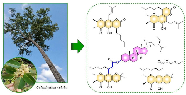
The genus Calophyllum (Calophyllaceae), distributed mainly in tropical regions, is rich in chromanone derivatives with diverse molecular structures that exhibit potential antimicrobial and anticancer effects. Phytochemical investigation of the stem bark of C. calaba collected in Thailand led to the discovery of eight previously undescribed chromanones, calabanones A–H (1‒8), and two known analogs, (‒)-isocalomembranone P (9) and (‒)-calomembranone P (10). The chemical structures of undescribed compounds were elucidated using spectroscopic analyses, particularly NMR and HRESIMS, while their absolute configurations were determined through ECD and NMR calculations combined with DP4+ probability analysis. Compounds 7 and 8 were identified as chromanone–steroid hybrids linked via an ester bond, representing the first report of such structures in plants. Cytotoxic evaluation of the isolated specialized metabolites revealed that compounds 6 and 9 displayed moderate activity against KB and HeLa S3 cancer cell lines, with IC50 values ranging from 12.71 to 25.50 μM, while compounds 3‒5 selectively inhibited the growth of KB cells, with IC50 values in the range of 18.77‒27.06 μM.Graphical Abstract
Keywords
Calophyllum calaba Calophyllaceae Chromanone Calabanone Cytotoxic1 Introduction
Chromanones are among the major specialized metabolites of Calophyllum plants (Calophyllaceae), alongside xanthones and coumarins. They are biosynthesized via Claisen condensation of isovaleryl-CoA with three malonyl-CoA units, yielding a 2,3-dimethylchromanone scaffold [1]. The incorporation of isoprene units and either cinnamic acid or short-chain fatty acids (C6 or C8) onto the chromanone skeleton generates diverse chemical architectures through esterification and cyclization, resulting in pyranochromanones and pyranocoumarins [2]. Several chromanone derivatives have demonstrated promising pharmacological activities, including (+)-calanolide A and (+)-inophyllum B, both known for their antiviral potential. The former, originally isolated from C. lanigerum, strongly inhibits HIV-1 reverse transcriptase and has advanced to phase Ⅱ clinical trials as an anti-HIV drug candidate, while the latter, the major constituent of C. inophyllum, is under preclinical evaluation, exhibiting a similar mechanism of action to calanolide A with inhibition at the nanomolar level [2, 3].
Calophyllum calaba L., locally known as "Thunghoon" in Thailand, grows in peat swamp and beach forests and is primarily distributed across Southeast Asia, Sri Lanka, and India [4]. Only limited phytochemical studies have been conducted on this species, including investigations of its stem bark, leaves, and roots from Sri Lanka, which revealed several bioactive metabolites such as chromanones, xanthones, biflavonoids, and triterpenoids [5–9]. In our previous work, cytotoxic prenylated xanthones were isolated from the roots of C. calaba collected in Thailand, with calaxanthone C exhibiting the strongest activity against five human cancer cell lines (KB, HeLa S3, HT-29, MCF-7, and HepG2), showing IC50 values below 5 μM [10]. Motivated by these results, we performed chemical screening using NMR analysis on extracts and fractions from other plant organs, which indicated that the stem bark extract contained characteristic proton and carbon signals of chromanone derivatives. Herein, we report the isolation, structure elucidation, and cytotoxic evaluation of chromanones obtained from the CH2Cl2 extract of C. calaba stem bark.
2 Results and discussion
Eight previously undercribed chromanones, calabanones A‒H (1–8), and two known analogs, (‒)-isocalomembranone P (9) and (‒)-calomembranone P (10) [1], were isolated from the CH2Cl2 extract of C. calaba stem bark through successive chromatographic separations, including silica gel, RP-C18, Sephadex LH-20, and radial chromatographic techniques. The structures of the previously undescribed specialized metabolites were elucidated by spectroscopic analyses (NMR and HRESIMS) and by comparison of their 1H and 13C NMR data with those of related compounds reported in the literature (Fig. 1).
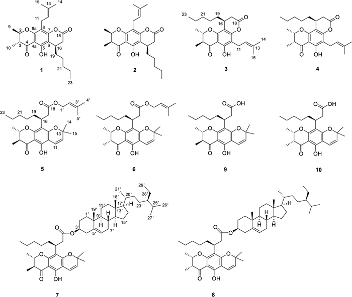
Chromanones 1 − 10 isolated from the stem bark of C. calaba
Calabanone A (1) was obtained as pale yellow gum. Its molecular formula, C24H32O5, was established from the sodium adduct ion at m/z 423.2159 [M + Na]+ (calcd for C24H32O5Na+, 423.2142) in the HRESIMS, indicating nine degrees of unsaturation. The UV absorption maxima at 215, 284, and 348 nm suggested the presence of a chromanone chromophore, consistent with those reported for Calophyllum-derived chromanones [1, 11]. The 1H NMR spectrum (Table 1) showed resonances for a hydrogen-bonded hydroxy proton at δH 12.26 (OH-5) and two methyl doublets at δH 1.53 (3H, d, J = 6.4 Hz, H-9) and 1.22 (3H, d, J = 6.8 Hz, H-10). The 1H–1H COSY spectrum revealed correlations of a contiguous spin system of H-10/H-3 (δH 2.59, dq, J = 12.4, 6.8 Hz)/H-2 (δH 4.19, dq, J = 12.4, 6.4 Hz)/H-9. The HMBC correlations of H-9 and H-10 to C-2 (δC 79.1) and C-3 (δC 46.2), as well as H-10 to the carbonyl C-4 (δC 200.3), confirmed the presence of a 2,3-dimethylchromanone scaffold (Fig. 2). An octanoic acid ester attached to the aromatic ring was deduced from the ester carbonyl C-18 (δC 167.6), together with proton signals of a methine at δH 3.33 (1H, q, J = 6.8 Hz, H-16), a terminal methyl at δH 0.86 (3H, t, J = 6.8 Hz, H-23), and five sets of methylenes at δH 2.83 (1H, d, J = 15.6 Hz, H-17a) and 2.64 (1H, dd, J = 15.6, 6.8 Hz, H-17b), 1.54 and 1.44 (each 1H, overlap, H-19), 1.41 and 1.28 (each 1H, overlap, H-20), 1.26 (2H, overlap, H-21), and 1.28 (2H, overlap, H-22). The COSY cross-peaks of H-17/H-16/H-19/H-20/H-21/H-22/H-23 supported this assignment. The HMBC correlations of H-16 to C-18 and C-20 (δC 26.5), H-17 to C-19 (δC 34.2), H-20 to C-21 (δC 31.8), and H-23 to C-21 and C-22 (δC 22.6) further confirmed the side chain. Additionally, correlations of OH-5 with C-4a (δC 104.2), C-5 (δC 157.6), and C-6 (δC 107.5), and of H-16 with C-5, C-6, and C-7 (δC 156.3), indicated cyclization of the side chain to C-6 and C-7, forming a pyran-2-one ring. The 1D NMR data also indicated a prenyl unit, as evidenced by resonances of a methine at δH 5.16 (1H, t, J = 7.6 Hz, H-12)/ δC 121.9 (C-12), a methylene at δH 3.28 (2H, d, J = 7.6 Hz, H-11)/ δC 21.7 (C-11), two methyl singlets at δH 1.66 (3H, s, H-14)/ δC 25.9 (C-14) and δH 1.77 (3H, s, H-15)/ δC 17.9 (C-15), and a sp2 quaternary carbon at δC 132.1 (C-13). The HMBC correlations of H-11 with C-7, C-8 (δC 108.9), and C-8a (δC 158.4) established the prenyl group at C-8.
1H (400 MHz) and 13C (100 MHz) NMR spectroscopic data of compounds 1‒4 in CDCl3
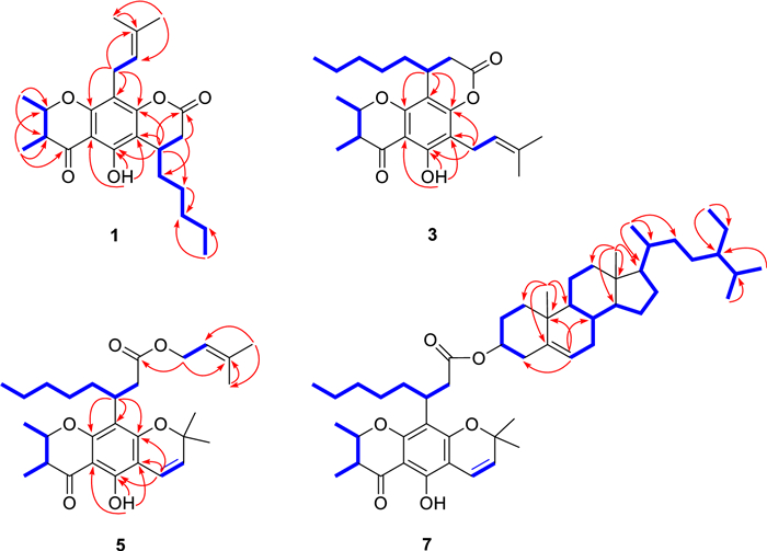
Key COSY and HMBC correlations of compounds 1, 3, 5, and 7
The large coupling constant between H-2 and H-3 (3J2,3 = 12.4 Hz) favored a trans-2,3-dimethyl configuration, with both protons was in an axial relationship (Table 1). The small 3J16,17 coupling (6.8 Hz) between H-16 and H-17 suggested that the n-amyl unit at C-16 was axially oriented in the half-chair conformation of the pyran-2-one ring. However, due to the spatial separation between H-16 and H-2/H-3, the relative configuration of C-16 could not be unambiguosly assigned. Thus, the NMR calculation coupled with DP4+ probability analysis were performed for two possible conformers (2R,3R,16R-1a and 2R,3R,16S-1b) using the GIAO method at the B3LYP/6-31++ G(d, p) level. DP4+ statistical analysis indicated isomer 1a (16R) as the predominant conformer, with probabilities of 99.91% (1H and 13C data) (Additional file 1: Table S1). The absolute configuration was confirmed by quantum ECD calculations for the two conformers 1a and 1b and their enantiomeric pairs 2S,3S,16S-1a* and 2S,3S,16R-1b*, with the predicted spectrum of 1a closely matching the experimental data (Fig. 3). Accordingly, the absolute configuration of compound 1 was assigned as 2R,3R,16R.
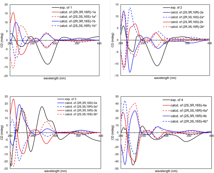
Experimental and calculated ECD spectra of 1‒4
Calabanone B (2) was assigned the molecular formula C24H32O5, based on the sodium adduct ion at m/z 423.2137 [M + Na]+ (calcd for C24H32O5Na+, 423.2142) in HRESIMS. Comparison of its 1H and 13C NMR data with those of 1 revealed that the structural differences were confined to the 2,3-dihydro-2,3-dimethyl-γ-pyran-4-one moiety. The small 3J value (3.2 Hz) between H-2 and H-3 preferred a cis-2,3-dimethyl configuration (Table 1). To determine the relative configuration at C-16, NMR calculations were performed for two possible diastereomers (2S,3R,16R-2a and 2S,3R,16S-2b). DP4+ statistical analysis of the calculated 1H and 13C NMR data supported 2b as the correct conformer, with an overall probability of 94.35% (Additional file 1: Table S3). To further corroborate the NMR/DP4+ results, quantum ECD calculations were carried out for 2a and 2b and their corresponding enantiomers (2R,3S,16S-2a* and 2R,3S,16R-2b*). The calculated ECD spectrum of model 2b* closely matched the experimental data (Fig. 3), thereby confirming the absolute configuration of compound 2 as 2R,3S,16R (Fig. 3).
Calabanone C (3) was obtained as a pale yellow gum. The (+)-HRESIMS spectrum displayed a protonated molecule [M + H]+ at m/z 401.2323 (calcd for C24H33O5+, 401.2323). The 1H and 13C NMR spectra of 3 closely resembled those of 1, except for variations in the aromatic carbon resoncances, suggesting a difference in side chain arrangement. In the HMBC spectrum, H-11 (δH 3.32, 2H, d, J = 7.2 Hz) correlated with C-5 (δC 159.9), C-6 (δC 110.4), and C-7 (δC 156.0), while H-16 (δH 3.27, 1H, q, J = 6.8 Hz) showed cross-peaks with C-7, C-8 (δC 105.8), and C-8a (δC 156.2). These correlations indicated that the prenyl unit was located at C-6 and the 2-pyranone moiety was fused at C-7 and C-8 of the chromamone skeleton (Fig. 2). The large coupling constant (3 J = 12.4 Hz) between H-2 and H-3 supported a trans-2,3-dimethyl configuration with the protons in an axial orientation. The relative configuration at C-16 was further resolved by NMR calculations, which identified conformer 2R,3R,16R-3b as the most possible candidate with a DP4+ probability of 99.46% in both 1H and 13C NMR data (Additional file 1: Table S5), compared with its diastereomer 2R,3R,16S-3a in DP4+ analysis. To confirm the absolte configuration, ECD calculations were performed for models 3a and 3b as well as their enantiomeric pairs (2S,3S,16R-3a* and 2S,3S,16S-3b*). The calculated ECD spectrum of the conformer 3b* matched the experimental data, thereby establishing the absolute configuration of 3 as 2S,3S,16S (Fig. 3).
The molecular formula of calabanone D (4), C24H32O5, was deduced from its protonated molecule at m/z 401.2319 [M + H]+ (calcd for C24H33O5+, 401.2323) in HRESIMS. Analysis of the 2D NMR data revealed key HMBC and COSY correlations similar to those of 3. However, the small coupling constant between H-2 and H-3 (3J = 3.2 Hz) indicated a cis-2,3-dimethyl configuration (Table 1). DP4+ statistical analysis supported conformer 2S,3R,16S-4a as the most probable structure, with an overall probability of 99.12% in all H and C data (Additional file 1: Table S7), compared with its diastereomer 2S,3R,16R-4b. The absolute configuration of 4 was further assessed by ECD calculations performed for 4a and 4b and their enantiomeric pairs 2R,3S,16R-4a* and 2R,3S,16S-4b*, respectively. The experimental ECD curve were similar with those calculated for 4a and 4b*, with 4a being slightly favored due to the presence of an additional minor positive CE at approximately 302 nm as well as similar CE amplitudes at ~ 227 and 206 nm that were also observed in the experimental spectrum. Taken together with the high DP4+ probability for 4a, these results established the absolute configuration of compound 4 as 2S,3R,16S (Fig. 3).
Calabanone E (5) was isolated as a pale yellow gum. Its molecular formula, C29H40O6, was determined from the sodium adduct ion at m/z 507.2739 [M + Na]+ (calcd for C29H40O6Na+, 507.2717) in the HRESIMS, indicating 10 degrees of unsaturation. The 1H and 13C NMR spectra (Table 2) showed resonances for two olefinic protons at δH 6.61 (1H, d, J = 10.0 Hz, H-11) and 5.46 (1H, d, J = 10.0 Hz, H-12), two gem-dimethyls at δH 1.44 (each 3H, s, H3-14 and H3-15), and an oxygenated sp3 carbon at δC 78.2 (C-13), supporting the presence of a pyran unit. HMBC correlations of H-11 to C-5 (δC 157.1), C-6 (δC 102.7), and C-7 (δC 160.3) placed the pyran ring at C-6 and C-7. The 1D NMR data closely resembled those of isocalomembranone P (9) from C. membranaceum, except for differences in the fatty acid side chain [1]. The more shielded ester carbonyl at δC 173.4 (C-18) in 5 suggested and ester bond formation. Additional resonances, including an olefinic proton at δH 5.20 (1H, t, J = 7.2 Hz, H-2′), diastereotopic oxymethylene protons at δH 4.49 and 4.43 (each 1H, dd, J = 12.4, 7.2 Hz, H2-1′), two methyl protons at δH 1.70 (3H, s, H3-4′) and 1.63 (3H, s, H3-5′), and a quaternary sp2 carbon at δC 138.5 (C-3′), indicated a prenyl unit linked to the ester carbonyl, as confirmed by the HMBC correlation of H2-1′ to C-18 (Fig. 2). The large coupling constant (J = 12.4 Hz) between H-2 (δH 4.12) and H-3 (δH 2.52) supported a trans-diaxial relationship, with both methyl groups H3-9 and H3-10 in equatorial orientations. The experimental ECD spectrum (Additional file 1: Fig. S49) and specific rotation ([α]D20= ‒46.2) of 5 was identical to that of (‒)-isocalomembranone P (9) [1], confirming the absolute configuration as 2S,3S,16S.
1H (400 MHz) and 13C (100 MHz) NMR spectroscopic data of compounds 5‒8 in CDCl3
Calabanone F (6) possessed the molecular formula C29H40O6, as indicated by the sodium adduct ion at m/z 507.2736 [M + Na]+ (calcd for C29H40O6Na+, 507.2717) in the (+)-HRESIMS. Its 1D NMR spectra differed from those of 5 mainly in the orientation of the methyl group at C-2. The small coupling constant (3J2,3 = 3.2 Hz) indicated a cis-2,3-dimethyl configuration (Table 2). The experimental ECD spectrum of 6 (Additional file 1: Fig. S49) closely matched that of (‒)-calomembranone P (10) [1], supported by their similar negative specific rotatations, thereby establishing the absolute configuration of 6 as 2S,3R,16S.
The molecular formula of calabanone G (7) was determined as C53H80O6 from the protonated molecule at m/z 813.6028 in (+)-HRESIMS (calcd for C53H81O6+, 813.6028). The 1H and 13C spectra indicated the same chromanone skeleton as in 5 but with a different ester moiety (Table 2). The absence of prenyl resonances, present in 5, was replaced by characteristic signals of β-sitosterol [12]. 2D NMR data confirmed the steroid unit, including its typical contiguous 1H‒1H spin system and HMBC correlations, such as olefinic proton H-6′ (δH 5.32, d, J = 6.0 Hz) to C-4′ (δC 38.3), C-8′ (δC 32.0), and C-10′ (δC 36.7) and six methyl resonances (H3-19′, H3-18′, H3-21′, H3-26′, H3-27′, and H3-29′) to the expected carbons of the steroid skeleton (Fig. 2). Although no direct HMBC correlation confirming the ester linkage between the 3′-OH of β-sitosterol and the chromanone carbonyl C-18, the downfield signals at δC 73.6 (C-3′) (ΔδC + 1.8 ppm) and the shielded carbonyl at δC 172.8 (C-18) (ΔδC ‒6.6 ppm), together with the HRESIMS data, supported a chromanone‒steroid hybrid linked via an ester bond [1, 12]. The ECD spectrum of 7 (Additional file 1: Fig. S49) closely matched that of (‒)-isocalomembranone P (9) [1], establishing the absolute configuration of 7 as 2S,3S,16S.
HRESIMS analysis of calabanone H (8) showed a sodium adduct ion at m/z 835.5810 [M + Na]+ (calcd for C53H80O6Na+, 835.5847), consistent with a chromanone‒steroid hybrid analogous to 7. 1D NMR data confirmed that 7 and 8 were diastereomers, differing only in the 2,3-dimethyl configuration. The small coupling constant between H-2 and H-3 (3 J = 3.2 Hz), supported a cis-relationship of the two protons (Table 2). Comparison of the experimental ECD spectrum of 8 (Additional file 1: Fig. S49) with that of (‒)-calomembranone P (10) [1] confirmed the absolute configuration of 8 as 2S,3R,16S.
Calophyllum chromanones are predominantly characterized by substitution with side chains such as C8-fatty acids, as observed in compounds 9 and 10, as well as cinnamic acid, as in calomembranones L–O isolated from C. membranaceum [1]. Intramolecular cyclization between the carboxylic acid group and a hydroxy group on the aromatic ring can further generate coumarin-like structures, as shown in compounds 1–4, which represent another major class of derivatives within this genus [2]. In addition to cyclization, esterification of the carboxylic acid moiety is also a frequent structural modification among Calophyllum metabolites [2]. Previously reported esterifications were largely limited to simple methyl chromanone esters, such as calomembranones E–G [2, 11]. The identification of chromanones bearing prenyl esters (5 and 6) and chromanone–steroid hybrids linked through an ester bond (7 and 8) therefore represents, to the best of our knowledge, the first report of such structural motifs in plants. In general, naturally occurring triterpenoid or stereoid hybrids connected to other metabolites via ester linkage are rare. Representative examples include myrtphlotritins, phloroglucinol–triterpenoid adducts isolated from Leptospermum scoparium and Xanthostemon chrysanthus (Myrtaceae) [13] and the triterpenoid–chromone hybrids cimitriteromones originally identified from the rhizomes of Actaea cimicifuga (Ranunculaceae) [14]. Ester functionalities in natural products can sometimes arise as artifacts, particularly when carboxylic acid–containing compounds undergo esterification in the presence of alcohols, such as the formation of methyl or ethyl esters from methanol or ethanol solvents during isolation [15]. As the crude plant extract in this study was entirely used during the isolation process and thus unavailable for direct verification, acid-catalyzed esterification reactions were carried out using β-sitosterol and (–)-calomembranone P (10) to assess whether compound 8 could be an artifact. These reaction mixtures were refluxed in methanol for 24 h using either acetic acid or H2SO4 as catalysts. TLC analysis revealed no formation of compound 8 under either condition, suggesting that the chromanone–steroid hybrids are unlikely to be artifacts of the isolation process (Additional file 1: Fig. S51).
Previous studies have shown that chromanones isolated from Calophyllum species exhibited potent cytotoxic activities against various cancer cell lines, including KB and HeLa cells. Notable examples included caloteysmannic acid, isocalolongic acid, and apetalic acid derivatives, which displayed IC50 values below 10 μM [16, 17]. Based on these reports, the cytotoxic activities of all isolated compounds in this work were evaluated using an MTT assay against KB and HeLa S3 cells, together with the HepG2 cancer cell line, and the results are summarized in Table 3. Compounds 6 and 9 exhibited moderate activity against KB and HeLa S3 cells, with IC50 values ranging from 12.71 to 25.50 μM. Compounds 3‒5 selectively inhibited the growth of KB cells, showing IC50 values between 18.77 and 27.06 μM. The remaining compounds displayed weak cytotoxic effects, whereas those containing a sitosterol moiety (7 and 8) were inactive, with IC50 values exceeding 100 μM. Notably, chromanones apetalic acid, isoapetalic acid, and their methyl esters isolated from C. blancoi exhibited stronger cytotoxic activities against KB and HeLa cells [17] despite their close structural similarity to compounds 5, 6, 9, and 10. The key structural difference lies in the length of the fatty acid side chain, with the former bearing C6-fatty acids, whereas compounds 5, 6, 9, and 10 contain C8-fatty acids. A similar trend was observed for the derivatives with cyclized fatty acid. Cyclization of the fatty acid moiety in apetalic acid and isoapetalic acid retained potent activity (IC50 < 10 μM) [17], in contrast to their analogs 3 and 4. These preliminary structure–activity relationship (SAR) observations suggest that shorter fatty acid side chains play a crucial role in enhancing cytotoxic activity, possibly due to increased hydrophilicity. This interpretation is further supported by the lack of cytotoxicity observed for compounds 7 and 8, which bear bulky and nonpolar steroidal moieties. Nevertheless, additional chromanones with related functional groups are required for cytotoxic screening to strengthen this preliminary SAR analysis, and further evaluations against a broader panel of cancer cell lines as well as noncancerous cells are necessary to assess both potency and selectivity.
Cytotoxic activitiesa of the isolated compounds after 72 h of treatment
3 Conclusions
In summary, eight previously undescribed chromanones, calabanones A–H (1–8), along with the known compounds (‒)-isocalomembranone P (9) and (‒)-calomembranone P (10), were isolated from the CH2Cl2 extract of the stem bark of C. calaba collected in Thailand. Notably, calabanones G and H represent the first naturally occurring chromanone–steroid hybrids, thereby expanding the chemical diversity and potential chemotaxonomic markers of Calophyllum species. In addition, calabanone F and (‒)-isocalomembranone P demonstrated moderate cytotoxicity against KB and HeLa S3 cell lines, with IC50 values below 30 μM, whereas the remaining compounds displayed weak or no activity against the tested cancer cell lines.
4 Experimental
4.1 General experimental procedures
Optical rotations were determined using a JASCO P-1010 polarimeter (JASCO Corporation, Easton, MD, USA). ECD spectra were obtained with a JASCO J-815 spectropolarimeter (JASCO Corporation). NMR spectra were measured on Bruker AVANCE 400 spectrometer (Bruker, Rheinstetten, Germany) using CDCl₃ as solvent (Merck, Darmstadt, Germany). HRMS data acquisition was performed on a Bruker MICROTOF model mass spectrometer (Bruker) and an Agilent 1260 series HPLC system coupled to a 6540 UHD accurate-mass QTOF mass spectrometer (Agilent Technologies, Waldbronn, Germany). Column chromatography was carried out using silica gel (70–230 mesh, Merck) and Sephadex LH-20 (25–100 μm, GE Healthcare Bio-Sciences AB, Uppsala, Sweden). Radial chromatography was performed on a Chromatotron model 7924 T (Harrison Research, Palo Alto, CA, USA) equipped with silica gel 60 GF2₅₄ containing gypsum (≤ 55 μm, Merck). TLC analysis was conducted on silica gel 60 F2₅₄ plates (0.2 mm, Merck). Spots were initially observed under UV light at 254 and 366 nm, followed by staining with p-anisaldehyde reagent and subsequent heating to enhance visualization.
4.2 Plant material
The stem bark of C. calaba was collected in April 2023 from Buachet District, Surin Province, Thailand (14°33′24″ N, 104°00′56″ E). The species was identified by Dr. Suttira Sedlak, and a voucher specimen (Khumkratok no. 1–13) was deposited at the Walai Rukhavej Botanical Research Institute, Mahasarakham University.
4.3 Extraction and isolation
The air-dried stem bark of C. calaba (5.0 kg) was extracted by maceration with CH2Cl2 (3 × 10 L) at room temperature for three consecutive 3-day periods. Removal of the solvent yielded a crude extract (125.0 g), which was suspended in MeOH and partitioned with hexanes to obtain a hexanes-soluble fraction (64.0 g). This fraction was subjected to silica gel column chromatography using a gradient of hexanes–CH2Cl2 (100:0 to 0:100), affording eight fractions (A–H). Fraction A (8.6 g) was further chromatographed on silica gel with hexanes–CH2Cl2 (100:0 to 0:100) to yield subfractions A1–A7. Subfraction A2 (511.2 mg) was separated on a Sephadex LH-20 column with CH2Cl2–MeOH (50:50) to provide A2.1–A2.3. Purification of subfraction A2.3 (103.0 mg) by Chromatotron with hexanes–acetone (95:5) afforded compounds 3 (9.5 mg) and 4 (10.2 mg). Subfraction A3 (3.8 g) was fractionated on Sephadex LH-20 with CH2Cl2–MeOH (50:50) to give A3.1–A3.5. Further separation of A3.2 (712.0 mg) on a silica gel column using an isocratic hexanes–CH2Cl2 (50:50) system produced A3.2.1–A3.2.6. Compounds 1 (12.6 mg) and 2 (13.2 mg) were purified from subfractions A3.2.1 (108.0 mg) and A3.2.2 (113.5 mg), respectively, using Chromatotron with hexanes–EtOAc (95:5). Subfraction A3.2.6 (201.6 mg) was also purified under the same conditions with hexanes–acetone (95:5), yielding compounds 5 (4.8 mg), 6 (4.3 mg), 9 (3.8 mg), and 10 (3.2 mg). Fraction B (4.4 g) was separated on silica gel column chromatography with a gradient of n-hexane–CH2Cl2 (100:0 to 0:100), affording B1–B3. Subfractions B3 (821.0 mg) was subjected to Sephadex LH-20 using CH2Cl2–MeOH (50:50), yieldeing B3.1–B3.3. Purification of subfraction B3.1 (150.3 mg) by Chromatotron with hexanes–EtOAc (90:10) afforded compounds 7 (6.3 mg) and 8 (7.2 mg).
Calabanone A (1): Pale yellow gum; [α]D20 ‒40.0 (c 0.10, CHCl3); UV (MeOH) λmax (log ε) 215 (3.3), 284 (1.7), 348 (0.9) nm; ECD (c 0.001, MeOH) λmax (Δɛ) 204 (+ 3.7), 225 (‒3.5), 246 (+ 2.1), 284 (‒13.4), 321 (+ 1.7) nm, 354 (‒0.1); 1H (400 MHz, CDCl3) and 13C NMR (100 MHz, CDCl3) spectroscopic data, see Table 1; and HRESIMS m/z 423.2159 [M + Na]+ (calcd for C24H32O5Na+, 423.2142).
Isocalabanone A (2): Pale yellow gum; [α]D20 ‒25.0 (c 0.10, CHCl3); UV (MeOH) λmax (log ε) 215 (2.6), 285 (1.3), 350 (0.8) nm; ECD (c 0.001, MeOH) λmax (Δɛ) 206 (+ 6.1), 226 (‒4.9), 242 (+ 3.8), 285 (‒10.6) nm; 1H (400 MHz, CDCl3) and 13C NMR (100 MHz, CDCl3) spectroscopic data, see Table 1; and HRESIMS m/z 423.2137 [M + Na]+ (calcd for C24H32O5Na+, 423.2142).
Calabanone B (3): Pale yellow gum; [α]D20 ‒72.7 (c 0.10, CHCl3); UV (MeOH) λmax (log ε) 219 (4.5), 279 (2.6), 349 (1.2) nm; ECD (c 0.001, MeOH) λmax (Δɛ) 202 (‒23.1), 226 (+ 17.0), 246 (‒13.2), 270 (+ 17.8), 311 (‒4.8), 345 (+ 0.6) nm; 1H (400 MHz, CDCl3) and 13C NMR (100 MHz, CDCl3) spectroscopic data, see Table 1; and HRESIMS m/z 401.2323 [M + H]+ (calcd for C24H33O5+, 401.2323).
Isocalabanone B (4): Pale yellow gum; [α]D20 ‒14.5 (c 0.10, CHCl3); UV (MeOH) λmax (log ε) 217 (4.5), 282 (2.5), 350 (1.1) nm; ECD (c 0.001, MeOH) λmax (Δɛ) 206 (+ 7.0), 227 (‒44.8), 247 (+ 9.2), 278 (‒9.8), 302 (‒1.9), 344 (‒12.4) nm; 1H (400 MHz, CDCl3) and 13C NMR (100 MHz, CDCl3) spectroscopic data, see Table 1; and HRESIMS m/z 401.2319 [M + H]+ (calcd for C24H33O5+, 401.2323).
Calabanone C (5): Pale yellow gum; [α]D20 ‒46.2 (c 0.10, CHCl3); UV (MeOH) λmax (log ε) 225 (2.5), 264 (2.9), 297 (1.8), 353 (0.9) nm; ECD (c 0.001, MeOH) λmax (Δɛ) 210 (‒4.6), 227 (+ 9.5), 268 (‒2.7), 308 (+ 6.9) nm; 1H (400 MHz, CDCl3) and 13C NMR (100 MHz, CDCl3) spectroscopic data, see Table 2; and HRESIMS m/z 507.2739 [M + Na]+ (calcd for C29H40O6Na+, 507.2717).
Isocalabanone C (6): Pale yellow gum; [α]D20 ‒24.5 (c 0.10, CHCl3); UV (MeOH) λmax (log ε) 227 (3.9), 251 (3.2), 318 (2.0), 359 (1.2) nm; ECD (c 0.001, MeOH) λmax (Δɛ) 205 (‒14.3), 214 (+ 4.0), 228 (‒7.9), 275 (+ 3.0), 303 (‒6.4) nm; 1H (400 MHz, CDCl3) and 13C NMR (100 MHz, CDCl3) spectroscopic data, see Table 2; and HRESIMS m/z 507.2736 [M + Na]+ (calcd for C29H40O6Na+, 507.2717).
Calabanone D (7): Pale yellow gum; [α]D20 ‒46.0 (c 0.10, CHCl3); UV (MeOH) λmax (log ε) 274 (1.9), 297 (1.2), 353 (0.7) nm; ECD (c 0.001, MeOH) λmax (Δɛ) 224 (‒6.0), 247 (‒3.4), 265 (‒5.6), 301 (+ 2.0) nm; 1H (400 MHz, CDCl3) and 13C NMR (100 MHz, CDCl3) spectroscopic data, see Table 2; and HRESIMS m/z 813.6028 [M + H]+ (calcd for C53H81O6+, 813.6028).
Isocalabanone D (8): Pale yellow gum; [α]D20 ‒47.4 (c 0.10, CHCl3); UV (MeOH) λmax (log ε) 274 (2.1), 298 (1.2), 363 (0.8) nm; ECD (c 0.001, MeOH) λmax (Δɛ) 224 (+ 6.8), 239 (‒0.3), 263 (+ 4.0), 295 (‒3.4), 317 (+ 2.9) nm; 1H (400 MHz, CDCl3) and 13C NMR (100 MHz, CDCl3) spectroscopic data, see Table 2; and HRESIMS m/z 835.5810 [M + Na]+ (calcd for C53H80O6Na+, 835.5847).
4.4 ECD and NMR chemical shift calculations
The 3D molecular structures of two diastereomers for each compound (1‒4), differing only in the configuration at C-16, were initially constructed using Chem3D 19.1 software. The diastereomeric pairs included 2R,3R,16R-1a and 2R,3R,16S-1b, 2S,3R,16R-2a and 2S,3R,16S-2b, 2R,3R,16S-3a and 2R,3R,16R-3b, and 2S,3R,16S-4a and 2S,3R,16R-4b). These structures were subsequently subjected to conformational analysis using MOE software with the MMFF94 force field and Monte Carlo search [18]. The lowest-energy conformers contributing to more than 1.0% of the Boltzmann population were optimized by DFT at the B3LYP/6-31++ G(d, p) level. NMR chemical shifts were calculated using the GIAO method at the same level of theory. Vibrational frequency analysis confirmed the absence of imaginary modes. Shielding tensors were evaluated by the DP4+ probability method with Boltzmann weighting [19]. Chemical shifts were further refined with a multi-standard approach, using methanol for sp3 carbons and benzene for sp2 carbons [20, 21]. The optimized conformers were also subjected to ECD calculations. ECD spectra were simulated using TD-DFT B3LYP/6-31++ G(d, p) incorporating polarizable continuum model (PCM) with methanol as solvent, considering 50 excited states. The spectra were visualized in SpecDis 1.71 (σ = 0.2) with final spectra obtained by Boltzmann-weighted averaging [22]. The theoretical ECD spectra of the related enantiomers (conformers 1a*, 1b*, 2a*, 2b*, 3a*, 3b*, 4a*, and 4b*) were generated by applying mirror-image inversion to the calculated spectra. All calculations were carried out with Gaussian16 program [23].
4.5 Cytotoxicity assay
All the isolated compounds (1‒10) were tested for cytotoxic activities against three human cancer cell lines, epidermoid carcinoma (KB), cervical carcinoma (HeLa S3), and hepatocellular carcinoma (HepG2), following a reported protocol using MTT assay [24]. Doxorubicin served as the positive control, while 0.1% DMSO was used for vehicle controls. IC50 values were derived from dose–response curves as the concentration reducing cell viability by 50% relative to untreated cells. Results are presented as mean ± S.D. from three independent experiments.
Notes
Acknowledgements
This research was funded by National Science, Research and Innovation Fund (NSRF), and King Mongkut's University of Technology North Bangkok (Project No. KMUTNB-FF-68-B-40). E.R.S acknowledges the funding by the Indonesian Endowment Fund for Education (LPDP) on behalf of the Indonesian Ministry of Higher Education, Science and Technology and managed under the EQUITY Program (Contract No. 4298/B3/DT.03.08/2025). M.R. and Y.S. acknowledge the MEXT Promotion of the Development of a Joint Usage/Research System Project: Coalition of Universities for Research Excellence (CURE) Program [Grant Number JPMXP1323015474] and the Multidisciplinary Cooperative Research Program at the Center for Computational Sciences, University of Tsukuba. Some of the computations were performed using computer facilities at the Research Center for Computational Science, Okazaki, Japan (Project: 25-IMS-C103).
Author contributions
Sutin Kaennakam: Conceptualization, Investigation, Project administration, Validation, Writing – review & editing, Funding acquisition. Fadjar Mulya: Investigation, Formal analysis. Manussada Ratanasak: Investigation, Formal analysis. Kitiya Rassamee: Investigation, Formal analysis. Pongpun Siripong: Validation, Resources, Writing – review & editing. Yasuteru Shigeta: Validation, Resources, Writing – review & editing, Supervision. Preecha Phuwapraisirisan: Resources, Writing – review & editing, Supervision. Edwin R. Sukandar: Investigation, Formal analysis, Validation, Writing – original draft, Supervision, Project administration. All authors read and approved the final manuscript.
Data availability
The datasets generated during and/or analysed during the current study are available from the corresponding author on reasonable request.
Declarations
Competing interests
The authors have no competing interests to declare that are relevant to the content of this article.
References
-
1.Shen W, Li L, Liu QH, Cui JM, Shi W, Shi XH, et al. Characteristic chromanone acids from Calophyllum membranaceum: determination of C-3 configuration and anti-inflammatory activity. Phytochemistry. 2024;217: 113902. CrossRef PubMed Google Scholar
-
2.Gupta S, Gupta P. The genus Calophyllum: review of ethnomedicinal uses, phytochemistry and pharmacology. In: Singh J, Meshram V, Gupta M, editors. Bioactive natural products in drug discovery. Singapore: Springer Nature; 2020. p. 215–42. https://doi.org/10.1007/978-981-15-1394-7_5. PubMed Google Scholar
-
3.Wang L, Ma T, Liu G. Recent progress in Calophyllum coumarins as potent anti-HIV agents. In: Liang XT, Fang WS, editors. Medicinal chemistry of bioactive natural products. Wiley: New Jersey; 2005. p. 325–55. https://doi.org/10.1002/0471739340.ch8. PubMed Google Scholar
-
4.Forest Botany Division, Department of National Parks, Wildlife and Plant Conservation. e-Flora of Thailand. 2025. https://botany.dnp.go.th/eflora/floraspecies.html?tdcode=07182. Accessed 28 September 2025. PubMed Google Scholar
-
5.Somanathan R, Sultanbawa MUS. Chemical investigation of ceylonese plants. Part Ⅰ. Extractives of Calophyllum calaba L. and Calophyllum bracteatum Thw. (Guttiferae). J Chem Soc Perkin Trans. 1972;1(0): 1935-43. CrossRef PubMed Google Scholar
-
6.Kumar V, Sotheeswaran S, Surendrakumar S, Balasubramaniam S. Calocalabaxanthone, the putative isoprenyl precursor of calabaxanthone from Calophyllum calaba. Phytochemistry. 1982;21(3): 807-9. CrossRef PubMed Google Scholar
-
7.Samaraweera U, Sotheeswaran S, Sultanbawa MUS, Balasubramaniam S. Bark acids of seven Calophyllum species (Guttiferae). J Chem Soc Perkin Trans. 1983;1(0): 703-6. CrossRef PubMed Google Scholar
-
8.Gunatilaka AAL, De Silva AMYJ, Sotheeswaran S, Balasubramaniam S, Wazeer MIM. Terpenoid and biflavonoid constituents of Calophyllum calaba and Garcinia spicata from Sri Lanka. Phytochemistry. 1984;23(2): 323-8. CrossRef PubMed Google Scholar
-
9.Dharmaratne HRW, Sotheeswaran S, Balasubramaniam S, Reisch J. Xanthones from roots of three Calophyllum species. Phytochemistry. 1986;25(8): 1957-9. CrossRef PubMed Google Scholar
-
10.Laopian F, Kaennakam S, Rassamee K, Siripong P, Tip-pyang S. Calaxanthones A-C, three new xanthones from the roots of Calophyllum calaba and the cytotoxicity. Nat Prod Res. 2019;33(11): 1584-90. CrossRef PubMed Google Scholar
-
11.Shen W, H XL, Li SY, Li L, Dong XW, Liu H, et al. Pyranochromones with anti-inflammatory activities in arthritis from Calophyllum membranaceum. J Nat Prod. 2022;85: 1374-87. CrossRef PubMed Google Scholar
-
12.Mekarunothai A, Bacher M, Buathong R, Intarasam S, Tayana N, Kongkiatpaiboon S, et al. β-Sitosterol isolated from the leaves of Trema orientalis (Cannabaceae) promotes viability and proliferation of BF-2 cells.". PeerJ. 2024;12: e16774. CrossRef PubMed Google Scholar
-
13.Zhang J, Liu F, Jin Q, Li X, Zhan Q, Chen M, et al. Discovery of unusual phloroglucinol–triterpenoid adducts from Leptospermum scoparium and Xanthostemon chrysanthus by building blocks-based molecular networking. Chin Chem Lett. 2024;35: 108881. CrossRef PubMed Google Scholar
-
14.Shi QQ, Gao Y, Lu J, Zhou L, Qiu MH. Two new triterpenoid-chromone hybrids from the rhizomes of Actaea cimicifuga L. (syn. Cimicifuga foetida L.) and their cytotoxic activities. Nat Prod Res. 2022;36: 193-9. CrossRef PubMed Google Scholar
-
15.Maltese F, van der Kooy F, Verpoorte R. Solvent derived artifacts in natural products chemistry. Nat Prod Commun. 2009;4: 447-54. CrossRef PubMed Google Scholar
-
16.Lim CK, Subramaniam H, Say YH, Jong VYM, Khaledi H, Chee CF. A new chromanone acid from the stem bark of Calophyllum teysmannii. Nat Prod Res. 2015;29: 1970-7. CrossRef PubMed Google Scholar
-
17.Shen YC, Wang LT, Khalil AT, Kuo YH. Chromanones and dihydrocoumarins from Calophyllum blancoi. Chem Pharm Bull. 2004;52: 402-5. CrossRef PubMed Google Scholar
-
18.Molecular Operating Environment (MOE) version 2009.10. Montreal, Canada: Chemical Computing Group, Inc., 2009. PubMed Google Scholar
-
19.Grimblat N, Zanardi MM, Sarotti AM. Beyond DP4: an improved probability for the stereochemical assignment of isomeric compounds using quantum chemical calculations of NMR shifts. J Org Chem. 2015;80(24): 12526-34. CrossRef PubMed Google Scholar
-
20.Sarotti AM, Pellegrinet SC. A multi-standard approach for GIAO 13C NMR calculations. J Org Chem. 2009;74(19): 7254-60. CrossRef PubMed Google Scholar
-
21.Sarotti AM, Pellegrinet SC. Application of the multi-standard methodology for calculating 1H NMR chemical shifts. J Org Chem. 2012;77(14): 6059-65. CrossRef PubMed Google Scholar
-
22.Bruhn T, Schaumlöffel A, Hemberger Y, Bringmann G. SpecDis: quantifying the comparison of calculated and experimental electronic circular dichroism spectra. Chirality. 2013;25: 243-9. CrossRef PubMed Google Scholar
-
23.Frisch MJ, Trucks GW, Schlegel HB, Scuseria GE, Robb MA, Cheeseman JR, Scalmani G, Barone V, Petersson GA, Nakatsuji H, Li X, Caricato M, Marenich AV, Bloino J, Janesko BG, Gomperts R, Mennucci B, Hratchian HP, Ortiz JV, Izmaylov AF, Sonnenberg JL, Williams, Ding F, Lipparini F, Egidi F, Goings J, Peng B, Petrone A, Henderson T, Ranasinghe D, Zakrzewski VG, Gao J, Rega N, Zheng G, Liang W, Hada M, Ehara M, Toyota K, Fukuda R, Hasegawa J, Ishida M, Nakajima T, Honda Y, Kitao O, Nakai H, Vreven T, Throssell K, Montgomery Jr. JA, Peralta JE, Ogliaro F, Bearpark MJ, Heyd JJ, Brothers EN, Kudin KN, Staroverov VN, Keith TA, Kobayashi R, Normand J, Raghavachari K, Rendell AP, Burant JC, Iyengar SS, Tomasi J, Cossi M, Millam JM, Klene M, Adamo C, Cammi R, Ochterski JW, Martin RL, Morokuma K, Farkas O, Foresman JB, Fox DJ. Gaussian 16 Rev. C.02. Wallingford, CT, 2019. PubMed Google Scholar
-
24.Sukandar ER, Kaennakam S, Raab P, Nöst X, Rassamee K, Bauer R, et al. Cytotoxic and anti-inflammatory activities of dihydroisocoumarin and xanthone derivatives from Garcinia picrorhiza. Molecules. 2021;26(21): 6626. CrossRef PubMed Google Scholar
Copyright information
© The Author(s) 2026
Open Access This article is licensed under a Creative Commons Attribution 4.0 International License, which permits use, sharing, adaptation, distribution and reproduction in any medium or format, as long as you give appropriate credit to the original author(s) and the source, provide a link to the Creative Commons licence, and indicate if changes were made. The images or other third party material in this article are included in the article’s Creative Commons licence, unless indicated otherwise in a credit line to the material. If material is not included in the article’s Creative Commons licence and your intended use is not permitted by statutory regulation or exceeds the permitted use, you will need to obtain permission directly from the copyright holder. To view a copy of this licence, visit http://creativecommons.org/licenses/by/4.0/.