
Phenazinoates A‒E, five pairs of phenazine conjugates from a mangrove soil-derived Streptomyces strain OUCMDZ-4923

Abstract
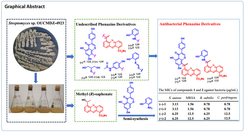
Phenazinoates A–E (1–5), comprising five pairs of methyl saphenate conjugates with genistein, o-aminophenol, p-acetaminophenol and glycerol, were isolated from the fermentation broth of mangrove soil-derived Streptomyces sp. OUCMDZ-4923. Their structures were determined through comprehensive one-dimensional and two-dimensional nuclear magnetic resonance spectroscopy, coupled with high-resolution electrospray ionization mass spectrometry. The absolute configurations of each isomer were established by comparing experimental electronic circular dichroism spectra with calculated counterparts. Based on the biosynthetic pathway analysis, compounds 1‒3 were semi-synthesized from the reactions of methyl (R)-saphenate with genistein, o-aminophenol, and o-formamidophenol, utilizing microwave-assisted solid acid catalysis. The compounds were resolved as enantiomerically pure forms and subsequently tested for antibacterial efficacy against six pathogenic bacteria. Phenazinoates A (1) and B (2) demonstrated bioactivity against four Gram-positive bacterial strains, with minimum inhibitory concentration values ranging from 0.78 to 3.13 μg/mL.Graphical Abstract
Keywords
Mangrove actinobacteria Streptomyces sp. Phenazine conjugates Structure elucidation Antibacterial activity1 Introduction
Natural phenazine derivatives, primarily synthesized by species of Pseudomonas and Streptomyces [1-3], have been shown to exhibit a broad spectrum of biological activities. These include antibacterial [4, 5], antitumor [6, 7], anti-inflammatory [8, 9], and neuroprotective effects [10, 11]. Our previous work also discovered six phenazine derivatives that demonstrated notable cytotoxicity against tumor cells, potent antifungal activity against Aspergillus fumigatus, and activity against the H1N1 virus. These compounds were derived from the marine sponge-associated strain Nocardiopsis dassonvillei OUCMDZ-4534 [12]. In our ongoing exploration of bioactive phenazine derivatives of microbial origin, we have been particularly drawn to the untapped metabolic potential of soil-dwelling microorganisms, especially endophytes and their rhizosphere counterparts [13]. Within this context, we focused on a mangrove soil-derived actinobacterial strain, Streptomyces sp. OUCMDZ-4923, isolated from mangrove sediment closely associated with the roots of Kandelia candel. Bioinformatics analysis confirmed the presence of a phenazine biosynthetic gene cluster (pnzBGC) in Streptomyces sp. OUCMDZ-4923 (GenBank accession No. CP170384), underscoring its potential to produce phenazine and its derivatives [14-16]. To pursue these bioactive compounds, a large-scale 60-L fermentation of Streptomyces sp. OUCMDZ-4923 was carried out. Chemical isolation of the ethyl acetate (EtOAc) extract resulted in the identification of sixteen enantiomerically pure phenazine dimers [16]. Further isolation efforts led to the discovery of five pairs of novel phenazine conjugates consisting of methyl saphenate linked with genistein, o-aminophenol, p-acetaminophenol and glycerol. These compounds were designated as phenazinoates A–E (1–5), along with the previously reported methyl (R)-saphenate (6) (Fig. 1) [16, 17]. Among the newly discovered compounds, phenazinoates A (1) and B (2), which are conjugates of genistein and o-aminophenol with methyl saphenate, demonstrated notable antibacterial activity against four strains of Gram-positive pathogenic bacteria, exhibiting minimum inhibitory concentration (MIC) values ranging from 0.78 to 3.13 μg/mL.
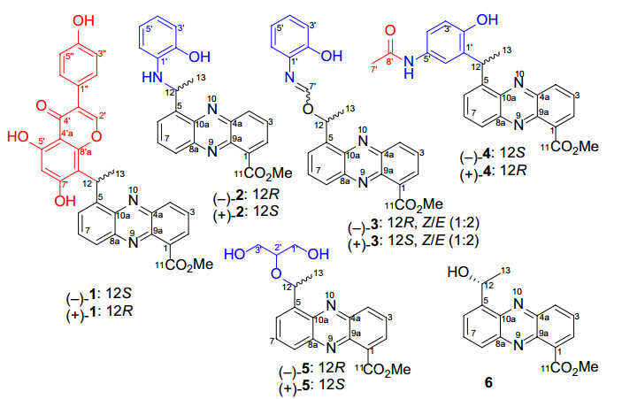
The chemical structures of compounds 1‒6
2 Results and discussion
2.1 Structural elucidation of phenazinoates A–E (1–5)
Phenazinoate A (1) was isolated as a racemic mixture in the form of a yellow powder. The molecular formula was established as C31H22O7N2 based on the high-resolution electrospray ionization mass spectrometry (HRESIMS) at m/z 535.1502 for the [M + H]+ ion (calcd for C31H23O7N2+, 535.1500). The UV–Visible absorption spectrum of the compound closely resembled those of known phenazine analogs, suggesting that the compound is indeed derivative of the phenazine family. The elucidation of the structure of compound 1 was further supported by the correlation spectroscopy (COSY) and heteronuclear multiple bond correlation (HMBC) experiments, as shown in Fig. 2. The COSY correlations revealed interactions from H-2 (δH 8.18) to H-4 (δH 8.23) through H-3 (δH 7.95), H-6 (δH 7.97) to H-8 (δH 8.06) through H-7 (δH 7.96), and between H-12 (δH 5.97) and H3-13 (δH 1.90), indicating the presence of 5-ethylidenephenazine-1-yl moiety. Key HMBC correlations provided further structural insights. The coupling of H3-13 to C-5 (δC 143.4), of H-12 to C-5, C-6 (δC 129.1), and C-10a (δC 141.6), as well as the interaction of the methoxy protons (δH 3.98) and H-2 with C-11 (δC 166.8), were indicative of a methyl 5-ethylidenephenazine-1-carboxylate moiety, akin to that found in methyl saphenate [17, 18]. Additionally, the 1H and 13C-NMR spectra hinted at the presence of an 8'-substituted genistein moiety within compound 1. This was further corroborated by crucial HMBC correlations, including interactions from proton H-2' (δH 8.44) to C-4' (δC 180.3), C-8'a (δC 155.4), and C-1'' (δC 121.8). Moreover, the proton of the 5'-OH group (δH 13.02) exhibited correlations to C-4'a (δC 104.1), C-5' (δC 159.9), and C-6' (δC 99.4). H-6' (δH 6.20) also correlated with C-4'a and C-8' (δC 110.1). The observed 1H-1H COSY correlations between H-3''/5'' (δH 6.82) and H-2''/6'' (δH 7.39) further supported the structure of the genistein moiety. The attachment of the genistein structure to the phenazine core was confirmed through HMBC correlations from H3-13 to C-8', and from H-12 to C-8', C-8'a, and C-7' (δC 164.4), indicating a single carbon–carbon bond connection between C-12 and C-8'. These NMR data and correlations provide compelling evidence for the structural integration of an 8'-substituted genistein moiety within the molecular architecture of the compound 1, specifically methyl 5-(1-(genistein-8-yl)ethyl)-phenazine-1-carboxylate.
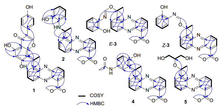
Key COSY and HMBC correlations for the structural assignment of compounds 1‒5
Phenazinoate B (2) was isolated as a racemic mixture and presented as a yellow powder. The molecular formula was determined to be C22H19O3N3, as indicated by the HRESIMS at m/z 374.1505 for the [M + H]+ ion. Analysis of one-dimensional (1D) and two-dimensional (2D) NMR spectra, as detailed in Table 1 and Fig. 2, revealed that the structure includes a methyl 12-deoxysaphenate unit, similar to the structural component identified in previously discussed compound 1. The spin system delineated by the chemical shifts from H-3' (δH 6.64) to H-6' (δH 6.17), passing through H-4' (δH 6.31) and H-5' (δH 6.33) in sequence, indicated the presence of an ortho-disubstituted phenyl group within the molecular structure. Critical HMBC interactions were observed, notably NH-12 (δH 5.38) showed correlations to C-5 (δC 144.1), C-2' (δC 144.3), and C-6' (δC 110.9). Additionally, the hydroxy proton HO-2' (δH 9.40) showed correlations with C-1' (δC 136.1), C-2', and C-3' (δC 113.7). These HMBC correlations confirmed the presence of an o-aminophenol moiety, which was connected to the C-12 atom via the amino nitrogen. Consequently, the molecular structure of phenazinoate B (2) was elucidated as methyl 5-(1-(2-hydroxyphenylamino)ethyl)phenazine-1-carboxylate.
1H (500 MHz) and 13C (125 MHz) NMR data for 1 and 2 in DMSO-d6
Phenazinoate C (3) was isolated as a yellow powder, presenting a complex mixture that is both racemic and an inseparable mix of E/Z-isomers. Its molecular formula was determined to be C23H19O4N3 based on the HRESIMS peak at m/z 402.1456 for [M + H]+ ion. When applying analytical high-performance liquid chromatography (HPLC) on an ND(2)-RH chiral column, it was feasible to separate compound 3 into enantiomerically pure entities, as illustrated in Fig. 3. The 1H-NMR and distortionless enhancement by polarization transfer including the detection of quaternary nuclei (DEPTQ) spectra for compound 3 revealed two distinct sets of signals, with an approximate ratio of 2:1 when measured in pyridine-d5. The structural elucidation of the major isomer within the mixture 3 began with the examination of its 1H-NMR spin systems, as depicted in Fig. 2. The identification of a methyl 5-(1-alkoxyethyl)-phenazine-1-carboxylate moiety was substantiated by delineating three distinct spin systems: one extending from H-2 (δH 8.21) to H-4 (δH 8.38), a second from H-6 (δH 8.09) to H-8 (δH 8.30), and a third connecting H-12 (δH 7.28) to H3-13 (δH 2.04). Additionally, pivotal HMBC interactions further clarified the structure, as illustrated in Fig. 2. Notable correlations including H3-13 to C-5 (δC 141.3), H-12 to both C-5 and C-6 (δC 129.7), as well as C-10a (δC 142.3). Correlations were also observed from both CH3O-11 (δH 4.04) and H-2 to C-11 (δC 167.9). The presence of an o-aminophenol moiety within the structure of compound 3 was suggested by a set of COSY correlations, which traced the continuous connectivity from H-3' (δH7.16) to H-6' (δH 7.09). The structure was further elucidated by analyzing the HMBC cross-peaks. These revealed significant correlations from H-7' (δH 8.63) to C-12 (δC 51.0) and C-1' (δC 128.5), indicating the formation of a C = N bond between the 12-alkoxy group and the 1'-NH2 group. These findings provided a clearer understanding of the compound's chemical constitution, identified as methyl 5-(1-((2-hydroxyphenylimino)methoxy)ethyl)phenazine-1-carboxylate. The structural assignment of the minor isomer of compound 3 was deduced to be the geometric isomer of its major counterpart, owing to the resemblance of their 2D NMR correlations, as presented in Fig. 2. The principal distinction arose from the 1D NMR data, particularly around the C = N moiety, detailed in Table 2. This assignment was further corroborated by the synthetic approach that involved combining methyl (R)-saphenate with o-formamidophenol or o-hydroxyphenyl formamide, which also aligned with the structural identification conclusions presented in Fig. 4a. Moreover, the fact that the mixture 3 remained an inseparable E/Z-isomeric blend was rationalized by the presence of a dynamic equilibrium between the E- and Z-geometric isomers in solution. This equilibrium is facilitated by two keto-enol tautomerizations, along with unrestricted rotation around the C-7'–N single bond, as depicted in Fig. 4b. These findings elucidated the dynamic behavior of the isomers and the complexity of their separation.
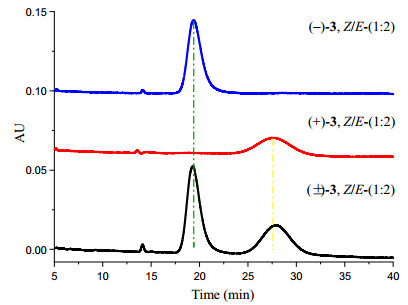
The mixture 3 was resolved into optically pure compounds (‒)-Z/E-3 and (+)-Z/E-3 using HPLC with a ND(2)-RH chiral column (4.6 × 250 mm) eluted with 45% MeCN-H2O at 1 mL/min
1H (600 MHz) and 13C (150 MHz) NMR data for 3 in pyridine-d6
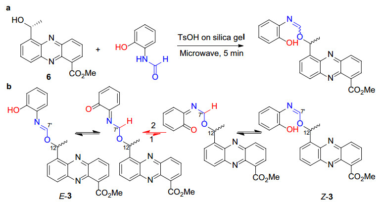
The synthesis of 3 from 6 and o-formamidophenol (a). Possible Z/E- dynamic equilibrium in solution via keto-enol tautomerization and single bond rotation at C-7'; this dynamic allows for the interconversion between the Z and E isomers, contributing to the complexity of their separation and characterization (b)
To ascertain the geometry of the C = N bond within the isomers of compound 3, a Nuclear Overhauser Enhancement difference (NOEdiff) experiment was employed. The results, illustrated in Fig. 5, revealed distinct NOE correlations for each isomer. For the major geometric isomer of 3, the NOE correlation observed between H-7' (δH 8.63) and H-6' (δH 7.09) allowed for the assignment of the E-geometry of the C = N bond Conversely, for the minor isomer of 3, the selective irradiation of H-7' (δH 9.38) led to an enhancement observed at H-12 (δH 6.71), which indicated the Z-geometry of the C = N bond.
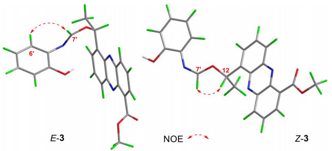
NOE correlations of Z-3 and E-3
The racemic phenazinoate D (4) was isolated as a yellow powder. Its molecular formula, C24H21O4N3, was deduced from the HRESIMS peak at m/z 416.1604 [M + H]+ (calcd for C24H21N3O4+, 416.1605). The UV–Visible spectral data of the compound matched those of known phenazine analogs, indicating that 4 is a phenazine derivative. The inclusion of a methyl 5-(1-hydroxyethyl)-phenazine-1-carboxylate moiety was substantiated by pivotal 2D-NMR correlations, as depicted in Fig. 2. The structure features a 1', 2', 5'-trisubstituted phenyl ring, as evidenced by the COSY correlation of H-3' (δH 6.76) with H-4' (δH 7.01), as well as essential HMBC correlations. These include the coupling of H-3' to C-1' (δC 136.1) and C-5' (δC 126.2), H-4' to C-2' (δC 146.2) and C-6' (δC 121.7), and H-6' (δH 7.72) to both C-2' and C-4' (δC 123.8). Additionally, the pivotal HMBC signals connecting H-12 (δH 5.56) to C-1', C-2', and C-6' suggest that C-12 is directly bound to C-1' through a single C‒C bond. The 1H and 13C NMR data (Table 3) revealed the presence of an additional methyl group at δH/C 2.03/23.5 (CH3-7'), a carbonyl carbon at δC 169.0 (C-8'), and a phenolic hydroxy proton at δH 9.35. The HMBC correlations, specifically between H3-7' and C-8', confirmed the presence of an acetamide moiety within the structure. The chemical shifts observed for C-2' and C-5' allowed for the definitive placement of the hydroxy and acetamide groups on the molecular framework. Consequently, the structures of these compounds were conclusively established as methyl 5-(1-(2-hydroxy-5-acetamidophenyl)ethyl)phenazine-1-carboxylate.
1H and 13C NMR data for compounds 4 and 5 in DMSO-d6
The racemic mixture of phenazinoate E (5) was obtained as a yellow powder and determined to have the molecular formula C19H20O5N2, as indicated by a HRESIMS peak at m/z 357.1451 [M + H]+. The UV–Visible and 1H, 13C-NMR spectral data for compound 5 suggested the presence of a methyl 5-(1-alkoxyethyl)phenazine-1-carboxylate moiety. The 1H-1H COSY correlations, as shown in Fig. 2, from HO-1' (δH 4.60) to HO-3' (δH 4.60) through H2-1' (δH 3.45/3.55), H-2' (δH 3.37) and H2-3' (δH 3.45/3.55) in sequence indicated a glycerin fragment, with substitution occurring at the 2'-hydroxy position. The connection between the glycerin and phenazine units was implied by an ether linkage between C-12 and C-2', supported by the HMBC correlation of H-2' to C-12 (δC 70.2). Consequently, the structure of compound 5 was conclusively identified as methyl 5-(1-((1, 3-dihydroxypropan-2-yl)oxy)ethyl) phenazine-1-carboxylate.
The new compounds 1–5 were successfully resolved into their optically pure enantiomers using HPLC equipped with chiral columns. To determine the absolute configurations (ACs) of these ten compounds, the electronic circular dichroism (ECD) curves for the (R)-isomers were computed using time-dependent density functional theory (TDDFT) with the B3LYP/6–31G(d) basis set [19, 20], as detailed in the Supporting Information. The comparison of the experimental ECD curves with the calculated ones for compounds (+)-1, (−)-2, (−)-3, (−)-4, and (−)-5 showed a good correlation with the curves of their respective (R)-isomers, as illustrated in Fig. 6. Consequently, the ACs of compounds (+)-1, (−)-2, (−)-3, (+)-4, and (−)-5 were assigned as (R)-, while the ACs of compounds (−)-1, (+)-2, (+)-3, (−)-4, and (+)-5 were unambiguously determined to be (S)-.
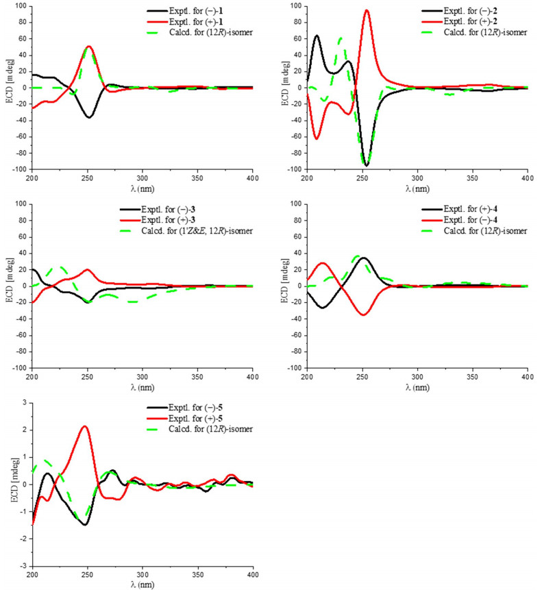
The experimental and calculated ECD curves of 1‒5
2.2 Physicochemical properties of 1–5
2.2.1 Phenazinoate A (1)
Yellow powder; UV (MeOH) λmax (log ε) 253 (3.65), 365 (2.87) nm; 1H and 13C NMR data (Table 1); HRESIMS m/z 535.1502 [M + H]+ (calcd for C31H23N2O7, 535.1500). (−)-1, [α]D28 −18.6 (c 0.1, MeOH); ECD (0.94 mM, MeOH) λmax (Δε) 251 (−11.7), 271 (+1.4) nm. (+)-1: [α]D28 +14.7 (c 0.1, MeOH); ECD (0.94 mM, MeOH) λmax (Δε) 251 (+16.5), 272 (−1.6) nm.
2.2.2 Phenazinoate B (2)
Yellow powder; UV (MeOH) λmax (log ε) 249 (2.93), 365 (2.17) nm; 1H and 13C NMR data (Table 1); HRESIMS m/z 374.1505 [M + H]+ (calcd for C22H20N3O3, 374.1499). (−)-2, [α]D28 −40.9 (c 0.1, MeOH); ECD (1.3 mM, MeOH) λmax (Δε) 209 (+14.4), 237 (+7.3), 254 (− 21.6) nm, 364 (−0.9). (+)-2, [α]D28 + 30.9 (c 0.1, MeOH); ECD (1.3 mM, MeOH) λmax (Δε) 209 (−14.0), 238 (−7.3), 254 (+21.6), 364 (+ 0.9) nm.
2.2.3 Phenazinoate C (3)
Yellow powder; UV (MeOH) λmax (log ε) 251 (3.69), 365 (2.99) nm; 1H and 13C NMR data (Table 2); HRESIMS m/z 402.1456 [M + H]+ (calcd for C23H20N3O4, 402.1488). (−)-3, [α]D28 −37.1 (c 0.1, MeOH); ECD (1.2 mM, MeOH) λmax (Δε) 229 (−1.9), 250 (−5.1), 306 (−0.6). (+)-3, [α]D28 + 36.5 (c 0.1, MeOH); ECD (1.2 mM, MeOH) λmax (Δε) 229 (+ 2.1), 251 (+ 5.0), 307 (+ 0.6) nm.
2.2.4 Phenazinoate D (4)
Yellow powder; UV (MeOH) λmax (log ε) 253 (3.46), 365 (2.77) nm; 1H and 13C NMR data (Table 3); HRESIMS m/z 416.1604 [M + H]+ (calcd for C24H21N3O4, 416.1605). (−)-4, [α]D28 −25.2 (c 0.1, MeOH); ECD (1.2 mM, MeOH) λmax (Δε) 214 (+7.2), 251 (−8.8), 286 (+0.3) nm. (+)-4, [α]D28 + 30.3 (c 0.1, MeOH); ECD (1.2 mM, MeOH) λmax (Δε) 214 (−6.6), 251 (+8.7), 284 (−0.2) nm.
2.2.5 Phenazinoate E (5)
Yellow powder; UV (MeOH) λmax (log ε) 252 (3.60), 366 (2.86) nm; 1H and 13C NMR data (Table 3); HRESIMS m/z 357.1451 [M + H]+ (calcd for C19H21N2O5, 357.1445). (−)-5, [α]D28 −9.3 (c 0.1, MeOH); ECD (1.4 mM, MeOH) λmax (Δε) 214 (+0.1), 248 (−0.3), 272 (+0.1) nm. (+)-5, [α]28 D + 9.6 (c 0.1, MeOH); ECD (1.4 mM, MeOH) λmax (Δε) 246 (+ 0.5), 272 (−0.1) nm.
2.3 Proposed biosynthetic pathway of compounds 1–5 and semi-synthetic production of compounds 1–3
Previously, we identified the enzymes Pnz16‒21 in Streptomyces sp. OUCMDZ-4923 as being responsible for the biosynthesis of the phenazine-1, 5-dicarboxylic acid core, which is consequently converted into (R)-saphenic acid by enzymes Pnz6‒12 and then O-methylated to form methyl (R)-saphenate (6) by the methyltransferase Pnz43 [16]. We now propose that methyl 5-vinylphenazine-1-carboxylate, produced by the dehydration of methyl (R)-saphenate (6) under the action of enzymes Pnz28/30 [16], serves as a critical intermediate. This intermediate undergoes non-enzymatic reactions with compounds such as genistein, o-aminophenol, o-formamidophenol, p-acetamidophenol, and glycerol through a nonenzymatic pathway, leading to the formation of phenazinoates A–E (1‒5) (Fig. 7). In line with this hypothesis, compounds 1‒3 were successfully semi-synthesized from methyl (R)-saphenate (6) by reacting it with genistein, o-aminophenol, or o-formamidophenol. This transformation was achieved using p-toluenesulfonic acid absorbed on silica gel as a catalyst under microwave irradiation.
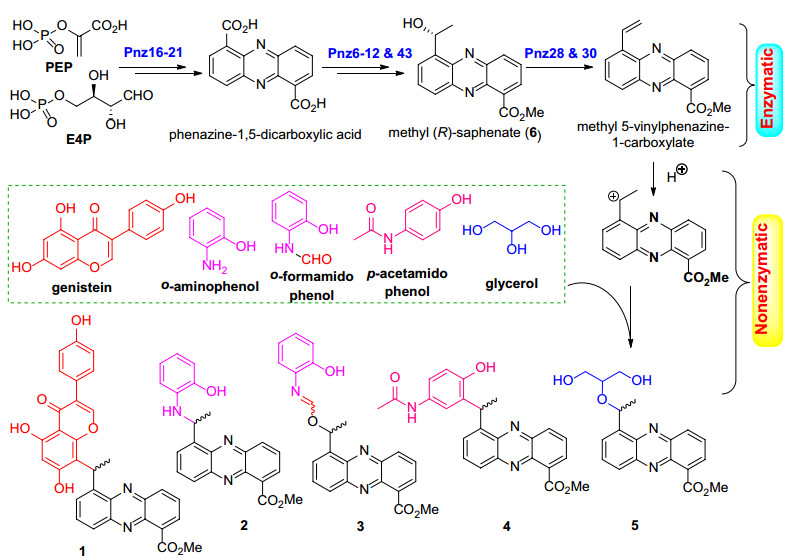
Proposed biosynthetic pathway of phenazinoates 1‒5 in Streptomyces sp. OUCMDZ-4923
2.4 Biological activity evaluation of compounds 1–5
The antibacterial efficacy of five pairs of enantiomerically pure compounds 1–5 was assessed as previously described [21]. The panel of test pathogens included four Gram-positive bacteria: Staphylococcus aureus ATCC 6538, methicillin-resistant S. aureus ATCC 43300 (MRSA), Clostridium perfringens CGMCC 1.0876, and Bacillus subtilis CGMCC 1.3376, along with two Gram-negative bacteria: Pseudomonas aeruginosa ATCC 10145 and Escherichia coli ATCC 11775. The minimum inhibitory concentrations (MICs), which are the lowest concentrations that inhibit microbial growth, were determined across a range of 25.0–0.78 μg/mL. Compounds 1 and 2 exhibited inhibitory activity against B. subtilis, C. perfringens, S. aureus, and MRSA, with MIC values ranging from 0.78 to 3.13 μg/mL, as detailed in Table 4. No antimicrobial activity was observed for the other tested bacteria, and compounds 3–5 did not exhibit inhibition against any of the test pathogens at the maximum concentration tested, which was 25.0 μg/mL. Additionally, it was noted that there was no difference in antibacterial activity between the two enantiomers. The results indicated that conjugating isoflavone or o-aminophenol moieties to the phenazine core can enhance the antibacterial efficacy of phenazine conjugates. However, both the Levo- and Dextro- enantiomers demonstrated the same level of efficacy against the four tested Gram-positive bacteria, suggesting that the chirality of these compounds does not influence their antibacterial activity. This observation could be explained by the possibility that the chiral center of the drug molecule may not located at the core position of the pharmacophore, or that the receptor's binding pocket is sufficiently spacious or flexible to accommodate either enantiomer, resulting in an equivalent biological effect.
The MICs of compounds 1 and 2 against bacteria (μg/mL)a
3 Conclusion
In summary, five novel phenazine derivatives, phenazinoates A–E (1–5), were isolated and characterized from the mangrove soil-derived Streptomyces sp. OUCMDZ-4923, representing five pairs of enantiomers. This marks the first study to report the conjugate of isoflavone, o-/p-aminophenol, or glycerol moieties to the phenazine core. Notably, compounds 1 and 2 which are conjugates of isoflavone or o-aminophenol and phenazine, exhibited antibacterial properties against a spectrum of four Gram-positive bacterial strains. Further insights were obtained from the analysis of biosynthetic pathways, leading to the successful semi-synthesis of phenazinoates A–C (1–3) from methyl (R)-saphenate using genistein, o-aminophenol, or o-acetamidophenol. This synthesis was facilitated by microwave-assisted solid acid catalysis, providing a method to produce these compounds in sufficient quantities for subsequent comprehensive studies on their antibacterial activities.
Notes
Acknowledgements
This work was financially supported by the National Key Research and Development Program of China (No. 2022YFC2804100), the National Natural Science Foundation of China (No. 82473838), and Guizhou Provincial Basic Research Program (Natural Science) (No. QKHJC MS[2025]117).
Author contributions
D. Wang performed the experiments and wrote the draft paper, P. Liu participated in the experiments, Y. Gao and L. Chen analyzed the data and discussed the result, L. Wang and N. Li reviewed the manuscript, and Weiming Zhu conceived the projects, revised manuscript, and provided financial support. All authors read and approved the final manuscript.
Data availability
The datasets used or analyzed during the current study are available from the corresponding author on reasonable request.
Declarations
Competing interests
The authors declare no competing interests.
References
-
1.Byng GS, Eustice DC, Jensen RA. Biosynthesis of phenazine pigments in mutant and wild-type cultures of Pseudomonas aeruginosa. J Bacteriol. 1979;138: 846-52. CrossRef PubMed Google Scholar
-
2.McDonald M, Wilkinson B, Van'T Land CW, Mocek U, Lee S, Floss HG. Biosynthesis of phenazine antibiotics in Streptomyces antibioticus: stereochemistry of methyl transfer from carbon-2 of acetate. J Am Chem Soc. 1999;121: 5619-24. CrossRef PubMed Google Scholar
-
3.Laursen JB, Nielsen J. Phenazine natural products: biosynthesis, synthetic analogues, and biological activity. Chem Rev. 2004;104: 1663-86. CrossRef PubMed Google Scholar
-
4.Kitahara M, Nakamura H, Matsuda Y, Hamada M, Naganawa H, Maeda K, et al. Saphenamycin, a novel antibiotic from a strain of streptomyces. J Antibiot. 1982;35: 1412-4. CrossRef PubMed Google Scholar
-
5.Liang Y, Chen L, Ye X, Anjum K, Lian XY, Zhang Z. New streptophenazines from marine Streptomyces sp. 182SMLY. Nat Prod Res. 2017;31: 411-7. CrossRef PubMed Google Scholar
-
6.Conda-Sheridan M, Marler L, Park EJ, Kondratyuk TP, Jermihov K, Mesecar AD, et al. Potential chemopreventive agents based on the structure of the lead compound 2-bromo-1-hydroxyphenazine, isolated from Streptomyces species, strain CNS284. J Med Chem. 2010;53: 8688-99. CrossRef PubMed Google Scholar
-
7.Moris M, Andrieu C, Rocchi P, Seillan C, Acunzo J, Brunel F, et al. 2, 3-Dialkoxyphenazines as anticancer agents. Tetrahedron Lett. 2015;56: 2695-8. CrossRef PubMed Google Scholar
-
8.Kondratyuk TP, Park EJ, Yu R, van Breemen RB, Asolkar RN, Murphy BT, et al. Novel marine phenazines as potential cancer chemopreventive and anti-inflammatory agents. Mar Drugs. 2012;10: 451-64. CrossRef PubMed Google Scholar
-
9.Lee HS, Kang JS, Choi BK, Lee HS, Lee YJ, Lee J, et al. Phenazine derivatives with anti-Inflammatory activity from the deep-sea sediment-serived yeast-like fungus Cystobasidium laryngis IV17-028. Mar Drugs. 2019;17: 482. CrossRef PubMed Google Scholar
-
10.Kim WG, Ryoo IJ, Yun BS, Shin-ya K, Seto H, Yoo ID. Phenazostatin C, a new diphenazine with neuronal cell protecting activity from Streptomyces sp. J Antibiot. 1999;52: 758-61. CrossRef PubMed Google Scholar
-
11.Kim WG, Ryoo IJ, Yun BS, Shin-ya K, Seto H, Yoo ID. New diphenazines with neuronal cell protecting activity, phenazostatins A and B, produced by Streptomyces sp. J Antibiot. 1997;50: 715-21. CrossRef PubMed Google Scholar
-
12.Liu H, Zhu G, Zhao S, Fu P, Zhu W. Bioactive natural products from the marine sponge-derived Nocardiopsis dassonvillei OUCMDZ-4534. Chin J Org Chem. 2019;39: 507-14. CrossRef PubMed Google Scholar
-
13.Wang D, Li M, Xu Y, Fu P, Zhu W, Wang LP. Dibohemamines I–O from Streptomyces sp. GZWMJZ-662, an endophytic actinomycete from the medicinal and edible plant Houttuynia cordata Thunb. Nat Prod Bioprospect. 2025;15: 9. CrossRef PubMed Google Scholar
-
14.Bauman KD, Li J, Murata K, Mantovani SM, Dahesh S, Nizet V, et al. Refactoring the cryptic streptophenazine biosynthetic gene cluster unites phenazine, polyketide, and nonribosomal peptide biochemistry. Cell Chem Biol. 2019;26: 724-736.e7. CrossRef PubMed Google Scholar
-
15.Rui Z, Ye M, Wang S, Fujikawa K, Akerele B, Aung M, et al. Insights into a divergent phenazine biosynthetic pathway governed by a plasmid-born esmeraldin gene cluster. Chem Biol. 2012;19: 1116-25. CrossRef PubMed Google Scholar
-
16.Wang D, Liu P, Xia Y, Wang L, Li N, Zhu W. Antibacterial dimeric phenazine derivatives from a marine-derived Streptomyces sp. OUCMDZ-4923. Mar Life Sci Technol. 2025;7: 925-36. CrossRef PubMed Google Scholar
-
17.Geiger A, Keller-Schierlein W, Brandl M, Zähner H. Metabolites of microorganisms. 247. Phenazines from Streptomyces antibioticus, strain TUE 2706. J Antibiot. 1988;41: 1542-51. CrossRef PubMed Google Scholar
-
18.Yun B, Ryoo I, Kim W, Kim J, Koshino H, Seto H, et al. Structures of phenazostatins A and B, neuronal cell protecting substances of microbial origin. Tetrahedron Lett. 1996;37: 8529-30. CrossRef PubMed Google Scholar
-
19.Du Y, Chen Z, Li H, Wang Y, Fu P, Zhu W. Pafuranones A and B, two dimeric polyketides from a rare marine algae-derived fungus Paraconiothyrium sp. Chin Chem Lett. 2019;30: 981-4. CrossRef PubMed Google Scholar
-
20.Zhu G, Kong F, Wang Y, Fu P, Zhu W. Cladodionen, a cytotoxic hybrid polyketide from the marine-derived Cladosporium sp. OUCMDZ-1635. Mar Drugs. 2018;16: 71-8. CrossRef PubMed Google Scholar
-
21.Wang D, Wang C, Gui P, Liu H, Khalaf SMH, Elsayed EA, et al. Identification, bioactivity, and productivity of actinomycins from the marine-derived Streptomyces heliomycini. Front Microbiol. 2017;8: 1147. CrossRef PubMed Google Scholar
Copyright information
© The Author(s) 2026
Open Access This article is licensed under a Creative Commons Attribution 4.0 International License, which permits use, sharing, adaptation, distribution and reproduction in any medium or format, as long as you give appropriate credit to the original author(s) and the source, provide a link to the Creative Commons licence, and indicate if changes were made. The images or other third party material in this article are included in the article’s Creative Commons licence, unless indicated otherwise in a credit line to the material. If material is not included in the article’s Creative Commons licence and your intended use is not permitted by statutory regulation or exceeds the permitted use, you will need to obtain permission directly from the copyright holder. To view a copy of this licence, visit http://creativecommons.org/licenses/by/4.0/.