Stingless bee propolis: a comprehensive review of chemical constituents and health efficacy

Abstract
Propolis, consisting of plant-derived materials, wax, and bee secretions, is abundant in bioactive constituents like flavonoids, phenolic compounds, and terpenes, which enhance its various biological functions. These encompass antioxidant, anti-inflammatory, antibacterial, anticancer, antidiabetic, and immunomodulatory properties. Propolis has demonstrated effectiveness in the prevention and treatment of multiple illnesses, including cardiovascular disease, atherosclerosis, infections, diabetes, wound healing, and burns. Its extensive health benefits endorse its application in medications, nutritional supplements, and cosmetics, where it is acknowledged as a safe and efficacious natural product. Propolis, whether utilized in its raw state, as extracts, or in conjunction with other products, exhibits considerable promise in alternative medicine and nutritional health. Propolis extracts are crucial to examine as a key component in health and wellness, offering prospective applications in disease prevention and therapeutic support Further research is necessary to clarify its molecular mechanisms, examine potential allergic reactions, and determine ideal dosages for various ages. This article provides a comprehensive comparative examination of various propolis types, emphasizing their distinct phytochemical contents and varying biological effects concurrently. It integrates results from both in vitro and in vivo investigations, enhancing the comprehension of health applications and mechanisms of action, grounded comparisons in pertinent prior studies.Graphical Abstract
Keywords
Antioxidant Nutritional supplements Chemical analysis Bioactive constituents Propolis Stingless bee1 Methods
1.1 Search strategy
The search strategy focused on literature directly addressing the primary topic, while studies with less relevant content were excluded. Key electronic databases, search engines, Web of Science, Google Scholar, and ScienceDirect for articles meeting the inclusion standards.
The search involved specific keyword combinations and Medical Subject Headings (MESH) in titles and abstracts, using terms such as: (Phytochemical, antioxidant, anti-cancer, bioactivity, inflammation, cardiovascular disease, antimicrobial, diabetes, wound healing, preclinical studies, and clinical studies), alongside non-MESH (bee propolis, phytochemicals, free radicals, stingless bee propolis). Search was refined with the “AND” operator to enhance relevance. This strategy yielded a broad selection of pertinent articles.
1.2 Inclusion criteria
Open-access publications from academic search engines were selected based on their direct relevance to the research topic and their ability to provide accurate, comprehensive, and up-to-date data. The inclusion criteria targeted observational and experimental studies examining the origins of propolis, its traditional uses, phytochemical composition, and biological efficacy, including both in vitro and animal studies. Non-original publications (e.g., reviews, letters, or comments), redundant studies, and those lacking full-text availability or addressing unrelated conditions were excluded. Only studies published in English were considered, and authorship rights were respected through proper citation. This systematic and selective approach ensured that the analysis was grounded in credible scientific evidence and aligned with ethical research standards.
2 Introduction
Stingless bees have a scientific name (Meliponini) and belong to the eusocial insects that are formed from a monophyletic group. The monophyletic group, originally from the Corbicolate bees (Hymenoptera: Apidae) is a huge group comprising around 550 species, with 61 genes [1]. The group of crappy bees includes bumblebees (Bombini), honeybees (Apini), and orchid bees (Euglossini) groups. This type of bee lives and breeds in tropical climates such as Malaysia, Australia, India, specific areas of tropical America, and parts of Africa, also in Mexico, Brazil, and Argentina. Stingless bees are not much different from other honeybees, as they live in a colony system and can produce honey, bee bread, and propolis, as shown in Fig. 1. However it lacks the property of stinging or a proper defence system, so it protects its colonies with wax-like substances that close the pores and holes [2].
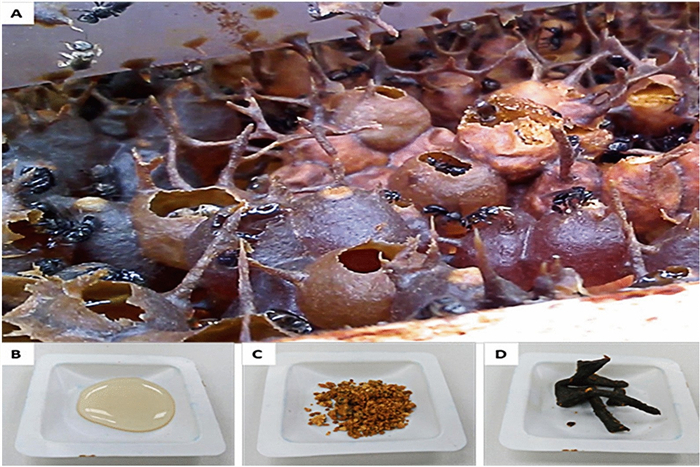
A selection of products derived from the nest of stingless bees. a The stingless bee nest; b Honey; c Bee bread and d Propolis [3]
Bee products, like other natural resources, are widely consumed and utilized in traditional medicine, with propolis being among the most significant. Derived from the Greek term meaning “entrance to the city,” propolis serves as a hive protector. It is a complex resinous substance partially digested by β-glycosidase in bee saliva and mixed with beeswax. Propolis exhibits temperature-dependent characteristics: it becomes soft and adhesive at higher temperatures, rigid when cooled, and transitions to liquid between 60 and 70 ℃. Its color varies from green to brown and reddish, depending on its botanical origin [4].
The chemical composition of propolis varies depending on the type of plants accessible to the bees. This article reviews the active ingredients and their biological activity in propolis, based on recent studies. The gathered data may contribute to studying the safety, toxicity, and potential development of new drugs or nutritional supplements from propolis.
3 Historical aspects, and traditional uses of propolis
Propolis was extensively used as a medicine by ancient civilizations such as the Greeks, Romans, and Egyptians [5]. In the Middle Ages, it lost its popularity a little, and despite this, knowledge of its medicinal properties continued in traditional folk medicine. In the Renaissance era in Europe, interest was restored in the study of natural alternatives in medicine and pharmacy, so researchers began to analyze and study the advantages of propolis, and scientists in the last century were able to prove its significance, as our forefathers had believed. Research on propolis’ chemical composition commenced during the early twentieth century and persisted beyond World War Ⅱ [6].
Propolis has been utilized as a conventional remedy, since 300 BC [7] with healing activities identified by Roman and Greek doctors [8] such as Dioscorides, Galen, Aristotle, and Pliny. In 1908, the initial scientific report on propolis, which outlines its phytochemical composition and chemical properties derived from this source, has been made available to the public. Previous literature indicated that the ancient Egyptians used propolis to keep corpses from decomposing, as well as to dress and muffle wounds, and it was noted that it prevents the formation of bacteria as well as inflammation [9]. Its use as antibacterial, antifungal and antiviral medicine has expanded in many regions around the world [10]. Propolis has also been known traditionally as a local anesthetic and pain reliever in first aid for injuries [11]. In complementary medicine, propolis extracts are used to treat various medical conditions such as stomach disorders, colds and flu, and asthma in the form of sprays, powders, and ointments [12, 13]. Furthermore, propolis, both in its raw form and as extracts, is utilized in nutritional supplements, and food products targeting specific health conditions, thanks to its versatile beneficial properties [14]. The significance and advantages of propolis have gained considerable attention, leading to its inclusion in beverages and food items as a strategy to enhance immunity, fortify the body, and safeguard against illnesses [15]. Propolis has garnered interest for its potential in treating purulent disorders, promoting wound healing, aiding in burn treatment, and addressing stomach ulcers [16]. Dentistry may also benefit from propolis, as it shows promise in relieving toothaches, treating caries, and potentially impacting tooth roots [13]. In China, Propolis has been acknowledged for its medicinal properties as an anti-infection and anti-cancer agent. In England, it was recognized and adopted as a superior treatment for wounds during the seventeenth century [6]. In Indian folk medicine, propolis is widely used as a treatment for stomach ulcers [17].
Extensive historical, traditional, and scientific evidence supports propolis as a natural drug. Its medicinal properties have been acknowledged across various cultures and eras, highlighting its therapeutic value. Modern research confirms its bioeffects, reinforcing its use as a natural remedy. Its incorporation into complementary medicine and health products further reflects its growing acceptance as an effective treatment.
4 Chemical analysis of propolis
Analysing the chemical composition of propolis extract is crucial for understanding its potential biological benefits and nutritional value. This information can provide valuable insights into how propolis extract may impact various health conditions.
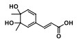
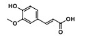
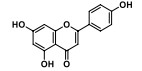
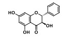
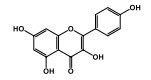
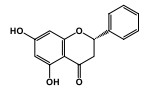
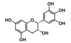
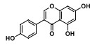
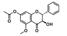
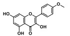
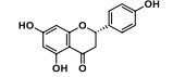
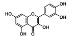
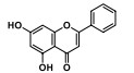
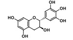
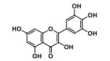
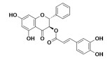
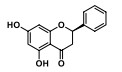
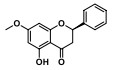
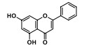
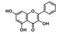
Studies has shown that more than 420 different compounds have been characterized so far in propolis [18, 19], including countries like the Netherlands, Malaysia, Australia, New Zealand, Italy and many more. Phenolic compounds, alkenylphenol, Flavanones, flavones, flavonol, and derivatives, terpenes, and flavonoids were identified as the active ingredients responsible for the significance and efficacy of propolis [20]. Table 1 presents several compounds revealing biological activity based on chemical classification, whereas Table S1 (Supplementary Materials) shows 257 other compounds have been identified in propolis, which shows other constituents along with their respective chemical structures, emphasizing the chemical variety of propolis.
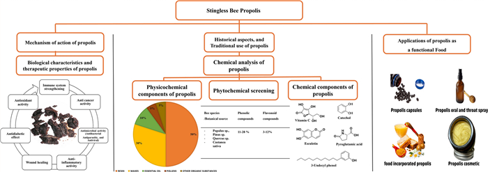
Chemical components of propolis
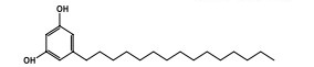


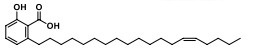
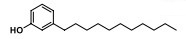
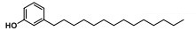
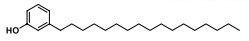


Medical benefits have been reported for propolis produced from Malaysian stingless bees, which are classified into 29 species Malaysian farms, and forests. The most famous species are (Tetrigona binghami, homotrigona fimbriata and tetrigona apicalis). These benefits are due to the richness of its composition of polyphenols, flavonoids, esters, terpenes, vitamins, and minerals, as well as enzymes, as it contains many phenols (5-pentadecyl and resorcinol), flavonoids (pinobanksin-5-methyl ether acetate and kaempferol- dimethyl ether), steroids (19-cyclolanost-24-en-3- ol, 9,19-cyclolanostane-3-ol, 24-methylene, and 9,19- cyclolanost-24-en-3-ol), terpenes such as (aromadendrene, α-eudesmol, caryophyllene oxide, and squalene), vitamins and minerals as well as enzymes [21]. In the same context, Brazilian propolis is characterized by its containing flavonoids, fatty acids, phenylpropanoids, and their derivatives, such as artibiline c, as well as chlorogenic acids [22]. Chi et al. (2020) reported that Chinese propolis contains phenolic alcohols, aldoketones, sesquiterpenoids, esters, and hydrocarbons [23]. Australian bee propolis has been shown to contain a high percentage of flavonoids (abyssinoflavanone, propolin, and nymphaeol) in addition to five-cyclic triterpenoids [24]. As for the content of bee propolis in the Mediterranean countries, it is characterized by its composition of phenols, esters, and flavonoids in its non-volatile part. The volatile part of propolis in Italy, Greece, Croatia, Egypt, Algeria and Libya was distinguished for containing benzoic acid and its esters, mono- and sesquiterpenes [25].
4.1 Physicochemical components
Nature and the original botanical type play an important role in propolis’ general and exact ingredient ratios and physical characteristics. Propolis typically comprises 40–50% resins, 10% essential oils, 20- 30% wax, 5% phenols, flavonoids, and 5% pollen-containing vitamins and minerals, Fig. 2 [103]. The percentage of moisture, carbohydrates, fats, protein, fibre, ash, and wax in different types of extracted bee propolis from different plant sources in various regions around the world show in Table 2.
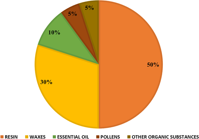
Main composition of propolis
Physicochemical components of propolis
It is worth mentioning that the carbohydrates source in the composition of propolis are still unknown, this may be since they contain sugar alcohols and acids. The main sources of sugars (glucose, fructose, and sucrose) are nectar and honey [13].
The wax in propolis is a stable, yellow-coloured, moisture-resistant, absorbent, and non-heat-resistant substance. Wax is composed of esters, acids, alcohol, and some free hydrocarbons. In addition, other components of propolis have been reported, including amino acids, pollen, and enzymes derived from bee glandular secretions like succinic dehydrogenase, adenosine triphosphatase, glucose-6-phosphatase, acid phosphatase, α-amylase, β-amylase, α-lactamase, β-lactamase, maltase, esterase, and transhydrogenase [104].
Based on absorption spectroscopy on propolis samples, literature has shown that propolis possesses vitamins as one of the most important micronutrients directly involved in the metabolism of the human body. vitamin B complex (Thiamine, riboflavin, nicotinamide, niacin, pyridoxine, and folic acid) [105], vitamin E,,vitamin C, and vitamin A. In the same context, Propolis contains a variety of minerals which are primarily as cofactors for enzymatic activities, as (sodium, potassium, copper, zinc, iron, calcium, phosphorus, manganese, selenium, and magnesium) [106].
4.2 Phytochemical screening
Numerous studies have shown the phytochemical makeup of propolis samples from diverse geographical regions and bee species as shown in Table 3. Portuguese propolis, sourced from Populus sp., Pinus sp., Quercus sp., and Castanea sativa, comprises phenolic components in concentrations of 11–28% and flavonoids at 3–12% [107]. Malaysian propolis, sourced from many stingless bee species, exhibits significant compositional heterogeneity. Tetrigona apicalis propolis contains 7.6 mg/mL of phenolics, 34.5 mg/mL of flavonoids, and 0.66 mg/mL of terpenoids; Tetrigona binghami contains 10.1 mg/mL of phenolics, 34.1 mg/mL of flavonoids, and 2.1 mg/mL of terpenoids; Heterotrigona fimbriata has 13.2 mg/mL of phenolics, 34.5 mg/mL of flavonoids, and 1.1 mg/mL of terpenoids; whereas Geniotrigona thoracica propolis contains 55.1 µM GAE/g of phenolics and 326.1 µM QE/g of flavonoids [2, 98]. Chilean propolis sourced from places including Rincon de Yaquil, Cuncumen, de la Araucanía, and Metropolitana has phenolic components ranging from 11.4 to 20.8 g GAE per 100 g of methanol extract and flavonoids between 1.7 and 14 g catechin equivalents per 100 g of methanol extract [83]. In Brazil (Paraná state), natural propolis was determined to contain 4.17% phenolics and 0.26 g/100 g flavonoids [109]. Propolis from India, sourced from places including Karna, Hamirpur, Sarangpur, Palladam, and Hubli, exhibited phenolic content of 10–18 µg/g, flavonoid levels ranging from 48–98 µg/g, and terpenoid concentrations of 15–38 µg/g [111].
Concentration of different phytochemical compounds in propolis
5 Mechanism of action of propolis
Phenolic compounds, which include phenolic acids (caffeic acid, salvianolic acid B, chlorogenic acid, and ferulic acid), tannins, and stilbenes, give propolis nutritional and medicinal curative traits, such as immune system strengthening, antioxidant, anti-inflammatory, and anti-cancer properties. It is noteworthy that the presence of flavonoids in propolis assesses its quality [112]. Flavonoids such as Apigenin and Galangin display notable biological activities as mentioned in the Table 4. Apigenin exhibits potent antioxidant and anti-inflammatory properties by promoting metal chelation, neutralizing free radicals, and augmenting phase Ⅱ detoxification enzymes in both cell culture and in vivo tumour models [113, 114]. It also contributes to cancer prevention by inducing apoptosis in various cell lines and animal models [115]. Galangin has shown potential in neuroprotection, particularly in Alzheimer’s and Parkinson’s disease, through the inhibition of pro-inflammatory cytokines (IL-1β, IL-6, IL-18, TNF-α) and reactive oxygen species (ROS) [116, 117]. Additionally, galangin improves insulin sensitivity by promoting glucose uptake and glycogen synthesis, increasing hexokinase (HK) and pyruvate kinase (PK) activity, and upregulating insulin receptor (IR), Akt, and GSK3β phosphorylation, while downregulating insulin receptor substrate and mTOR phosphorylation [118, 119].
Mechanism of bioactive compounds in propolis
Terpenoids are approved as the most common volatile compounds in Greece propolis, among others (alpha-pinene, junipene, and δ-cadinene). Isocupressic acid, pimaric acid, communic acid, and 14,15-dinor-13-oxo-8(17)-labden-19-oic acid as the most dominant terpenoids in propolis extracts. Two diterpenes were identified from extracts of propolis in Malta and diterpene esters of hydroxybenzoic acids were isolated and bound to the plant source [120]. The extract compounds showed high antibacterial activity against Staphylococcus aureus [121]. Zhao et al. (2017) reported that non-stingless tropical bee propolis, such as Heterotrigona itama propolis in Malaysia, contains terpenoids as major biologically active compounds within 28 compounds, including (phenolic acids, flavones, terpenoids, and phytosterols), the two major terpenoids being (24(E)-cycloart)-24-ene-26-ol-3-one and 20-hydroxy-24-dammaren-3-one) [122]. Propolis from Geniotrigona breastacica bees in Kelantan, Malaysia, contained other derivatives of terpenoids, including (fren-9(11)-en-2-alpha-ol, lup-20(29)-ene-3,21-dione, and beta-amyrenol) [123]. Furthermore, Pujirahayu et al. (2019), linked the difference in terpenoids to the plant source when identifying different types of triterpenes isolated from propolis extracts in Indonesia (mangiferolic acid, cycloartenol, and ambolic acid), from Mangifera indica as the plant source [124].
6 Biological characteristics and therapeutic properties of propolis
The identification of the phytochemical constituents present in propolis extracts has aided in inferring the biotic and biological effects of propolis. Figure 3 shows that propolis exhibits multiple biological effects, as supported by various in vitro and in vivo studies as summarized in Table 5. The extent of its biological activity depends on factors such as the propolis chemical composition, the concentration of phytochemical compounds, as well as the extraction methods and treatments used.
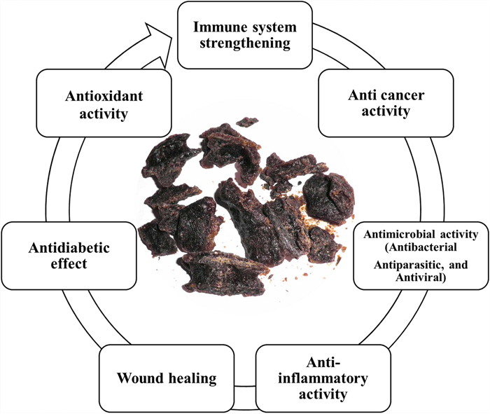
Main biological properties of propolis
Summary of the biological characteristics and therapeutic properties of propolis
6.1 Antimicrobial activity
Given the urgent need to discover new natural remedies against infectious diseases, especially since some pathogens can be resistant to repeated antibiotics, propolis has been shown to be effective against Gram-positive and Gram-negative bacterial strains, as well as anaerobic and aerobic bacteria. The activity of propolis on bacterial strains occurs in two ways: firstly, by direct action on bacteria, and secondly, by stimulating the immune system (the immune system of host cells) and thus activating natural defence mechanisms. Possible mechanisms of action include inhibition of bacterial adhesion and division, reduction of their motility, disruption of membrane potential, and increased permeability of the cell membrane [145].
Studies indicate that propolis exhibits stronger activity against Gram-positive bacteria compared to Gram-negative bacteria. Its effects on bacterial strains such as (Escherichia coli, and Bacillus subtilis) include altering membrane permeability and disrupting potential, ultimately inhibiting bacterial movement [146]. A phenolic extract of propolis has been suggested as a supportive agent for antibiotics in treating Gram-positive bacterial infections. Propolis enhanced the effectiveness of ampicillin, gentamicin, and streptomycin, reducing the required antibiotic concentrations. A study on French propolis demonstrated varying effects of ethanolic extracts on 12 strains of methicillin-sensitive Staphylococcus aureus (MSSA) when combined with 10 anti-staphylococcal drugs [147]. Propolis showed the same results on S. aureus. As well as, it is proven the efficacy of Brazilian nanohydroxyapatite (nanoHA) surfaces infused in propolis extracts in preventing the growth and formation of biofilms of S. aureus bacteria, and staphylococcal biofilm formation reported [148].
In a comparative study between 48 ethanolic extracts of Turkish propolis against bacterial strains (Streptococcus sanguinis, Strep- tococcus pyogenes, Streptococcus mutans, and C. albicans) in addition to chemical analysis and examination of other biological activities. It was observed that the highest antimicrobial activity was due to samples with a higher content of flavonoid, phenols, cinnamic, ferulic, caffeic, chlorogenic and coumaric acids [145]. In evaluating the effect of Malaysian ethanol and n-hexane propolis extracts from the plant source Heterotrigona itama, Disc diffusion and broth dilution methods were used; the extracts showed significant activity against all studied bacterial strains (B. subtilis, S. aureus, E. coli and Salmonella) [149]. Ramón-Sierra et al. (2019) reported significant antifungal activity (against C. albicans ATCC 10,231) of ethanol extracts of propolis originating from two species of stingless bees, Apis mellifera and Melipona beecheii [150]. The ethanolic extract of Polish propolis manifest antifungal activity against several classes of fungi, such as (C. albicans, C. glabrata, and C. krusei) [151]. Moreover, a French study indicated the effectiveness of propolis extract dissolved in methanol and dichloromethane that are active against C. albicans and C. glabrata [152]. The antifungal activity of Argentine propolis has been attributed to the isolated compounds Galangin and Penocymphrene [153]. Inhibitory activity of Brazilian red propolis has been reported for oral pathogens as well as for all oral pathogens (S. mutans ATCC 25,175, S). The results of this study were taken into consideration as a preparation for applications of natural alternatives to oral rinses and toothpaste [154].
Moreover, research has demonstrated that Brazilian green propolis extract can effectively hinder the growth and formation of biofilms by Candida albicans, a fungus responsible for vulvovaginal candidiasis [155]. On other hand, propolis has fungicidal properties, including propolis ethanolic extract, propolis wax extract, propolis dry extract, and propolis microparticle, against three different morphotypes of C. albicans (yeast, pseudo hyphae, and hyphae) [156].
6.2 Immune system strengthening
In laboratory tests, propolis demonstrated its immunomodulatory properties on macrophages, while in live mice, it increased the ratio of CD4+/CD8+ T-cells [157]. The results shed light on why propolis is used to treat different ailments like respiratory tract inflammations (both acute and chronic), skin ulcers, pharyngotracheitis, and periodontitis [14]. Studies have shown that propolis, used as a vaccine adjuvant, enhances immune defense by boosting phagocytic activity and promoting sustained antibody production and mucosal immunity. It stimulates lymphocyte proliferation, augments leukocyte response, reduces required dosage, extends vaccine efficacy, initiates early protection, and improves non-specific immunity [158]. According to B Li et al. (2015), the combination of propolis and Taishan Pinus massoniana pollen polysaccharide had a beneficial impact on the immune system and reduced the viral load in study samples suffering from immunosuppressive viral infections. This result emphasises the potential of employing natural remedies to enhance prevention and treatment strategies for immunosuppressive diseases [159].
6.3 Anti-inflammatory capacity
Inflammation is a complex process involving chemical signals that initiate and sustain healing after tissue damage, progressing through acute and chronic phases. During the acute phase, immune cells are activated and migrate to the affected site, releasing growth factors, reactive oxygen/nitrogen species, and cytokines (Pahwa, Goyal et al., 2021). Uncontrolled acute inflammation can transition into chronic inflammation, contributing to the development of various diseases [160].
Propolis has a significant anti-inflammatory activity due to its role in regulating inflammatory mediators. Several chemical compounds isolated from propolis have proven anti-inflammatory efficacy, where the biological activity of flavonoids, including quercetin, flavanols and flavones has been shown to modulate inflammatory cell function [161]. Chrysin, cearoin, (4-methoxydalbergion), and (3′,4′-dihydroxy-4-methoxydalbergione) inhibit expression of inflammatory gene expression in bone marrow-derived cells (BMMC) such as (IL-6, TNF-α, and IL-13), in addition to inhibiting IKK stimulation, result in IκBα degradation and inactivation of nuclear factor-κB (NF-κB) [162]. These results are in agreement with Franchin et al. (2018); the biological components isolated from Brazilian propolis, vestitol, artepillin C, neovestitol significantly affect the body’s immunity and its response to inflammation by inhibiting inflammatory cytokines (TNF-α) and chemokines (CXCL1/KC and CXCL2/MIP2), inhibition of NF-κB; in addition to suppressing neutrophil attachment and movement across barriers (ICAM-1, VCAM-1 and E-selectin expression) [163]. In a similar vein, Green propolis water extracts were found to have anti-inflammatory effects in LPS-induced pulmonary inflammation. They decreased macrophages and neutrophils and reduced TNF-α and IL-6 secretion, while increasing IL-10, and TGF-β levels [164]. Another study on red propolis reported its significant efficacy in reducing renal macrophage infiltration in laboratory animals (mice) with chronic kidney disease [165]. The presence of caffeic acid and polyethylene compounds derived from propolis effectively hinders the generation of nitric oxide (NO) caused by lipopolysaccharide (LPS) when acted upon by RAW264.7 macrophages. These compounds exert their influence on the transcriptional level, thereby exhibiting anti-inflammatory properties through the suppression of NF-κB and p38 MAP kinase [166]. Fesetitol and neovestitol compounds from Brazilian propolis exhibited comparable effects in a study using laboratory mice to induce inflammation with lipopolysaccharide (LPS). These compounds hindered neutrophil transmigration by blocking calcium influx, consequently inhibiting the levels of CXCL1/KC and CXCL2/MIP2 and impeding neutrophil chemotaxis [167].
Propolis exerts its effectiveness by modulating inflammatory mechanisms and transforming the environment into an anti-inflammatory state. Its anti-inflammatory effects involve multiple mechanisms, including the inhibition of TLR4, MyD88, NLRP inflammasomes, NF-κB, as well as pro-inflammatory cytokines like IL-1β, IL-6, and TNF-α. Additionally, propolis regulates CXCL9 and CXCL10, limiting the migration of neutrophils and macrophages [168]. A study showed propolis to modulate the anti-inflammatory conditions by promoting the differentiation of M1 macrophages to D11b +, Gr-1 + myeloid-derived suppressor cells (MDSCs) in visceral adipose tissue and the peritoneal cavity of mice [169]. Moreover, Hsieh et al. (2019) reported that propolis reduced the excessive entry of neutrophils caused by injecting monosodium urate (MSU) into the abdominal cavity. Additionally, propolis suppressed the production of caspase-1, IL-1β IL-6, and MCP-1 in the fluid used for peritoneal dialysis, which was induced by MSU [170].
Croatian propolis extract shows potential in improving psoriasis that are caused by irritant substances. It achieves this by reducing skin lipid damage, decreasing the presence of inflammatory cells in the injured skin and peritoneal cavity, and specifically suppressing the performance of macrophages [171]. Zamarrenho et al. (2023) investigated the effectivness of different propolis extracts formulations on cytokine production in macrophages, focusing on IL-6, TNF-α, and IL-10. The extracts showed varied immune-modulating effects [172]. Furthermore, the application of Thai propolis extract in non-toxic amounts inhibited COX-2 expression and PGE2 synthesis in IL-1β-treated human dental pulp cells through NF-kB activation. This anti-inflammatory effect highlights its potential as a therapeutic material for pulp capping [173]. The potent elements present in propolis encourage the generation of a high quantity of antibodies and promote the growth of lymphocytes, which is associated with its ability to minimize inflammation. This connection strengthens the potential application of propolis in vaccines [174].
On other hand, Propolis, with its rich chemical composition, has demonstrated significant effects on atherosclerosis. Studies indicate that propolis extracts enhance the plasma lipid profile, stabilize atherosclerotic plaques, and mitigate macrophage cell death, vascular smooth muscle proliferation, and metalloproteinase activity [175]. Additionally, Chinese propolis extracts have been shown to lower blood pressure and improve myocardial function in hypertensive rats through mechanisms involving catecholamine synthesis inhibition, endothelium-dependent vasodilation, and vascular anti-inflammatory activity [176].
A study involving male rats shown that 30 days of propolis extract administration decreased LDL levels and elevated HDL levels, hence alleviating variables associated with atherosclerosis [177]. Mice treated with propolis demonstrated reduced LDL levels compared to untreated mice, underscoring the significance of oxidized LDL (Ox-LDL) in atherosclerosis [178]. Yigit et al. (2024) found that propolis diminishes LDL oxidation in mice by activating Nrf2 and augmenting antioxidant enzymes. Moreover, propolis suppressed inflammation-associated enzymes ADAM10 and ADAM17 in mice, markedly diminishing atherosclerosis and dyslipidaemia [179].
Propolis reduces foam cell formation in macrophages by limiting ox-LDL uptake, and cholesterol ester accumulation, thereby lowering pro-inflammatory cytokines and promoting lipid degradation. Malaysian propolis extract further inhibits lipid buildup in ox-LDL-treated macrophages, reducing total cholesterol and cytokines linked to atherosclerosis [180]. It additionally inhibits LDL oxidation by activating Nrf2 and augmenting the activity of antioxidant enzymes, encompassing phase Ⅱ detoxification and GSH metabolism [181]. Furthermore, caffeic acid phenethyl ester (CAPE) from Polish propolis protects the cardiovascular system by modulating cytokines in a dose-dependent manner [182].
6.4 Antioxidant activity
Oxidative stress assumes an important function in the development of numerous human ailments, including neurodegenerative disorders, cardiovascular conditions, cancer, and diabetes [178].
Propolis extracts efficiently neutralized free radicals and safeguarded red blood cells from oxidative injury, as evidenced by reduced a biomarkers of lipid peroxidation [146]. Participants taking 15 propolis drops twice daily for 90 days showed a 67% decrease in lipid peroxidation markers, a 175% increase in glutathione levels, and improved HDL levels compared to the placebo group. These findings suggest propolis enhances redox balance and may reduce cardiovascular risk [183]. Another study investigated the effects of propolis supplementation (900 mg/day/8 weeks) on antioxidant status in individuals with type 2 diabetes. Propolis increased glutathione (GSH), decreased markers of protein oxidation, and decreased lactate dehydrogenase activity. Serum TNF-α levels decreased, while IL-1β and IL-6 levels increased, but no changes were observed in glucose, HbA1c, insulin, aldose reductase, or adiponectin levels [184].
The relationship between polyphenolic derivatives and antioxidant activity using water and ethanolic solutions. UHPLC-MS identified 21 polyphenolic derivatives, with percentages varying by solvent. Ethanolic extracts showed a broader range of polyphenolics and higher antioxidant capacity in ABTS and DPPH assays [185]. Furthermore, the cardioprotective potential of Malaysian propolis was demonstrated using DPPH and FRAP assays and an isoproterenol-induced myocardial infarction rat model. Administering 100 mg/kg/day of propolis for 30 days significantly reduced cardiac enzyme markers, troponin I, and lipid peroxides, while enhancing antioxidant defence enzymes [186]. Likewise, the antioxidant properties of propolis are linked to compounds like flavonoids, phenolic acids, terpenoids, where propolis from three Malaysian stingless bee species (Tetrigona apicalis, Heterotrigona itama, and Geniotrigona thoracica) showed significant antioxidant activity in vitro assays. A positive correlation was found between polyphenol concentration and antioxidant efficiency [2].
6.5 Anti-cancer activity
Cancer is described as a complex disease in which there is uncontrolled growth of abnormal cells, and it’s therapies still have several limitations, such as the occurrence of side effects and unintended effects on areas of the body that were not the intended target [187].
Bioactive compounds in propolis exhibit indirect anti-cancer effects by inhibiting angiogenesis, reducing cell proliferation, and promoting apoptosis, thereby affecting disease progression [188]. It modulates the tumour microenvironment, combats drug resistance, and acts as a chemo preventive agent by reducing tumour growth and oxidative stress while enhancing antioxidant enzyme activity. Additionally, propolis serves as a nutritional supplement to alleviate side effects during chemotherapy and radiotherapy [189]. Bonamigo et al. (2017) studied Brazilian propolis extracts from Plebeia droryana and Apis mellifera, finding significant antioxidant and cytotoxic effects, by suppressed lipid peroxidation caused by AAPH, protecting erythrocytes from oxidative haemolysis and reducing MDA levels [190]. Two Brazilian propolis species, bipunctata and anthidioides, exhibited notable cytotoxicity against human melanoma cells (SK-MEL-28) [191]. The propolis from Malaysian (Tetrigona apicalis) demonstrated apoptosis-inducing effects in MCF7 breast cancer cells, ascribed to phenolic chemicals [192]. Moreover Trigona Sirindhorn propolis diminished the viability of head and neck cancer cells (HN30) in viability compared to the control [193].
The effects of Tetragonula pagdeni propolis were evaluated on human cancer cell lines (KB, HepG2, CacoEL-2, SK), revealed strong anticancer activity with low damage to normal cells, possibly attributable to gamma and alpha mangosteen components [194]. Likewise, the ethanolic extract of Trigona laeviceps shown significant cytotoxicity towards ChaGo, KATO-Ⅲ, SW620, and HepG2 cancer cell lines while preserving the integrity of normal cells [195]. The anticancer capabilities of Populus nigra propolis demonstrated a concentration-dependent decrease in cancer cell volume, endorsing its application in functional food supplements for pharmacological advantages [196]. Furthermore, Altabbal et al. (2023) established that propolis elicits cytotoxic effects on cancer cells, halts the cell cycle, and initiates apoptosis and autophagy by modulating signalling pathways including β-catenin, p53, NF-κB, MAPK, and ERK1, thereby impeding tumour advancement [197].
6.6 Wound healing properties
Propolis, has emerged as a promising option for burn management due to its skin-friendly nature, minimal risk of allergic reactions, and lack of toxicity. Additionally, its ability to stimulate skin cell reproduction, activation, and growth contributes to various biological effects that expedite the healing process, which makes it an excellent choice for this purpose.
Chrysin and kaempferol, flavonoids found in propolis, diminish cytokine production in mast cells, the remodeling phase, macrophages recruited by mast cells stimulate fibroblast proliferation and tissue remodeling [198]. According to Olczyk et al. (2013), propolis has been shown to accelerate the healing of burnt tissues by promoting the accumulation of glycosaminoglycans in the wound area, which are important in the processes of granulation, tissue growth, and wound closure [199]. The topical application of propolis is an efficacious approach for managing diabetic foot ulcers, resulting in substantial wound healing in a brief timeframe, and is effective in mitigating skin infections in individuals with chronic wound infections [200]. Additionally, the propolis formulation (3.6%) was determined is optimum for wound healing in Wistar rats, exhibiting antibacterial efficacy against pathogens such as P. aeruginosa, K. pneumoniae, E. coli, S. aureus, and S. epidermidis [201].
A polyurethane composite porous foam created and infused with nano lignin and coated with green propolis for use as a wound dressing. The propolis coating improved mechanical characteristics, chemical stability, and antibacterial efficacy against Staphylococcus aureus and Escherichia coli, while facilitating wound healing in live animal experiments [202]. In the context, Marquele-Oliveira et al. (2019) developed bacterial cellulose membranes including propolis through a self-emulsifying formulation, exhibiting facilitating wound healing within one week [203].
6.7 Anti-diabetes effect of propolis
Diabetes is a metabolic disorder characterized by insufficient insulin production, leading to elevated blood glucose levels. Propolis extract show antihyperglycemic properties due to its caffeoylquinic acid (CQA) concentration, which inhibits both beta-glucosidase and alpha-amylase [204]. Clinical study demonstrated that propolis extracts reduced glucose levels and protected against lipid peroxidation in diabetic rats, compared to Nigella sativa [205].
Zinc oxide nanoparticles integrated with propolis (Pro-ZnO NPs) demonstrate promise in diabetes management by neutralizing free radicals and inhibiting the enzymes α-amylase and α-glucosidase [206]. Farida et al. (2023) emphasized the effectiveness of different propolis extracts in inhibiting α-glucosidase. Key components such as genistein, apigenin, kaempferol, chrysin, and luteolin are recognized as active constituents in propolis for diabetes, whilst flavones like naringenin replicate insulin actions and diminish resistance [207]. Iranian propolis reduces blood sugar, insulin, and glycosylated haemoglobin (HbA1c) levels by blocking α-glucosidase activity during carbohydrate metabolism and enhancing insulin secretion through the activation of pancreatic beta cells [208]. Furthermore, the consumption of propolis supplements by individuals with diabetes for a period of two months resulted in a significant decrease in both fasting blood glucose levels and glycosylation [209].
7 Applications of propolis as a functional food
Consumers shift toward natural products, the demand for functional foods has risen, positioning propolis as a key ingredient due to its biological and therapeutic properties. Propolis supplements, available globally in capsules, sprays, powders, and cosmetics (Fig. 4), have shown efficacy in managing chronic conditions like metabolic syndrome [210]. Furthermore, Yazgan et al. (2020 demonstrated the effectiveness of water- and alcohol-based propolis extracts in extending the shelf life of vacuum-packed sardines [211], while Pobiega et al. (2019) highlighted its ability to reduce lipid oxidation in meat products [212]. In dairy, red propolis extract enhanced yogurt preservation as an alternative to potassium sorbate [213]. Alvarez et al. (2017) reported that combining propolis extract with heat and ultrasound preserved the quality of fresh-cut vegetables [214].
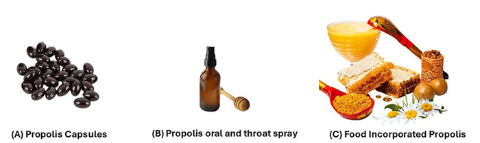
Propolis applications
Propolis poses challenges for food applications due to its strong sensory properties such as smell, colour, and taste. Ray et al. (2016) proposed encapsulating its active components to mask flavour and protect antioxidants, using nanoparticles formed by enclosing the active agent within a wall substance [215]. Spray drying, enhanced by additives like maltodextrin and Arabic gum, has proven effective for improving properties of encapsulated propolis. As well as propolis-based protective layers on fruit surfaces improve texture preservation, reduce industrial packaging needs, and extend shelf life while preventing microbial contamination [216].
8 Discussion
Propolis has garnered significant attention due to its diverse phytochemical constituents and extensive biological activities, supported by numerous in vitro and in vivo studies. Rich in bioactive compounds such as flavonoids, phenolic acids, and terpenes, propolis demonstrates antioxidant, anti-inflammatory, antibacterial, anticancer, antidiabetic, and immunomodulatory properties, making it highly valuable for disease prevention and therapeutic support.
Comparative analyses of Brazilian, Chinese, Malaysian, and Australian propolis have shown that variations in botanical sources, environmental conditions, and bee species significantly influence chemical composition and biological efficacy. Brazilian green propolis, characterized by high levels of artepillin C and caffeic acid phenethyl ester (CAPE), exhibits strong anti-inflammatory and antioxidant properties. Malaysian propolis is abundant in flavonoids and terpenes, enhancing its antimicrobial and wound-healing capacities. Chinese propolis contains unique phenolic alcohols and sesquiterpenes, providing notable cardioprotective and anti-inflammatory effects, while Australian propolis, rich in flavonoids and triterpenoids, possesses significant antimicrobial and anticancer potential.
Propolis effectively modulates inflammatory pathways, including NF-κB, reducing cytokines such as TNF-α and IL-6, thus demonstrating efficacy in managing chronic inflammatory conditions like atherosclerosis and rheumatoid arthritis. Additionally, its immunomodulatory properties—such as promoting macrophage differentiation and enhancing antibody production—highlight its promising application in vaccine development and immune therapy. Its antioxidant activity, attributed mainly to flavonoids and phenolic acids, neutralizes reactive oxygen species, reducing oxidative stress and protecting against cardiovascular diseases and cancer. The antibacterial properties of propolis further extend their therapeutic relevance, especially against antibiotic-resistant pathogens, indicating potential as an alternative or adjunctive treatment.
Despite these promising findings, critical issues remain. Variability in chemical composition among different propolis types necessitates standardized extraction and formulation protocols to ensure consistency and reproducibility. Additionally, the bioavailability and pharmacokinetics of propolis are challenging due to its poor solubility and rapid metabolism. Advanced delivery systems, including nanotechnology-based encapsulation, have been proposed, though clinical validation is still required. Further research into potential allergic reactions and long-term toxicity is also essential for ensuring safety.
Moreover, the dual effects of propolis, acting as an antioxidant at low concentrations and a pro-oxidant at high concentrations, complicate therapeutic applications and require precise dose determination. The promising anticancer potential of propolis, involving apoptosis induction and modulation of the tumor microenvironment, demands comprehensive evaluation in controlled clinical settings due to variability in response among different cancers and possible interactions with conventional treatments.
Clinical trials have yielded mixed outcomes, emphasizing the necessity for large-scale, rigorously designed studies to conclusively establish efficacy, optimal dosing, and safety propolis. Establishing regulatory frameworks and standardized guidelines will further enhance the credibility, safety, and widespread acceptance of propolis-based therapeutic products in modern medical practice.
9 The main research findings
Propolis demonstrates various biological actions, encompassing antioxidants, anti-inflammatory, antibacterial, anticancer, antidiabetic, and immunomodulatory properties. Green Brazilian propolis, abundant in artepillin C and CAPE, exhibits potent anti-inflammatory and antioxidant characteristics. Malaysian propolis, rich in flavonoids and terpenes, augments antibacterial and wound-healing efficacy. Chinese propolis, which comprises phenolic alcohols and sesquiterpenes, exhibits cardioprotective and anti-inflammatory properties. Australian propolis, rich in flavonoids and triterpenoids, exhibits antibacterial and anticancer properties. Inflammatory pathways (like NF-κB) are controlled by propolis, which also boosts the immune system, lowers oxidative stress, speeds up glucose metabolism, and kills cancer cells. Its antibacterial activities are efficacious against antibiotic-resistant pathogens.
10 Conclusion
In conclusion, propolis is an important resource in integrative medicine and nutrition, with bioactive compounds contributing to its potential in disease prevention, and therapeutic support for several conditions. Whether used raw, as extracts, or with other products, it holds promise in supplements and cosmetics. Further research is needed to address allergies, clarify molecular pathways, and determine appropriate dosages, ensuring its full therapeutic potential is realized.
The main research findings are that Propolis demonstrates various biological actions, encompassing antioxidant, anti-inflammatory, antibacterial, anticancer, antidiabetic, and immunomodulatory properties. Green Brazilian propolis, abundant in artepillin C and CAPE, exhibits potent anti-inflammatory and antioxidant characteristics. Malaysian propolis, rich in flavonoids and terpenes, augments antibacterial and wound-healing efficacy. Chinese propolis, which comprises phenolic alcohols and sesquiterpenes, exhibits cardioprotective and anti-inflammatory properties. Australian propolis, rich in flavonoids and triterpenoids, exhibits antibacterial and anticancer properties. Inflammatory pathways (like NF-κB) are controlled by propolis, which also boosts the immune system, lowers oxidative stress, speeds up glucose metabolism, and kills cancer cells. Its antibacterial activities are efficacious against antibiotic-resistant pathogens.
Future studies should focus on standardizing extraction and formulation methods to reduce compositional variability. Enhancing bioavailability with advanced delivery technologies, like nanotechnology-based encapsulation (e.g., liposomes, nanoparticles) can improve efficacy. Clinical studies are crucial for establishing the optimal dosage, safety, and long-term effects. Understanding the dual antioxidant and pro-oxidant characteristics of propolis will improve its therapeutic use. The medical potential could be enhanced by establishing regulatory standards and investigating its interaction with current treatments.
It should explore the environmental impact on propolis composition and develop sustainable harvesting practices to ensure long-term availability and consistency in therapeutic efficacy. The development of propolis-based products tailored to specific health conditions, combined with an improved understanding of its molecular and cellular mechanisms, will enable the creation of more effective and targeted treatments.
Notes
Acknowledgements
We want to thank our colleagues for their helpful discussions and comments. The work reported in this study is funded by the Universiti Sains Malaysia (USM), RESEARCH UNIVERSITY (RU) GRANT (No. R502-KR-ARU001-0008012332-K134). The authors would like to thank USM for its invaluable support of this study.
Author contributions
All the listed authors contributed to this study and agreed to publish it. Nosiba A. Alsarayrah: methodology, investigation, validation, resources, visualization, and writing—original draft. Rafeezul Mohamed: validation, writing—review and editing. Eshaifol A. Omar: writing—review & editing, visualization, supervision and project administration. The author(s) read and approved the final manuscript.
Funding
Universiti Sains Malaysia, R502-KR-ARU001-0008012332-K134, Eshaifol A. Omar
Data availability
Data sharing is not applicable to this article, as no datasets were generated or analysed during the current study; all the articles analysed in this study are cited with its DOI (when available).
Declarations
Ethics approval and consent to participate
Not applicable.
Competing interests
The authors declare that there are no competing interests associated with this work.
References
-
1.Ga MELO. Stingless bees (meliponini). Switzerland: Springer, 2020. PubMed Google Scholar
-
2.Asem N, Abdul Gapar NA, Abd Hapit NH, et al. Correlation between total phenolic and flavonoid contents with antioxidant activity of Malaysian stingless bee propolis extract. J Apic Res 2020;59(4): 437-42. CrossRef PubMed Google Scholar
-
3.Hamzah SA, Zawawi N, Sabri S. A review on the association of bacteria with stingless bees. Sains Malaysiana 2020. CrossRef PubMed Google Scholar
-
4.Ristivojević P, Trifković J, Andrić F, et al. Poplar-type propolis: chemical composition, botanical origin and biological activity. Nat Prod Commun 2015;10(11): 1934578X1501001117. CrossRef PubMed Google Scholar
-
5.Machado B, Pulcino TN, Silva AL, et al. Propolis as an alternative in prevention and control of dental cavity. J Apither 2017. CrossRef PubMed Google Scholar
-
6.Kuropatnicki AK, Szliszka E, Krol W. Historical aspects of propolis research in modern times. Evid Complemen Altern Med 2013. CrossRef PubMed Google Scholar
-
7.Sung S-H, Choi G-H, Lee N-W, et al. External use of propolis for oral, skin, and genital diseases: a systematic review and meta-analysis. Evid Complement Altern Med 2017. CrossRef PubMed Google Scholar
-
8.Elnakady YA, Rushdi AI, Franke R, et al. Characteristics, chemical compositions and biological activities of propolis from Al-Bahah, Saudi Arabia. Sci Rep 2017. CrossRef PubMed Google Scholar
-
9.Martinotti S, Ranzato E. Propolis: a new frontier for wound healing. Burns Trauma 2015;3(1): 1-7. CrossRef PubMed Google Scholar
-
10.Vd WAGH. Propolis: a wonder bees product and its pharmacological potentials. Adv Pharmacol Pharm Sci 2013. CrossRef PubMed Google Scholar
-
11.Stawiarz E, Dyduch J. The use of honey bee products of plant origin in apitherapy. Episteme 2014;25: 111-27. PubMed Google Scholar
-
12.Matuszewska E, Klupczynska A, Maciołek K, et al. Multielemental analysis of bee pollen, propolis, and royal jelly collected in west-central Poland. Molecules 2021;26(9): 2415. CrossRef PubMed Google Scholar
-
13.Ahangari Z, Naseri M, Vatandoost F. Propolis: chemical composition and its applications in endodontics. Iran Endod J 2018;13(3): 285. CrossRef PubMed Google Scholar
-
14.Pasupuleti VR, Sammugam L, Ramesh N, et al. Honey, propolis, and royal jelly: a comprehensive review of their biological actions and health benefits. Oxid Med Cell Long 2017. CrossRef PubMed Google Scholar
-
15.Hayriye A. Effects of propolis on immune system. Anadolu Ege Tarımsal Araştırma Enstitüsü Dergisi, 2018. 28(2): 99–104. https://dergipark.org.tr/en/pub/anadolu/issue/41816/504495. PubMed Google Scholar
-
16.Krol W, Scheller S, Czuba Z, et al. Inhibition of neutrophils’ chemiluminescence by ethanol extract of propolis (EEP) and its phenolic components. J Ethnopharmacol 1996;55(1): 19-25. CrossRef PubMed Google Scholar
-
17.Kapare HS, Sathiyanarayanan L. Nutritional and therapeutic potential of propolis: a review. Res J Pharm Technol 2020;13(7): 3545-9. CrossRef PubMed Google Scholar
-
18.Hodel KVS, Machado BAS, Santos NR, et al. Metal content of nutritional and toxic value in different types of brazilian propolis. Sci World J 2020;2020(1): 4395496. CrossRef PubMed Google Scholar
-
19.Pobiega K, Kot AM, Przybył JL, et al. Comparison of the chemical composition and antioxidant properties of propolis from urban apiaries. Molecules 2023;28(18): 6744. CrossRef PubMed Google Scholar
-
20.Moskwa J, Naliwajko SK, Markiewicz-Żukowska R, et al. Propolis from poland versus propolis from new zealand-chemical composition and antiproliferative properties on glioblastoma cell lines. 2020. PubMed Google Scholar
-
21.Maroof K, Gan SH. A review on chemical compositions, biological activity and formulation techniques of Malaysian honey bee and meliponine propolis. J Biol Act Prod Nat 2020;10(6): 507-23. CrossRef PubMed Google Scholar
-
22.Pilario KE, Tielemans A, Mojica E-RE. Geographical discrimination of propolis using dynamic time warping kernel principal components analysis. Expert Syst App 2022. CrossRef PubMed Google Scholar
-
23.Chi Y, Luo L, Cui M, et al. Chemical composition and antioxidant activity of essential oil of Chinese propolis. Chem Biodivers 2020. CrossRef PubMed Google Scholar
-
24.Massaro CF, Simpson JB, Powell D, et al. Chemical composition and antimicrobial activity of honeybee (Apis mellifera ligustica) propolis from subtropical eastern Australia. Sci Nat 2015;102: 1-11. CrossRef PubMed Google Scholar
-
25.El-Guendouz S, Lyoussi B, Miguel MG. Insight on propolis from mediterranean countries: chemical composition, biological activities and application fields. Chem Biodiv 2019. CrossRef PubMed Google Scholar
-
26.Oliveira L, Macedo M, Rodrigues J, et al. Plant metabolite 5-pentadecyl resorcinol is produced by the amazonian fungus penicillium sclerotiorum lm 5679. Braz J Biol 2021. CrossRef PubMed Google Scholar
-
27.Romagnoli C, Baldisserotto A, Vicentini CB, et al. Antidermatophytic action of resorcinol derivatives: ultrastructural evidence of the activity of phenylethyl resorcinol against microsporum gypseum. Molecules 2016;21(10): 1306. CrossRef PubMed Google Scholar
-
28.Zhang Y-J, Chen X, Zhang L, et al. Protective effects of 3, 4-dihydroxyphenylethanol on spinal cord injury-induced oxidative stress and inflammation. NeuroReport 2019;30(15): 1016-24. CrossRef PubMed Google Scholar
-
29.Shehata MG, Ahmad FT, Badr AN, et al. Chemical analysis, antioxidant, cytotoxic and antimicrobial properties of propolis from different geographic regions. Ann Agric Sci 2020;65(2): 209-17. CrossRef PubMed Google Scholar
-
30.Harbatsevich H, Loginova N, Nabebina K, et al. Nickel (ⅱ) complexes with ‘non innocent’ligands–cycloaminomethyl derivatives of 1, 2-dihydroxybenzene: sod-like and antimicrobial activity. RAD Assoc J 2017;2(2): 129-33. CrossRef PubMed Google Scholar
-
31.Krishna CM, Liebmann JE, Kaufman D, et al. The catecholic metal sequestering agent 1, 2-dihydroxybenzene-3, 5-disulfonate confers protection against oxidative cell damage. Arch Biochem Biophys 1992;294(1): 98-106. CrossRef PubMed Google Scholar
-
32.Kerdsomboon K, Chumsawat W, Auesukaree C. Effects of moringa oleifera leaf extracts and its bioactive compound gallic acid on reducing toxicities of heavy metals and metalloid in saccharomyces cerevisiae. Chemosphere 2021;270: 128659. CrossRef PubMed Google Scholar
-
33.Wu Y, Li K, Zeng M, et al. Serum metabolomics analysis of the anti-inflammatory effects of gallic acid on rats with acute inflammation. Front Pharmacol 2022. CrossRef PubMed Google Scholar
-
34.Ismail T, Sulaiman SA, Ponnuraj KT, et al. Chemical constituents of malaysian apis mellifera propolis. Sains Malays 2018. CrossRef PubMed Google Scholar
-
35.Deng Z, Li C, Luo D, et al. A new cinnamic acid derivative from plant-derived endophytic fungus pyronema sp. Nat Prod Res 2017;31(20): 2413-9. CrossRef PubMed Google Scholar
-
36.Lan J-S, Hou J-W, Liu Y, et al. Design, synthesis and evaluation of novel cinnamic acid derivatives bearing n-benzyl pyridinium moiety as multifunctional cholinesterase inhibitors for alzheimer’s disease. J Enzyme Inhib Med Chem 2017;32(1): 776-88. CrossRef PubMed Google Scholar
-
37.Amalan V, Vijayakumar N, Ramakrishnan A. P-coumaric acid regulates blood glucose and antioxidant levels in streptozotocin induced diabetic rats. J Chem Pharm Res 2015;7(7): 831-9. CrossRef PubMed Google Scholar
-
38.Muhammad N, Saeed M, Adhikari A, et al. Isolation of a new bioactive cinnamic acid derivative from the whole plant of viola betonicifolia. J Enzyme Inhib Med Chem 2013;28(5): 997-1001. CrossRef PubMed Google Scholar
-
39.Keshari AK, Verma AK, Kumar T, et al. Oxidative stress: a review. Int J Sci Technol 2015;3(7): 155. PubMed Google Scholar
-
40.Singh P, Grewal AS, Pandita D, et al. Synthesis and evaluation of a series of caffeic acid derivatives as anticancer agents. Future J Pharm Sci 2018;4(2): 124-30. CrossRef PubMed Google Scholar
-
41.Sun J, Ran Y, Wang Y, et al. Synthesis of bioisosteres of caffeic acid phenethyl ester: 1, 3, 4-oxadiazole derivatives containing a catechol fragment with anti-inflammatory activities in vitro and in vivo. Bioorg Chem 2025;155: 108-23. CrossRef PubMed Google Scholar
-
42.Falcão SI, Vale N, Gomes P, et al. Phenolic profiling of portuguese propolis by lc–ms spectrometry: uncommon propolis rich in flavonoid glycosides. Phytochem Anal 2013;24(4): 309-18. CrossRef PubMed Google Scholar
-
43.Adeyemi OS, Atolani O, Banerjee P, et al. Computational and experimental validation of antioxidant properties of synthesized bioactive ferulic acid derivatives. Int J Food Prop 2018;21(1): 86-98. CrossRef PubMed Google Scholar
-
44.Wu Z, Zhang J, Chen J, et al. Design, synthesis, antiviral bioactivity and three-dimensional quantitative structure–activity relationship study of novel ferulic acid ester derivatives containing quinazoline moiety. Pest Manag Sci 2017;73(10): 2079-89. CrossRef PubMed Google Scholar
-
45.Stompor-Gorący M, Machaczka M. Recent advances in biological activity, new formulations and prodrugs of ferulic acid. Int J Mol Sci 2021;22(23): 12889. CrossRef PubMed Google Scholar
-
46.Salehi B, Venditti A, Sharifi-Rad M, et al. The therapeutic potential of apigenin. Int J Mol Sci 2019;20(6): 1305. CrossRef PubMed Google Scholar
-
47.Ibrahim N, Niza N, Rodi MM, et al. Chemical and biological analyses of malaysian stingless bee propolis extracts. Malays J Anal Sci 2016. CrossRef PubMed Google Scholar
-
48.Sabatier S, Amiot M, Tacchini M, et al. Identification of flavonoids in sunflower honey. J Food Sci 1992;57(3): 773-4. CrossRef PubMed Google Scholar
-
49.Dabeek WM, Marra MV. Dietary quercetin and kaempferol: bioavailability and potential cardiovascular-related bioactivity in humans. Nutrients 2019;11(10): 2288. CrossRef PubMed Google Scholar
-
50.Rasul A, Millimouno FM, Ali Eltayb W, et al. Pinocembrin: a novel natural compound with versatile pharmacological and biological activities. BioMed Res Int 2013. CrossRef PubMed Google Scholar
-
51.Lambert JD, Sang S, Hong J, et al. Peracetylation as a means of enhancing in vitro bioactivity and bioavailability of epigallocatechin-3-gallate. Drug Metab Dispos 2006;34(12): 2111-6. CrossRef PubMed Google Scholar
-
52.Rasheed S, Rehman K, Shahid M, et al. Therapeutic potentials of genistein: new insights and perspectives. J Food Biochem 2022. CrossRef PubMed Google Scholar
-
53.Okińczyc P, Widelski J, Nowak K, et al. Phytochemical profiles and antimicrobial activity of selected Populus spp. bud extracts. Molecules 2024;29(2): 437. CrossRef PubMed Google Scholar
-
54.Nwiloh BI, Monago-Ighorodje CC, Onwubiko GN. Analyses of bioactive compounds in fiddleheads of pteridium aquilinum l. Kuhn collected from khana, southern nigeria, using gas chromatography-flame ionization detector. J Pharmacogn Phytochem 2020;9(2): 1079-86. PubMed Google Scholar
-
55.Stabrauskiene J, Kopustinskiene DM, Lazauskas R, et al. Naringin and naringenin: their mechanisms of action and the potential anticancer activities. Biomedicines 2022;10(7): 1686. CrossRef PubMed Google Scholar
-
56.Liu W, Zheng W, Cheng L, et al. Citrus fruits are rich in flavonoids for immunoregulation and potential targeting ACE2. Nat Prod Bioprospect 2022;12(1): 4. CrossRef PubMed Google Scholar
-
57.Silva CCFD, Salatino A, Motta LBD, et al. Chemical characterization, antioxidant and anti-HIV activities of a Brazilian propolis from Ceará state. Rev Bras Farmacogn 2019;29: 309-18. CrossRef PubMed Google Scholar
-
58.Gharacheh RH, Eslami M, Amani P, et al. Tacrine-flavonoid quercetin hybride as a mtdl ligand against alzheimer’s disease with metal chelating and ache, bche, ache-induced aβ aggregation inhibition properties: a computational study. J Arch 2020. CrossRef PubMed Google Scholar
-
59.Xiao Z-P, Wang X-D, Wang P-F, et al. Design, synthesis, and evaluation of novel fluoroquinolone–flavonoid hybrids as potent antibiotics against drug-resistant microorganisms. Eur J Med Chem 2014;80: 92-100. CrossRef PubMed Google Scholar
-
60.Unver T. Isorhamnetin as a promising natural bioactive flavonoid: in vitro assessment of its antifungal property. Int J Agric Environ Food Sci 2024;8(1): 54-61. CrossRef PubMed Google Scholar
-
61.Zangade SB, Dhulshette BS, Patil PB. Flavonoid-metal ion complexes as potent anticancer metallodrugs: a comprehensive review. Mini Rev Med Chem 2024;24(10): 1046-60. CrossRef PubMed Google Scholar
-
62.Kim D-S, Lim S-B. Subcritical water extraction of rutin from the aerial parts of common buckwheat. J Supercrit Fluids 2019;152: 104561. CrossRef PubMed Google Scholar
-
63.Bangar SP, Sharma N, Sanwal N, et al. Bioactive potential of beetroot (beta vulgaris). Food Res Int 2022;158: 111556. CrossRef PubMed Google Scholar
-
64.Elangovan B. A review on pharmacological studies of natural flavanone: pinobanksin. 3 Biotech 2024;14(4): 111. CrossRef PubMed Google Scholar
-
65.Song Y, Wu W, Sheng L, et al. Chrysin ameliorates hepatic steatosis induced by a diet deficient in methionine and choline by inducing the secretion of hepatocyte nuclear factor 4α-dependent very low-density lipoprotein. J Biochem Mol Toxicol 2020. CrossRef PubMed Google Scholar
-
66.Xie Y, Peng X. Effects of chrysin on the apoptosis in oral squamous carcinoma kb cell line and the underlying mechanisms. Zhong nan da xue xue bao Yi xue ban J Central South Univ Med Sci 2019. CrossRef PubMed Google Scholar
-
67.Li H-J, Wu N-L, Pu C-M, et al. Chrysin alleviates imiquimod-induced psoriasis-like skin inflammation and reduces the release of ccl20 and antimicrobial peptides. Sci Rep 2020;10(1): 2932. CrossRef PubMed Google Scholar
-
68.Zhang S, Mao B, Cui S, et al. Absorption, metabolism, bioactivity, and biotransformation of epigallocatechin gallate. Crit Rev Food Sci Nutr 2024;64(19): 6546-66. CrossRef PubMed Google Scholar
-
69.Taheri Y, Suleria HAR, Martins N, et al. Myricetin bioactive effects: moving from preclinical evidence to potential clinical applications. Bmc Complement Med Ther 2020. CrossRef PubMed Google Scholar
-
70.Okińczyc P, Widelski J, Ciochoń M, et al. Phytochemical profile, plant precursors and some properties of georgian propolis. Molecules 2022;27(22): 7714. CrossRef PubMed Google Scholar
-
71.Puertas-Bartolomé M, Włodarczyk-Biegun MK, del Campo A, et al. Development of bioactive catechol functionalized nanoparticles applicable for 3D bioprinting. Mater Sci Eng 2021. CrossRef PubMed Google Scholar
-
72.Puertas-Bartolomé M, Vázquez-Lasa B, San Román J. Bioactive and bioadhesive catechol conjugated polymers for tissue regeneration. Polymers 2018;10(7): 768. CrossRef PubMed Google Scholar
-
73.dos Santos AN, de L Nascimento TR, Gondim BL, et al. Catechins as model bioactive compounds for biomedical applications. Curr Pharm Des 2020. CrossRef PubMed Google Scholar
-
74.Kimura Y, Sumiyoshi M. Antitumor and antimetastatic actions of dihydroxycoumarins (esculetin or fraxetin) through the inhibition of m2 macrophage differentiation in tumor-associated macrophages and/or g1 arrest in tumor cells. Eur J Pharmacol 2015;746: 115-25. CrossRef PubMed Google Scholar
-
75.Yang L, Ding W, Xu Y, et al. New insights into the antibacterial activity of hydroxycoumarins against Ralstonia solanacearum. Molecules 2016;21(4): 468. CrossRef PubMed Google Scholar
-
76.Paraschiv C, Gosav S, Burlacu CM, et al. Exploring the inhibitory efficacy of resokaempferol and tectochrysin on pi3kα protein by combining dft and molecular docking against wild-type and h1047r mutant forms. Inventions 2024;9(5): 96. CrossRef PubMed Google Scholar
-
77.Ijaz MU, Alvi K, Hamza A, et al. Curative effects of tectochrysin on paraquat-instigated testicular toxicity in rats: a biochemical and histopathological based study. Heliyon 2024. CrossRef PubMed Google Scholar
-
78.Pillaiyar T, Manickam M, Namasivayam V. Skin whitening agents: medicinal chemistry perspective of tyrosinase inhibitors. J Enzyme Inhib Med Chem 2017;32(1): 403-25. CrossRef PubMed Google Scholar
-
79.Ogunleye FA, Fapohunda O, Nwangwu S. A review on medicinal uses and pharmacological activities of african star apple (chrysophyllum albidum). Acta Scientific Cancer Biology, 2020. 1(4). https://actascientific.com/ASPC/pdf/ASPC-01-0023.pdf. PubMed Google Scholar
-
80.Jia A, Liu F, Fan S-Y. In vivo antihyperuricemic activities of 3, 4, 5-tri-o-caffeoylquinic acid, 4, 4’, 6’-trihydroxy-2’-methoxychalcone, and caffeic acid from the aerial parts of Gnaphalium affine. Pharm Front 2023;5(02): e77-83. CrossRef PubMed Google Scholar
-
81.Fu Y, Chen J, Li Y-J, et al. Antioxidant and anti-inflammatory activities of six flavonoids separated from licorice. Food Chem 2013;141(2): 1063-71. CrossRef PubMed Google Scholar
-
82.Sinyeue C, Matsui M, Oelgemöller M, et al. Synthesis and investigation of flavanone derivatives as potential new anti-inflammatory agents. Molecules 2022;27(6): 1781. CrossRef PubMed Google Scholar
-
83.Nina N, Quispe C, Jiménez-Aspee F, et al. Antibacterial activity, antioxidant effect and chemical composition of propolis from the región del Maule, central Chile. Molecules 2015;20(10): 18144-67. CrossRef PubMed Google Scholar
-
84.Bai M, Zheng C-J, Wu L-J, et al. Bioactive flavonoid derivatives from scutellaria luzonica. Chem Nat Compd 2018;54: 350-3. CrossRef PubMed Google Scholar
-
85.Chang LS, Li CB, Qin N, et al. Synthesis and antidiabetic activity of 5, 7-dihydroxyflavonoids and analogs. Chem Biodivers 2012;9(1): 162-9. CrossRef PubMed Google Scholar
-
86.Afolayan A, Meyer J. The antimicrobial activity of 3, 5, 7-trihydroxyflavone isolated from the shoots of helichrysum aureonitens. J Ethnopharmacol 1997;57(3): 177-81. CrossRef PubMed Google Scholar
-
87.Schultz DJ, Wickramasinghe NS, Klinge CM. Anacardic acid biosynthesis and bioactivity. Amsterdam: Elsevier, 2006. PubMed Google Scholar
-
88.Nguyen HX, VAN Do TN, Nguyen MTT, et al. A new alkenylphenol from the propolis of stingless bee trigona minor. Nat Prod Commun 2018. CrossRef PubMed Google Scholar
-
89.Alen Y, Nakajima S, Nitoda T, et al. Two antinematodal phenolics from knema hookeriana, a Sumatran rainforest plant. Z Naturforsch C 2000;55(3–4): 300-4. CrossRef PubMed Google Scholar
-
90.Kardar M, Zhang T, Coxon G, et al. Characterisation of triterpenes and new phenolic lipids in Cameroonian propolis. Phytochemistry 2014;106: 156-63. CrossRef PubMed Google Scholar
-
91.Ts S, Senthilraja P, Manivel G. Molecular docking, admet property analysis and antibacterial potency of bioactive compounds from marine bacillus cereus against espf (e. Coli). World J Pharm Res 2024. CrossRef PubMed Google Scholar
-
92.Negri G, Silva CCF, Coelho GR, et al. Cardanols detected in non-polar propolis extracts from scaptotrigona aff. postica (hymenoptera, apidae, meliponini). Braz J Food Technol 2019;22: e2018265. CrossRef PubMed Google Scholar
-
93.Knödler M, Conrad J, Wenzig EM, et al. Anti-inflammatory 5-(11′ z-heptadecenyl)-and 5-(8′ z, 11′ z-heptadecadienyl)-resorcinols from mango (Mangifera indica L.) peels. Phytochemistry 2008;69(4): 988-93. CrossRef PubMed Google Scholar
-
94.Montanari RM, Barbosa LC, Demuner AJ, et al. Chemical composition and antibacterial activity of essential oils from verbenaceae species: alternative sources of (E)-caryophyllene and germacrene-D. Quim Nova 2011;34: 1550-5. CrossRef PubMed Google Scholar
-
95.Kim TD, Lee JY, Cho BJ, et al. The analgesic and anti-inflammatory effects of 7-oxosandaracopimaric acid isolated from the roots of aralia cordata. Arch Pharm Res 2010;33: 509-14. CrossRef PubMed Google Scholar
-
96.Jerz G, Elnakady YA, Braun A, et al. Preparative mass-spectrometry profiling of bioactive metabolites in saudi-arabian propolis fractionated by high-speed countercurrent chromatography and off-line atmospheric pressure chemical ionization mass-spectrometry injection. J Chromatogr A 2014;1347: 17-29. CrossRef PubMed Google Scholar
-
97.Proteggente AR, Pannala AS, Paganga G, et al. The antioxidant activity of regularly consumed fruit and vegetables reflects their phenolic and vitamin C composition. Free Radic Res 2002;36(2): 217-33. CrossRef PubMed Google Scholar
-
98.Salleh SNAS, Hanapiah NAM, Johari WLW, et al. Analysis of bioactive compounds and chemical composition of Malaysian stingless bee propolis water extracts. Saudi J Biol Sci 2021;28(12): 6705-10. CrossRef PubMed Google Scholar
-
99.Traber MG, Atkinson J. Vitamin E, antioxidant and nothing more. Free Radic Biol Med 2007;43(1): 4-15. CrossRef PubMed Google Scholar
-
100.Guerrini A, Bruni R, Maietti S, et al. Ecuadorian stingless bee (meliponinae) honey: a chemical and functional profile of an ancient health product. Food Chem 2009;114(4): 1413-20. CrossRef PubMed Google Scholar
-
101.Gang F-L, Zhu F, Li X-T, et al. Synthesis and bioactivities evaluation of l-pyroglutamic acid analogues from natural product lead. Bioorg Med Chem 2018;26(16): 4644-9. CrossRef PubMed Google Scholar
-
102.Tania Ad, Suoth E, Fatimawali F, et al. Molecular docking of bioactive compounds of nut grass (cyperus rotundus l.) tuber against sars-cov-2. New York: AIP Publishing, 2023. DOI:10.1063/5.0103882 CrossRef PubMed Google Scholar
-
103.ÖZER. Propolis and potential use in food products. Turkish J Agriculture-Food Sci Technol 2020;8(5): 1139-44. CrossRef PubMed Google Scholar
-
104.Gavanji S, Larki B. Comparative effect of propolis of honey bee and some herbal extracts on Candida albicans. Chin J Integr Med 2017;23: 201-7. CrossRef PubMed Google Scholar
-
105.Mulyati AH, Sulaeman A, Marliyati SA, et al. Macro and micronutrient content of raw propolis collected from different regions in Indonesia. J Gizi Pangan 2021;16(1): 109-14. PubMed Google Scholar
-
106.Ristivojević P, Nešić J, Andrić F, et al. Elemental profile of propolis from different areas of Serbia. Chem Biodivers 2023. CrossRef PubMed Google Scholar
-
107.Dias LG, Pereira AP, Estevinho LM. Comparative study of different portuguese samples of propolis: pollinic, sensorial, physicochemical, microbiological characterization and antibacterial activity. Food Chem Toxicol 2012;50(12): 4246-53. CrossRef PubMed Google Scholar
-
108.Afata TN, Nemo R, Ishete N, et al. Phytochemical investigation, physicochemical characterization, and antimicrobial activities of Ethiopian propolis. Arab J Chem 2022;15(7): 103931. CrossRef PubMed Google Scholar
-
109.Kunrath CA, Savoldi DC, Mileski JPF, et al. Application and evaluation of propolis, the natural antioxidant in italian-type salami. Brazilian J Food Technol 2017. CrossRef PubMed Google Scholar
-
110.Pant K, Thakur M, Chopra H, et al. Characterization and discrimination of Indian propolis based on physico-chemical, techno-functional, thermal and textural properties: a multivariate approach. J King Saud Univ-Sci 2021;33(4): 101405. CrossRef PubMed Google Scholar
-
111.Godhi BS, Beeraka NM, Buddi JSHP, et al. Updates in the analytical isolation of Indian propolis chemical constituents and their role in dental pharmacology - a review. Nat Prod J 2022;12(7): 77-88. CrossRef PubMed Google Scholar
-
112.Cui-Ping Z, Shuai H, Wen-Ting W, et al. Development of high-performance liquid chromatographic for quality and authenticity control of chinese propolis. J Food Sci 2014;79(7): C1315-22. CrossRef PubMed Google Scholar
-
113.Singh P, Mishra SK, Noel S, et al. Acute exposure of apigenin induces hepatotoxicity in swiss mice. PLoS ONE 2012;7(2): e31964. CrossRef PubMed Google Scholar
-
114.Middleton E Jr, Kandaswami C, Theoharides TC. The effects of plant flavonoids on mammalian cells: implications for inflammation, heart disease, and cancer. Pharmacol Rev 2000;52(4): 673-751. CrossRef PubMed Google Scholar
-
115.Kaur P, Shukla S, Gupta S. Plant flavonoid apigenin inactivates akt to trigger apoptosis in human prostate cancer: an in vitro and in vivo study. Carcinogenesis 2008;29(11): 2210-7. CrossRef PubMed Google Scholar
-
116.Liu G, Bao X, Jiang Y, et al. Identifying the association between Alzheimer’s disease and Parkinson’s disease using genome-wide association studies and protein-protein interaction network. Mol Neurobiol 2015;52: 1629-36. CrossRef PubMed Google Scholar
-
117.Choi M-J, Lee E-J, Park J-S, et al. Anti-inflammatory mechanism of galangin in lipopolysaccharide-stimulated microglia: critical role of ppar-γ signaling pathway. Biochem Pharmacol 2017;144: 120-31. CrossRef PubMed Google Scholar
-
118.Sivakumar AS, Viswanathan P, Anuradha CV. Dose-dependent effect of galangin on fructose-mediated insulin resistance and oxidative events in rat kidney. Redox Rep 2010;15(5): 224-32. CrossRef PubMed Google Scholar
-
119.Liu Y, Liang X, Zhang G, et al. Galangin and pinocembrin from propolis ameliorate insulin resistance in hepg2 cells via regulating akt/mtor signaling. Evid Complement Altern Med 2018;2018(1): 7971842. CrossRef PubMed Google Scholar
-
120.Melliou E, Stratis E, Chinou I. Volatile constituents of propolis from various regions of Greece–antimicrobial activity. Food Chem 2007;103(2): 375-80. CrossRef PubMed Google Scholar
-
121.Popova M, Trusheva B, Antonova D, et al. The specific chemical profile of Mediterranean propolis from Malta. Food Chem 2011;126(3): 1431-5. CrossRef PubMed Google Scholar
-
122.Zhao L, Yu M, Sun M, et al. Rapid determination of major compounds in the ethanol extract of geopropolis from Malaysian stingless bees, Heterotrigona itama, by UHPLC-Q-TOF/MS and NMR. Molecules 2017;22(11): 1935. CrossRef PubMed Google Scholar
-
123.Nazir H, Shahidan WNS, Ibrahim HA, et al. Chemical constituents of malaysian geniotrigona thoracica propolis. Pertanika J Trop Agri Sci 2018;41(3): 955. PubMed Google Scholar
-
124.Pujirahayu N, Suzuki T, Katayama T. Cycloartane-type triterpenes and botanical origin of propolis of stingless Indonesian bee tetragonula sapiens. Plants 2019;8(3): 57. CrossRef PubMed Google Scholar
-
125.Du W-Y, Xiao Y, Yao J-J, et al. Involvement of NADPH oxidase in high-dose phenolic acid-induced pro-oxidant activity on rat mesenteric venules. Exp Ther Med 2017;13(1): 17-22. CrossRef PubMed Google Scholar
-
126.Michalak A. Phenolic compounds and their antioxidant activity in plants growing under heavy metal stress. Pol j environ stud 2006;15(4): 523-30. PubMed Google Scholar
-
127.Kurek-Górecka A, Rzepecka-Stojko A, Górecki M, et al. Structure and antioxidant activity of polyphenols derived from propolis. Molecules 2013;19(1): 78-101. CrossRef PubMed Google Scholar
-
128.Jordan LG, Booth BW. Her2+ breast cancer cells undergo apoptosis upon exposure to tannic acid released from remodeled cross-linked collagen type Ⅰ. J Biomed Mater Res A 2018;106(1): 26-32. CrossRef PubMed Google Scholar
-
129.Chung K-T, Wong TY, Wei C-I, et al. Tannins and human health: a review. Crit Rev Food Sci Nutr 1998;38(6): 421-64. CrossRef PubMed Google Scholar
-
130.Dvorakova M, Landa P. Anti-inflammatory activity of natural stilbenoids: a review. Pharmacol Res 2017;124: 126-45. CrossRef PubMed Google Scholar
-
131.el Khawand T, Courtois A, Valls J, et al. A review of dietary stilbenes: sources and bioavailability. Phytochem Rev 2018;17(5): 1007-29. CrossRef PubMed Google Scholar
-
132.Nikzad B, Vang O. Synthesis of prenylated pterostilbene and testing of its effect on human colon epithelial cells. Roskilde: Roskilde University; 2016. PubMed Google Scholar
-
133.Riviere C, Pawlus AD, Merillon J-M. Natural stilbenoids: distribution in the plant kingdom and chemotaxonomic interest in vitaceae. Nat Prod Rep 2012;29(11): 1317-33. CrossRef PubMed Google Scholar
-
134.da Silva Rivas AC, Lopes PM, de Azevedo Barros MM, et al. Biological activities of α-pinene and β-pinene enantiomers. Molecules 2012. CrossRef PubMed Google Scholar
-
135.Grover M, Behl T, Virmani T, et al. Exploration of cytotoxic potential of longifolene/junipene isolated from chrysopogon zizanioides. Molecules 2022;27(18): 5764. CrossRef PubMed Google Scholar
-
136.Manley-Harris M, Grainger M, Peters LM, et al. Composition and bioactivity of propolis derived from New Zealand native forest. Fitoterapia 2025. CrossRef PubMed Google Scholar
-
137.Suh S-J, Kwak C-H, Chung T-W, et al. Pimaric acid from aralia cordata has an inhibitory effect on TNF-α-induced MMP-9 production and HASMC migration via down-regulated NF-κB and AP-1. Chem Biol Interact 2012;199(2): 112-9. CrossRef PubMed Google Scholar
-
138.Smith EC, Williamson EM, Wareham N, et al. Antibacterials and modulators of bacterial resistance from the immature cones of chamaecyparis lawsoniana. Phytochemistry 2007;68(2): 210-7. CrossRef PubMed Google Scholar
-
139.Barrero AF, Herrador MM, Arteaga P, et al. Communic acids: occurrence, properties and use as chirons for the synthesis of bioactive compounds. Molecules 2012;17(2): 1448-67. CrossRef PubMed Google Scholar
-
140.Popova MP, Chinou IB, Marekov IN, et al. Terpenes with antimicrobial activity from cretan propolis. Phytochemistry 2009;70(10): 1262-71. CrossRef PubMed Google Scholar
-
141.Leong KH, Looi CY, Loong X-M, et al. Cycloart-24-ene-26-ol-3-one, a new cycloartane isolated from leaves of aglaia exima triggers tumour necrosis factor-receptor 1-mediated caspase-dependent apoptosis in colon cancer cell line. PLoS ONE 2016. CrossRef PubMed Google Scholar
-
142.Han G, Lee DG. Antibacterial mode of action of β-amyrin promotes apoptosis-like death in escherichia coli by producing reactive oxygen species. J Microbiol Biotechnol 2022;32(12): 1547. CrossRef PubMed Google Scholar
-
143.Meemongkolkiat T, Puthong S, Khongkarat P, et al. In vitro cytotoxic activity on kato-ⅲ cancer cell lines of mangiferolic acid purified from Thai Tetragonula laeviceps propolis. Heliyon 2024. CrossRef PubMed Google Scholar
-
144.Nair ANS, Nair RVR, Nair APR, et al. Antidiabetes constituents, cycloartenol and 24-methylenecycloartanol, from ficus krishnae. PLoS ONE 2020. CrossRef PubMed Google Scholar
-
145.Ristivojević P, Dimkić I, Guzelmeric E, et al. Profiling of Turkish propolis subtypes: comparative evaluation of their phytochemical compositions, antioxidant and antimicrobial activities. LWT 2018;95: 367-79. CrossRef PubMed Google Scholar
-
146.Silva-Carvalho R, Baltazar F, Almeida-Aguiar C. Propolis: a complex natural product with a plethora of biological activities that can be explored for drug development. Evid Complement Altern Med 2015. CrossRef PubMed Google Scholar
-
147.Wojtyczka RD, Dziedzic A, Idzik D, et al. Susceptibility of Staphylococcus aureus clinical isolates to propolis extract alone or in combination with antimicrobial drugs. Molecules 2013;18(8): 9623-40. CrossRef PubMed Google Scholar
-
148.Grenho L, Barros J, Ferreira C, et al. In vitro antimicrobial activity and biocompatibility of propolis containing nanohydroxyapatite. Biomed Mater 2015. CrossRef PubMed Google Scholar
-
149.Akhir RAM, Bakar MFA, Sanusi SB. Antioxidant and antimicrobial activity of stingless bee bread and propolis extracts. In AIP conference proceedings. AIP Publishing LLC 2017. CrossRef PubMed Google Scholar
-
150.Ramón-Sierra J, Peraza-López E, Rodríguez-Borges R, et al. Partial characterization of ethanolic extract of melipona beecheii propolis and in vitro evaluation of its antifungal activity. Rev Bras Farmacogn 2019;29: 319-24. CrossRef PubMed Google Scholar
-
151.Szweda P, Gucwa K, Kurzyk E, et al. Essential oils, silver nanoparticles and propolis as alternative agents against fluconazole resistant Candida albicans, Candida glabrata and Candida krusei clinical isolates. Indian J Microbiol 2015;55: 175-83. CrossRef PubMed Google Scholar
-
152.Boisard S, Le Ray A-M, Landreau A, et al. Antifungal and antibacterial metabolites from a french poplar type propolis. Evid Complement Altern Med 2015. CrossRef PubMed Google Scholar
-
153.Quiroga EN, Sampietro DA, Soberon JR, et al. Propolis from the northwest of Argentina as a source of antifungal principles. J Appl Microbiol 2006;101(1): 103-10. CrossRef PubMed Google Scholar
-
154.Martins ML, Monteiro ASN, Guimarães JEC, et al. Cytotoxic and antibacterial effect of a red propolis mouthwash, with or without fluoride, on the growth of a cariogenic biofilm. Arch Oral Biol 2019. CrossRef PubMed Google Scholar
-
155.Capoci IRG, Bonfim-Mendonça PDS, Arita GS, et al. Propolis is an efficient fungicide and inhibitor of biofilm production by vaginal Candida albicans. Evid Complement Altern Med 2015. CrossRef PubMed Google Scholar
-
156.Berretta AA, de Castro PA, Cavalheiro AH, et al. Evaluation of mucoadhesive gels with propolis (epp-af) in preclinical treatment of candidiasis vulvovaginal infection. Evid Complement Altern Med 2013. CrossRef PubMed Google Scholar
-
157.Conte FL, Tasca KI, Santiago KB, et al. Propolis increases foxp3 expression and lymphocyte proliferation in HIV-infected people: a randomized, double blind, parallel-group and placebo-controlled study. Biomed Pharmacother 2021;142: 111984. CrossRef PubMed Google Scholar
-
158.Yuan J, Liu J, Hu Y, et al. The immunological activity of propolis flavonoids liposome on the immune response against nd vaccine. Int J Biol Macromol 2012;51(4): 400-5. CrossRef PubMed Google Scholar
-
159.Li B, Wei K, Yang S, et al. Immunomodulatory effects of Taishan Pinus massoniana pollen polysaccharide and propolis on immunosuppressed chickens. Microb Pathog 2015;78: 7-13. CrossRef PubMed Google Scholar
-
160.Pahwa R, Goyal A, Jialal I. Chronic inflammation. StatPearls. 2021. https://www.ncbi.nlm.nih.gov/books/NBK493173/ PubMed Google Scholar
-
161.Chagas MDSS, Behrens MD, Moragas-Tellis CJ, et al. Flavonols and flavones as potential anti-inflammatory, antioxidant, and antibacterial compounds. Oxid Med Cell Longev 2022. CrossRef PubMed Google Scholar
-
162.Funakoshi-Tago M, Okamoto K, Izumi R, et al. Anti-inflammatory activity of flavonoids in Nepalese propolis is attributed to inhibition of the IL-33 signaling pathway. Int Immunopharmacol 2015;25(1): 189-98. CrossRef PubMed Google Scholar
-
163.Franchin M, Freires IA, Lazarini JG, et al. The use of Brazilian propolis for discovery and development of novel anti-inflammatory drugs. Eur J Med Chem 2018;153: 49-55. CrossRef PubMed Google Scholar
-
164.Machado JL, Assunçao AKM, da Silva MCP, et al. Brazilian green propolis: anti-inflammatory property by an immunomodulatory activity. Evid Complement Altern Med 2012. CrossRef PubMed Google Scholar
-
165.Teles F, da Silva TM, da Cruz Junior FP, et al. Brazilian red propolis attenuates hypertension and renal damage in 5/6 renal ablation model. PLoS ONE 2015. CrossRef PubMed Google Scholar
-
166.Naito Y, Yasumuro M, Kondou K, et al. Antiinflammatory effect of topically applied propolis extract in carrageenan-induced rat hind paw edema. Phytother Res 2007;21(5): 452-6. CrossRef PubMed Google Scholar
-
167.Franchin M, Colon DF, Castanheira FV, et al. Vestitol isolated from Brazilian red propolis inhibits neutrophils migration in the inflammatory process: elucidation of the mechanism of action. J Nat Prod 2016;79(4): 954-60. CrossRef PubMed Google Scholar
-
168.Zulhendri F, Lesmana R, Tandean S, et al. Recent update on the anti-inflammatory activities of propolis. Molecules 2022;27(23): 8473. CrossRef PubMed Google Scholar
-
169.Kitamura H, Saito N, Fujimoto J, et al. Brazilian propolis ethanol extract and its component kaempferol induce myeloid-derived suppressor cells from macrophages of mice in vivo and in vitro. BMC Complement Altern Med 2018;18: 1-11. CrossRef PubMed Google Scholar
-
170.Hsieh CY, Li LH, Rao YK, et al. Mechanistic insight into the attenuation of gouty inflammation by Taiwanese green propolis via inhibition of the NLRP3 inflammasome. J Cell Physiol 2019;234(4): 4081-94. CrossRef PubMed Google Scholar
-
171.Oršolić N, Skurić J, Đikić D, et al. Inhibitory effect of a propolis on di-n-propyl disulfide or n-hexyl salycilate-induced skin irritation, oxidative stress and inflammatory responses in mice. Fitoterapia 2014;93: 18-30. CrossRef PubMed Google Scholar
-
172.Zamarrenho LG, de Lima MHF, Hori JI, et al. Effects of three different Brazilian green propolis extract formulations on pro-and anti-inflammatory cytokine secretion by macrophages. Appl Sci 2023;13(10): 6247. CrossRef PubMed Google Scholar
-
173.Kantrong N, Kumtawee J, Damrongrungruang T, et al. An in vitro anti-inflammatory effect of Thai propolis in human dental pulp cells. J Appl Oral Sci 2023. CrossRef PubMed Google Scholar
-
174.Wolska K, Gorska A, Antosik K, et al. Immunomodulatory effects of propolis and its components on basic immune cell functions. Indian J Pharm Sci 2019;81(4): 575-88. CrossRef PubMed Google Scholar
-
175.Silva H, Francisco R, Saraiva A, et al. The cardiovascular therapeutic potential of propolis—a comprehensive review. Biology 2021;10(1): 27. CrossRef PubMed Google Scholar
-
176.Zhou H, Wang H, Shi N, et al. Potential protective effects of the water-soluble Chinese propolis on hypertension induced by high-salt intake. Clin Transl Sci 2020;13(5): 907-15. CrossRef PubMed Google Scholar
-
177.Oršolić N, Landeka Jurčević I, Đikić D, et al. Effect of propolis on diet-induced hyperlipidemia and atherogenic indices in mice. Antioxidants 2019;8(6): 156. CrossRef PubMed Google Scholar
-
178.Nader MA, El-Agamy DS, Suddek GM. Protective effects of propolis and thymoquinone on development of atherosclerosis in cholesterol-fed rabbits. Arch Pharm Res 2010;33: 637-43. CrossRef PubMed Google Scholar
-
179.Yigit E, Deger O, Korkmaz K, et al. Propolis reduces inflammation and dyslipidemia caused by high-cholesterol diet in mice by lowering ADAM10/17 activities. Nutrients 2024;16(12): 1861. CrossRef PubMed Google Scholar
-
180.Aziz MY, Omar EA, Zulkafli NES, et al. Therapeutic potential of malaysian stingless bee propolis extract targeting macrophages in atherogenesis: a review: therapeutic potential of malaysian stingless bee propolis extract. J Trop Life Sci 2024. CrossRef PubMed Google Scholar
-
181.Mohd Suib MS, Wan Omar WA, Omar EA, et al. Ethanolic extract of propolis from the Malaysian stingless bee geniotrigona thoracica inhibits formation of THP-1 derived macrophage foam cells. J Apic Res 2021;60(3): 478-90. CrossRef PubMed Google Scholar
-
182.Kurek-Górecka A, Kłósek M, Pietsz G, et al. Ethanolic extract of propolis and cape as cardioprotective agents against lps and ifn-α stressed cardiovascular injury. Nutrients 2024;16(5): 627. CrossRef PubMed Google Scholar
-
183.Mujica V, Orrego R, Pérez J, et al. The role of propolis in oxidative stress and lipid metabolism: a randomized controlled trial. Evid Complement Altern Med 2017. CrossRef PubMed Google Scholar
-
184.Zhao L, Pu L, Wei J, et al. Brazilian green propolis improves antioxidant function in patients with type 2 diabetes mellitus. Int J Environ Res Public Health 2016;13(5): 498. CrossRef PubMed Google Scholar
-
185.Nichitoi MM, Josceanu AM, Isopescu RD, et al. Polyphenolics profile effects upon the antioxidant and antimicrobial activity of propolis extracts. Sci Rep 2021;11(1): 1-12. CrossRef PubMed Google Scholar
-
186.Ahmed R, Tanvir E, Hossen MS, et al. Antioxidant properties and cardioprotective mechanism of malaysian propolis in rats. Evid Complement Altern Med 2017. CrossRef PubMed Google Scholar
-
187.Sung H, Ferlay J, Siegel RL, et al. Global cancer statistics 2020: Globocan estimates of incidence and mortality worldwide for 36 cancers in 185 countries. CA Cancer J Clin 2021;71(3): 209-49. CrossRef PubMed Google Scholar
-
188.Doi K, Fujioka M, Sokuza Y, et al. Chemopreventive action by ethanol-extracted brazilian green propolis on post-initiation phase of inflammation-associated rat colon tumorigenesis. In Vivo 2017. CrossRef PubMed Google Scholar
-
189.Oršolić N, Car N, Lisičić D, et al. Synergism between propolis and hyperthermal intraperitoneal chemotherapy with cisplatin on e hrlich ascites tumor in mice. J Pharm Sci 2013;102(12): 4395-405. CrossRef PubMed Google Scholar
-
190.Bonamigo T, Campos JF, Oliveira AS, et al. Antioxidant and cytotoxic activity of propolis of plebeia droryana and apis mellifera (hymenoptera, apidae) from the Brazilian cerrado biome. PLoS ONE 2017. CrossRef PubMed Google Scholar
-
191.Cisilotto J, Sandjo LP, Faqueti LG, et al. Cytotoxicity mechanisms in melanoma cells and UPLC-QTOF/MS2 chemical characterization of two Brazilian stingless bee propolis: uncommon presence of piperidinic alkaloids. J Pharm Biomed Anal 2018;149: 502-11. CrossRef PubMed Google Scholar
-
192.Mohamed WAS, Ismail NZ, Muhamad M, et al. Q-tof lc-ms compounds evaluation of propolis extract derived from malaysian stingless bees, tetrigona apicalis, and their bioactivities in breast cancer cell, mcf7. Saudi J Biol Sci 2022;29(10): 103403. CrossRef PubMed Google Scholar
-
193.Utispan K, Chitkul B, Koontongkaew S. Cytotoxic activity of propolis extracts from the stingless bee trigona sirindhornae against primary and metastatic head and neck cancer cell lines. Asian Pac J Cancer Prev 2017;18(4): 1051. CrossRef PubMed Google Scholar
-
194.Vongsak B, Chonanant C, Machana S. In vitro cytotoxicity of thai stingless bee propolis from chanthaburi orchard. Walailak J Sci Technol 2017;14(9): 741-7. PubMed Google Scholar
-
195.Umthong S, Phuwapraisirisan P, Puthong S, et al. In vitro antiproliferative activity of partially purified trigona laeviceps propolis from thailand on human cancer cell lines. BMC Complement Altern Med 2011;11: 1-8. CrossRef PubMed Google Scholar
-
196.Guzelmeric E, Sipahi H, Özhan Y, et al. Comprehensive estrogenic/anti-estrogenic, anticancer, mutagenic/anti-mutagenic, and genotoxic/anti-genotoxic activity studies on chemically characterized black poplar and eurasian aspen propolis types. J Pharm Biomed Anal 2023;226: 115241. CrossRef PubMed Google Scholar
-
197.Altabbal S, Athamnah K, Rahma A, et al. Propolis: a detailed insight of its anticancer molecular mechanisms. Pharmaceuticals 2023;16(3): 450. CrossRef PubMed Google Scholar
-
198.Komi DEA, Khomtchouk K, Santa Maria PL. A review of the contribution of mast cells in wound healing: involved molecular and cellular mechanisms. Clin Rev Allergy Immunol 2020. CrossRef PubMed Google Scholar
-
199.Olczyk P, Komosinska-Vassev K, Winsz-Szczotka K, et al. Propolis induces chondroitin/dermatan sulphate and hyaluronic acid accumulation in the skin of burned wound. Evid Complement Altern Med 2013. CrossRef PubMed Google Scholar
-
200.Kwiecińska-Piróg J, Skowron K, Śniegowska A, et al. The impact of ethanol extract of propolis on biofilm forming by proteus mirabilis strains isolated from chronic wounds infections. Nat Prod Res 2019;33(22): 3293-7. CrossRef PubMed Google Scholar
-
201.Berretta AA, Nascimento AP, Bueno PCP, et al. Propolis standardized extract (epp-af®), an innovative chemically and biologically reproducible pharmaceutical compound for treating wounds. Int J Biol Sci 2012;8(4): 512. CrossRef PubMed Google Scholar
-
202.Pahlevanneshan Z, Deypour M, Kefayat A, et al. Polyurethane-nanolignin composite foam coated with propolis as a platform for wound dressing: synthesis and characterization. Polymers 2021;13(18): 3191. CrossRef PubMed Google Scholar
-
203.Marquele-Oliveira F, DA Silva Barud H, Torres EC, et al. Development, characterization and pre-clinical trials of an innovative wound healing dressing based on propolis (epp-af®)-containing self-microemulsifying formulation incorporated in biocellulose membranes. Int J Biol Macromol 2019;136: 570-8. CrossRef PubMed Google Scholar
-
204.Karagecili H, YıLMAZ MA, Ertürk A, et al. Comprehensive metabolite profiling of berdav propolis using lc-ms/ms: determination of antioxidant, anticholinergic, antiglaucoma, and antidiabetic effects. Molecules 2023;28(4): 1739. CrossRef PubMed Google Scholar
-
205.el Rabey HA, Al-Seeni MN, Bakhashwain AS. The antidiabetic activity of nigella sativa and propolis on streptozotocin-induced diabetes and diabetic nephropathy in male rats. Evid Complement Altern Med 2017. CrossRef PubMed Google Scholar
-
206.Pd DA, Plashintania DR, Putri RM, et al. Synthesis of zinc oxide nanoparticles using methanol propolis extract (pro-zno nps) as antidiabetic and antioxidant. PLoS ONE 2023. CrossRef PubMed Google Scholar
-
207.Farida S, Pratami DK, Sahlan M, et al. In vitro study on antidiabetic and antihypertensive activities of ethanolic extract of propolis of Indonesian stingless bee Tetragonula sapiens. J King Saud University-Science 2023. CrossRef PubMed Google Scholar
-
208.Zakerkish M, Jenabi M, Zaeemzadeh N, et al. The effect of Iranian propolis on glucose metabolism, lipid profile, insulin resistance, renal function and inflammatory biomarkers in patients with type 2 diabetes mellitus: a randomized double-blind clinical trial. Sci Rep 2019. CrossRef PubMed Google Scholar
-
209.Afsharpour F, Javadi M, Hashemipour S, et al. Propolis supplementation improves glycemic and antioxidant status in patients with type 2 diabetes: a randomized, double-blind, placebo-controlled study. Complement Ther Med 2019;43: 283-8. CrossRef PubMed Google Scholar
-
210.Zampini IC, Salas AL, Maldonado LM, et al. Propolis from the Monte region in Argentina: a potential phytotherapic and food functional ingredient. Metabolites 2021;11(2): 76. CrossRef PubMed Google Scholar
-
211.Yazgan H, Burgut A, Durmus M, et al. The impacts of water and ethanolic extracts of propolis on vacuum packaged sardine fillets inoculated with morganella psychrotolerans during chilly storage. J Food Saf 2020. CrossRef PubMed Google Scholar
-
212.Pobiega K, Kraśniewska K, Gniewosz M. Application of propolis in antimicrobial and antioxidative protection of food quality–a review. Trends Food Sci Technol 2019;83: 53-62. CrossRef PubMed Google Scholar
-
213.Santos MS, Estevinho LM, de Carvalho CAL, et al. Rheological and sensorial evaluation of yogurt incorporated with red propolis. J Food Sci Technol 2020;57: 1080-9. CrossRef PubMed Google Scholar
-
214.Alvarez MV, Ponce AG, Goyeneche R, et al. Physical treatments and propolis extract to enhance quality attributes of fresh-cut mixed vegetables. J Food Process Preserv 2017. CrossRef PubMed Google Scholar
-
215.Ray S, Raychaudhuri U, Chakraborty R. An overview of encapsulation of active compounds used in food products by drying technology. Food Biosci 2016;13: 76-83. CrossRef PubMed Google Scholar
-
216.Moreno MA, Vallejo AM, Ballester A-R, et al. Antifungal edible coatings containing Argentinian propolis extract and their application in raspberries. Food Hydrocoll 2020;107: 105973. CrossRef PubMed Google Scholar
Copyright information
© The Author(s) 2025
Open Access This article is licensed under a Creative Commons Attribution 4.0 International License, which permits use, sharing, adaptation, distribution and reproduction in any medium or format, as long as you give appropriate credit to the original author(s) and the source, provide a link to the Creative Commons licence, and indicate if changes were made. The images or other third party material in this article are included in the article’s Creative Commons licence, unless indicated otherwise in a credit line to the material. If material is not included in the article’s Creative Commons licence and your intended use is not permitted by statutory regulation or exceeds the permitted use, you will need to obtain permission directly from the copyright holder. To view a copy of this licence, visit http://creativecommons.org/licenses/by/4.0/.