鞘脂(sphingolipids,SLs) 是普遍存在于真核细胞细胞膜上的一类脂质分子。近年来,越来越多的研究发现鞘脂类分子不仅是细胞膜的组成成份,其亦具有重要的生物学功能,参与了多种生理过程,包括细胞增殖、分化、衰老和凋亡等。其中研究较多的包括神经酰胺 (ceramide,Cer)、鞘氨醇 (sphingosine)、1-磷酸神经酰胺 (ceramide-1-phosphate,C-1-P) 和1-磷酸鞘氨醇 (sphingosine-1-phosphate,S-1-P)。二氢神经酰胺 (dihydroceramide,dhCer) 作为鞘脂类成分,在较长一段时间内却受到了相对较少的关注。在过往的研究中,一直认为二氢神经酰胺是无生物学活性的。在细胞生长抑制实验[1]、线粒体修复复合体抑制实验[2]等体外实验中,二氢神经酰胺常被当作神经酰胺的阴性对照。但近年来越来越多的研究表明,二氢神经酰胺可能也具有重要生物学活性,其在多个生理病理过程中发挥重要作用,如参与线粒体活性氧产生[3]、抑制蛋白磷酸酶2A的活性[4]及细胞自噬等。在肿瘤[5]、人类免疫缺陷病毒 (human immunodeficiency virus,HIV) 感染[6]以及囊性纤维化[7]等疾病 中,参与二氢神经酰胺代谢的关键酶——二氢神经酰胺去饱和酶 (dihydroceramide desaturase,DES),已成为一个新的治疗靶点。本文就二氢神经酰胺的代谢、DES及其抑制剂和二氢神经酰胺的生物学活性等方面进行总结与讨论。
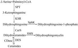
1 二氢神经酰胺的代谢与关键酶 1.1 二氢神经酰胺的代谢过程二氢神经酰胺是神经酰胺从头合成的中间产物。神经酰胺的从头合成 起始于内质网 (图 1),L-丝氨酸和棕榈酰CoA通过丝 氨酸棕榈酰转移酶 (serine palmitoyltransferase,SPT) 合成3-酮基二氢鞘氨醇,然后酮基还原酶通过辅酶Ⅰ的作用,还原3-酮基鞘氨醇产生二氢鞘氨醇,在神经酰胺合酶 (ceramide synthase,CerS) 的作用下再生成二氢神经酰胺。二氢神经酰胺由DES引入双键合成神经酰胺。在从头合成通路中二氢鞘氨醇还能在鞘氨醇激酶 (sphingosine kinase,SphK) 的作用下生成磷酸二氢鞘氨醇; 二氢神经酰胺也可以在鞘磷脂合成酶 (sphingomyelin synthase,SMS) 的作用下生成二氢鞘磷脂。

|
Figure 1 Metabolism of dihydroceramides. CDase: Ceramidase; CerS: Ceramide syhthase; DES: Dihydroceramide desaturase; KSR: 3-Ketosphinganine reductase; SMS: Sphingomyelin synthase; SphK: Sphingosine kinase; SPT: Serine palmitoyltransferase |
与二氢神经酰胺合成密切相关且研究较多的酶为SPT、CerS和DES。SPT是鞘脂类物质从头合成途径的第1个酶,也是限速酶。哺乳动物的SPT由 2个亚基组成——SPT1和SPT2,其中SPT2为主要的催化亚基[8]。研究发现CerS存在6种亚型,分别为CerS1、CerS2、CerS3、CerS4、CerS5和CerS6,每种CerS的组织分布不同,对不同碳链长度的乙酰CoA具有不同的优先选择性[9],这是合成的二氢神经酰胺和神经酰胺多样性的基础。DES是与二氢神经酰胺含量密切相关的酶,二氢神经酰胺生物学活性的发现也是基于对该酶及其抑制剂的研究。
1.2 二氢神经酰胺去饱和酶1993年,Castrillon 等[10]在研究果蝇雄性不育突变体时,第一次描述了DES基因。DES基因突变的果蝇,在精子减数分裂 过程中,细胞核中的染色体不能凝集。1996年,Endo等[11]第一次克隆了果蝇中负责编码DES的基因,并对DES基因进行了定位,发现该基因位于二号染色体左臂的26A位点,同时还发现了两种剪接体—— DES1和DES2,但仅DES1在精子发生过程中发挥作用。之后,Endo等[12]又在小鼠中发现了该基因的同 源基因。
DES负责在二氢神经酰胺的鞘氨醇骨架上引入4,5-反式双键合成神经酰胺[13]。DES1表现出较高的Δ4-DES活性和极低的C-4羟化酶活性,而DES2既具有Δ4-DES活性,又具有C-4羟化酶的活性[14],同时还控制着植物鞘氨醇的合成[15]。两种DES酶的组织分布也有差别,DES1普遍存在于各组织中,但在肝脏、哈氏腺、肾脏以及肺中较高表达且具有较高活 性[15, 16]。而DES2在皮肤、小肠以及肾脏中较高表达,这些部位含有较高含量的植物鞘氨醇[15, 17]。利用生物信息学的方法,Ternes等[14]对比了不同组织中可能的DES序列,发现它们包含3个高度保守的组氨酸序列盒: HX(3-4)H、HX(2-3)HH和H/QX(2-3)HH。一些细胞膜脂肪酸去饱和酶和碳氢化合物羟化酶同样也含有这些富含组氨酸的序列[18]。
DES亦具有一定的底物特异性,其活性受底物立体化学特性的影响,对D-赤-异构体的去饱和作用优于L-苏-异构体。鞘氨醇骨架上连接的烷基碳链长度也会影响DES的活性,大鼠肝微粒体实验显示DES的活性随着碳链长度的增加而降低[13]。该酶在pH 6.5~9内均具有活性,其最适pH值为8.5。
1.3 二氢神经酰胺去饱和酶抑制剂DES抑制剂的发现为研究二氢神经酰胺生物学活性提供了工具。2003年,Triola等[19]通过引入环丙烯单元替代神经酰胺上的双键,第一次合成了DES抑制剂GT11,这种包含环丙烯的鞘脂通过和底物 (D-赤-N-辛酰基二氢鞘氨醇) 竞争结合DES达到抑制酶活性的作用。GT11能以剂量依赖的方式抑制DES,当底物浓度为50 μmol∙L-1时,其半数抑制率 (IC50) 为20 μmol∙L-1。随后,Triola等[20]以GT11为母核合成了一系列衍生物,结果表明,2S和3R的立体化学构象、C1位点的羟基以及在神经酰胺双键位置的环丙烯环是维持GT11活性的基本保证。
2008年,Munoz-Olaya等[21]根据GT11的合成原理以及已有的去饱和酶抑制剂的结构特点,合成了一种新型的DES抑制剂XM462。研究显示,XM462在大鼠肝微粒体和人白血病Jurket A3细胞中均能抑制DES的活性,IC50分别为8.2和0.43 μmol∙L-1。利用LC-MS的方法,发现XM462能够引起内源性二氢神经酰胺含量的增加。随后,Camacho等[22]合成了3种XM462类似物,这些化合物不仅是DES,同时还是酸性神经酰胺酶的抑制剂。
1979次合成了芬维A胺 [fenretinide,N-(4-hydroxyphenyl) retinamide,4-HPR]。4-HPR是一种维生素A的类似物,能够抑制细胞增殖,抑制肿瘤组织中的血管生成[24],提高糖尿病小鼠糖耐量[25]。近年来,有研究显示4-HPR能够降低神经酰胺的合成、升高二氢神经酰胺及二氢鞘氨醇的含量[26]。2011年,Rahmaniyan等[27]发现,4-HPR通过干扰脱氢过程中的电子传递链,不可逆地抑制DES活性。随后,有研究证实[28],4-HPR通过直接作用于DES,降低神经酰胺的含量,同时升高二氢神经酰胺的含量来发挥提高胰岛素敏感性的作用。
2 二氢神经酰胺的生物学活性 2.1 二氢神经酰胺与细胞凋亡细胞凋亡是主动的细胞程序性死亡。神经酰胺被认为是一种能够引起 细胞凋亡的脂质分子[29, 30]。在凋亡的早期,神经酰胺在线粒体外膜形成通道,增加线粒膜的通透性,引起细胞色素C等释放,进而激活内源性凋亡通路[31]。二氢神经酰胺在细胞凋亡中的作用一直饱受争议。有研究[32]显示在人结肠癌细胞中加入C2-二氢神经酰胺不能引起凋亡,而C2-神经酰胺则能引起细胞凋亡。另外一项研究[33]表明,在人鳞状细胞癌细胞中加入C2-二氢神经酰胺和C2-神经酰胺,也得到类似结果。然而这些实验利用的都是短链、可渗透的外源性二氢神经酰胺,而不是内源性、长链的二氢神经酰胺。近年来,利用DES抑制剂、siRNA或基因敲除等方法抑制DES的活性,提高细胞内内源性二氢神经酰胺的含量,均显示内源性二氢神经酰胺的升高能够抵抗多种原因引起的细胞凋亡[34, 35]。如Stiban等[36]证实二氢神经酰胺能干扰线粒体中神经酰胺通道的形成,加入二氢神经酰胺能显著降低线粒体外膜的通透性,抑制神经酰胺引起的细胞凋亡,因此推测线粒体途径介导的凋亡发生不仅需要神经酰胺含量的升高,还需要二氢神经酰胺含量的降低。但也有研究[37]显示,DES抑制剂4-HRP引起的细胞凋亡与抑制DES进而引起内质网中二氢神经酰胺蓄积有关。二氢神经酰胺对凋亡的作用是抑制还是促进尚有待进一步研究,可能与不同的细胞类型有关。
2.2 二氢神经酰胺与细胞自噬自噬是真核生物进化上高度保守的、细胞进行自我保护的一种重要机制,在维持细胞存活、更新、物质再利用和内环境稳定中发挥重要作用。在生理状况下,细胞通过自噬清除受损的大分子,以维持细胞正常的生存。在缺氧以及饥饿等条件下,细胞内的自噬水平迅速上升,并可能引发细胞过度自噬。过度自噬会引起细胞不可逆的损伤,最终导致细胞的死亡。Holliday等[38]在急性淋巴母细胞性白血病细胞系中加入外源性C22∶0和C24∶0的二氢神经酰胺,证实这种特定长链的二氢神经酰胺能通过不依赖caspase的机制增加细胞自噬,引起细胞死亡。另外一项研究[39]也表明在胃癌细胞中加入白藜芦醇能抑制DES1活性,从而引起二氢神经酰胺的堆积,导致肿瘤细胞自噬,发挥抗肿瘤作用。
二氢神经酰胺引起自噬的机制还不清楚,但是有研究[34]表明,DES1基因敲除的小鼠胚胎成纤维母细胞中二氢神经酰胺的堆积可引起电子传递链复合体蛋白的表达发生改变,特别是复合体IV表达下降及活性丢失,导致AMP-依赖的蛋白激酶激活,进一步激活unc-51样激酶 (unc-51-like kinase 1,Ulk1),引起自噬小体的形成,增加细胞自噬。在该DES1基因敲除的小鼠胚胎成纤维母细胞中加入SPT抑制剂myriocin或CerS抑制剂fumonisin B1,阻断二氢神 经酰胺的合成,使ATP合成得以恢复,进一步证实二氢神经酰胺的堆积而不是神经酰胺的缺失导致自噬的增加。
2.3 二氢神经酰胺与细胞周期阻滞研究表明,二氢神经酰胺含量增加能够引起细胞周期阻滞。Kraveka等[40]利用siRNA干扰SMS-KCNR人成神经细胞瘤细胞中DES1,引起细胞中二氢神经酰胺含量显著增高,细胞生长周期停滞在G0/G1期。进一步的研究显示,利用siRNA干扰DES1活性能够引起磷酸化的成视网膜细胞瘤蛋白 (phosphorylated retinoblastoma protein,pRb) 含量显著降低,而pRb对于控制G1/S期转化、调控细胞周期发挥着关键作用。同样,Gagliostro等[41]在HGC27人胃癌细胞中加入DES1抑制剂XM462,结果显示,二氢神经酰胺的增加可引发细胞周期调节蛋白D1 (cyclin D1) 表达降低,细胞周期G1/S期转化延迟,同时引起自噬体相关蛋白表达升高,自噬增加。XM462还可以通过翻译启动因子2α (eukaryotic initiation factor 2α,eIF2α) 和X-盒结合蛋白1 (X-box- binding protein 1,Xbp1) 引发内质网应激。加入外源性的二氢神经酰胺类似物 (d2C8dhCer) 得到了类似结果。另外的研究[35]发现,从DES1基因敲除的小鼠中分离的胚胎成纤维细胞增殖缓慢,但这一现象是否与二氢神经酰胺的堆积直接相关,尚需进一步证实。
2.4 磷酸化的二氢神经酰胺与固有免疫固有免疫 (innate immunity) 是机体抵抗微生物入侵的第一道防线,参与维持机体自稳,并参与特异性免疫应答的启动、进程和效应。有研究表明,二氢神经酰胺的衍生物可能参与了牙周病的固有免疫应答。Nichols等[42]测定了牙周病的病原体——牙龈卟啉单胞菌中的鞘脂类分子成分,以考察这些脂质分子是否在自身免疫疾病中发挥作用,结果显示磷酸化的二氢神经酰胺 (phosphorylated dihydroceramide,PDHC) 能够增强自身免疫应答,增加树突状细胞白介素-6的释放,并且这一作用与脂多糖无关,因此他们推测PDHC可能通过作用于Toll样受体2 (Toll-like receptor 2,TLR-2) 的配体而发挥作用。随后他们又发现小肠和口腔中的其他种属的微生物也可以合成此类脂质,PDHCs不仅存在于牙龈组织中,也存在于血液、血管组织以及大脑中。这种TLR-2激活的脂质在人体组织中的分布与组织的位置及疾病的状态密切相关,提示PDHCs可能在人类疾病中扮演着重要角色[43]。
3 总结与展望在很长一段时间内,二氢神经酰胺一直被认为仅是神经酰胺的前体,本身无生物活性。近年来随着质谱技术的应用、分子生物学技术的进步以及DES抑制剂的出现,二氢神经酰胺具有生物学活性这一事实得到了验证,如参与凋亡、诱导自噬及引起细胞周期阻滞等。本研究团队最近的研究结果提示二氢神经酰胺可能参与了肝病慢性化的过程,二氢神经酰胺 (d18∶0/24∶0) 水平也有作为乙肝相关慢加急性肝衰竭预后生物标记物的潜能[44],相关的验证和机制研究正在开展。
作为二氢神经酰胺代谢通路的关键酶,DES近年来亦成为关注的重点,其调节二氢神经酰胺和神经酰胺的含量,参与疾病的发生发展。在肿瘤、HIV和囊性纤维化等疾病中,DES已成为一个新的治疗靶点。很多肿瘤化疗药,如4-HPR、白藜芦醇等都能够降低DES,升高内源性二氢神经酰胺及其衍生物的含量。在HIV研究中发现[6],利用siRNA或DES抑制剂等抑制DES活性,引起二氢神经酰胺含量升高,增加二氢鞘磷脂含量,后者可进入细胞膜的脂伐,使细胞膜的硬度增加,从而抑制gp41蛋白的插入,抑制病毒与宿主细胞细胞膜的融合,进而抑制HIV感染。在囊性纤维化治疗方面,DES也具有成为新的治疗靶点的潜能[7],抑制DES活性,不仅能降低神经酰胺的含量,增加二氢神经酰胺的含量,降低上皮细胞的凋亡,还能够通过改变脂伐成分降低感染。
目前对二氢神经酰胺的研究尽管已取得了一些进展,但尚处于起步阶段,多个可能的生物学活性均只是有所发现,尚未深入探究。不同碳链长度的二氢神经酰胺可能具有不同的生物学活性[38],而关于糖基化二氢神经酰胺以及磷酸化二氢神经酰胺的生物学研究更鲜见报道。对二氢神经酰胺类鞘脂生物学活性的研究任重而道远,而二氢神经酰胺类鞘脂生物学活性的阐明将对解释疾病的发生发展 (如肝病) 具有重要意义。
| [1] | Bielawska A, Crane HM, Liotta D, et al. Selectivity of ceramide-mediated biology. Lack of activity of erythro-dihydroceramide[J]. J Biol Chem , 1993, 268 :26226–26232. |
| [2] | Gudz TI, Tserng KY, Hoppel CL. Direct inhibition of mitochondrial respiratory chain complex III by cell-permeable ceramide[J]. J Biol Chem , 1997, 272 :24154–24158. DOI:10.1074/jbc.272.39.24154 |
| [3] | Garcia-Ruiz C, Colell A, Mari M, et al. Direct effect of ceramide on the mitochondrial electron transport chain leads to generation of reactive oxygen species. Role of mitochondrial glutathione[J]. J Biol Chem , 1997, 272 :11369–11377. DOI:10.1074/jbc.272.17.11369 |
| [4] | Chalfant CE, Szulc Z, Roddy P, et al. The structural require-ments for ceramide activation of serine-threonine protein phosphatases[J]. J Lipid Res , 2004, 45 :496–506. |
| [5] | Devlin CM, Lahm T, Hubbard WC, et al. Dihydrocermide-based response to hypoxia[J]. J Biol Chem , 2011, 286 :38069–38078. DOI:10.1074/jbc.M111.297994 |
| [6] | Vieira CR, Munoz-Olaya JM, Sot J, et al. Dihydrosphingomyelin impairs HIV-1 infection by rigidifying liquid-ordered membrane domains[J]. Chem Biol , 2010, 17 :766–775. DOI:10.1016/j.chembiol.2010.05.023 |
| [7] | Zaidi T, Bajmoczi M, Zaidi T, et al. Disruption of CFTR-dependent lipid rafts reduces bacterial levels and corneal disease in a murine model of Pseudomonas aeruginosa keratitis[J]. Invest Ophthalmol Vis Sci , 2008, 49 :1000–1009. DOI:10.1167/iovs.07-0993 |
| [8] | Hornemann T, Richard S, Rütti MF, et al. Cloning and initial characterization of a new subunit for mammalian serine-palmitoyltransferase[J]. J Biol Chem , 2006, 281 :37275–37281. DOI:10.1074/jbc.M608066200 |
| [9] | Wang SY, Zhang JL, Zhang D, et al. Recent advances in study of sphingolipids on liver disease[J]. Acta Pharm Sin (药学学报) , 2015, 50 :1551–1558. |
| [10] | Castrillon DH, Gönczy P, Alexander S, et al. Toward a molecular genetic analysis of spermatogenesis in Drosophila melanogaster: characterization of male-sterile mutants generated by single P element mutagenesis[J]. Genetics , 1993, 135 :489–505. |
| [11] | Endo K, Akiyama T, Kobayashi S, et al. Degenerative spermatocyte, a novel gene encoding a transmembrane protein required for the initiation of meiosis in Drosophila spermato-genesis[J]. Mol Gen Genet , 1996, 253 :157–165. DOI:10.1007/s004380050308 |
| [12] | Endo K, Matsuda Y, Kobayashi S. Mdes, a mouse homolog of the Drosophila degenerative spermatocyte gene is expressed during mouse spermatogenesis[J]. Dev Growth Differ , 1997, 39 :399–403. DOI:10.1046/j.1440-169X.1997.00015.x |
| [13] | Michel C, van Echten-Deckert G, Rother J, et al. Characteri-zation of ceramide synthesis. A dihydroceramide desaturase introduces the 4,5-trans-double bond of sphingosine at the level of dihydroceramide[J]. J Biol Chem , 1997, 272 :22432–22437. DOI:10.1074/jbc.272.36.22432 |
| [14] | Ternes P, Franke S, Zähringer U, et al. Identification and characterization of a sphingolipid delta 4-desaturase family[J]. J Biol Chem , 2002, 277 :25512–25518. DOI:10.1074/jbc.M202947200 |
| [15] | Omae F, Miyazaki M, Enomoto A, et al. DES2 protein is responsible for phytoceramide biosynthesis in the mouse small intestine[J]. Biochem J , 2004, 379 :687–695. DOI:10.1042/bj20031425 |
| [16] | Causeret C, Geeraert L, Van der Hoeven G, et al. Further characterization of rat dihydroceramide desaturase: tissue distribution, subcellular localization, and substrate specificity[J]. Lipids , 2000, 35 :1117–1125. DOI:10.1007/s11745-000-0627-6 |
| [17] | Mizutani Y, Kihara A, Igarashi Y. Identification of the human sphingolipid C4-hydroxylase, hDES2, and its up-regulation during keratinocyte differentiation[J]. FEBS Lett , 2004, 563 :93–97. DOI:10.1016/S0014-5793(04)00274-1 |
| [18] | Shanklin J, Cahoon EB. Desaturation and related modifica-tion of fatty acids[J]. Annu Rev Plant Physiol Plant Mol Biol , 1998, 49 :611–641. DOI:10.1146/annurev.arplant.49.1.611 |
| [19] | Triola G, Fabriãs G, Llebaria A. Synthesis of a cyclopropene analogue of ceramide, a potent inhibitor of dihydroceramide desaturase[J]. Angew Chem Int Ed Engl , 2001, 40 :1960–1962. DOI:10.1002/(ISSN)1521-3773 |
| [20] | Triola G, Fabriãs G, Casas J, et al. Synthesis of cyclopropene analogues of ceramide and their effect on dihydroceramide desaturase[J]. J Org Chem , 2003, 68 :9924–9932. DOI:10.1021/jo030141u |
| [21] | Munoz-Olaya JM, Matabosch X, Bedia C, et al. Synthesis and biological activity of a novel inhibitor of dihydroceramide desaturase[J]. ChemMedChem , 2008, 3 :946–953. DOI:10.1002/(ISSN)1860-7187 |
| [22] | Camacho L, Simbari F, Garrido M, et al. 3-Deoxy-3,4-dehydro analogs of XM462. Preparation and activity on sphingolipid metabolism and cell fate[J]. Bioorg Med Chem , 2012, 20 :3173–3179. DOI:10.1016/j.bmc.2012.03.073 |
| [23] | Moon RC, Thompson HJ, Becci PJ, et al. N-(4-Hydroxy-phenyl)retinamide, a new retinoid for prevention of breast cancer in the rat[J]. Cancer Res , 1979, 39 :1339–1346. |
| [24] | Rodriguez-Cuenca S, Barbarroja N, Vidal-Puig A. Dihydro-ceramide desaturase 1, the gatekeeper of ceramide induced lipotoxicity[J]. Biochim Biophys Acta , 2015, 1851 :40–50. DOI:10.1016/j.bbalip.2014.09.021 |
| [25] | Yang Q, Graham TE, Mody N, et al. Serum retinol binding protein 4 contributes to insulin resistance in obesity and type 2 diabetes[J]. Nature , 2005, 436 :356–362. DOI:10.1038/nature03711 |
| [26] | Valsecchi M, Aureli M, Mauri L, et al. Sphingolipidomics of A2780 human ovarian carcinoma cells treated with synthetic retinoids[J]. J Lipid Res , 2010, 51 :1832–1840. DOI:10.1194/jlr.M004010 |
| [27] | Rahmaniyan M, Curley RW Jr, Obeid LM, et al. Identifica-tion of dihydroceramide desaturase as a direct in vitro target for fenretinide[J]. J Biol Chem , 2011, 286 :24754. DOI:10.1074/jbc.M111.250779 |
| [28] | Bikman BT, Guan Y, Shui G, et al. Fenretinide prevents lipid-induced insulin resistance by blocking ceramide biosyn-thesis[J]. J Biol Chem , 2012, 287 :17426–17437. DOI:10.1074/jbc.M112.359950 |
| [29] | Pastore D, Della-Morte D, Coppola A, et al. SGK-1 protects kidney cells against apoptosis induced by ceramide and TNF-α[J]. Cell Death Dis , 2015, 6 :e1890. DOI:10.1038/cddis.2015.232 |
| [30] | Tsukamoto S, Huang Y, Kumazoe M, et al. Sphingosine kinase-1 protects multiple myeloma from apoptosis driven by cancer-specific inhibition of RTKs[J]. Mol Cancer Ther , 2015, 14 :2303–2312. DOI:10.1158/1535-7163.MCT-15-0185 |
| [31] | Abou-Ghali M, Stiban J. Regulation of ceramide channel formation and disassembly: insights on the initiation of apoptosis[J]. Saudi J Biol Sci , 2015, 22 :760–772. DOI:10.1016/j.sjbs.2015.03.005 |
| [32] | Ahn EH, Schroeder JJ. Induction of apoptosis by sphingosine, sphinganine, and C2-ceramide in human colon cancer cells, but not by C2-dihydroceramide[J]. Anticancer Res , 2010, 30 :2881–2884. |
| [33] | Sugiki H, Hozumi Y, Maeshima H, et al. C2-ceramide induces apoptosis in a human squamous cell carcinoma cell line[J]. Br J Dermatol , 2000, 143 :1154–1163. DOI:10.1046/j.1365-2133.2000.03882.x |
| [34] | Siddique MM, Li Y, Wang L, et al. Ablation of dihydroceramide desaturase 1, a therapeutic target for the treatment of metabolic diseases, simultaneously stimulates anabolic and catabolic signaling[J]. Mol Cell Biol , 2013, 33 :2353–2369. |
| [35] | Siddique MM, Bikman BT, Wang L, et al. Ablation of dihydroceramide desaturase confers resistance to etoposide-induced apoptosis in vitro[J]. PLoS One , 2012, 7 :e44042. DOI:10.1371/journal.pone.0044042 |
| [36] | Stiban J, Fistere D, Colombini M. Dihydroceramide hinders ceramide channel formation: implications on apoptosis[J]. Apoptosis , 2006, 11 :773–780. DOI:10.1007/s10495-006-5882-8 |
| [37] | Boppana NB, Stochaj U, Kodiha M, et al. Enhanced killing of SCC17B human head and neck squamous cell carcinoma cells after photodynamic therapy plus fenretinide via the de novo sphingolipid biosynthesis pathway and apoptosis[J]. Int J Oncol , 2015, 46 :2003–2010. |
| [38] | Holliday MW Jr, Cox SB, Kang MH, et al. C22∶0-and C24∶0-dihydroceramides confer mixed cytotoxicity in T-cell acute lymphoblastic leukemia cell lines[J]. PLoS One , 2013, 8 :e74768. DOI:10.1371/journal.pone.0074768 |
| [39] | Signorelli P, Munoz-Olaya JM, Gagliostro V, et al. Dihydro-ceramide intracellular increase in response to resveratrol treatment mediates autophagy in gastric cancer cells[J]. Cancer Lett , 2009, 282 :238–243. DOI:10.1016/j.canlet.2009.03.020 |
| [40] | Kraveka JM, Li L, Szulc ZM, et al. Involvement of dihydro-ceramide desaturase in cell cycle progression in human neuro-blastoma cells[J]. J Biol Chem , 2007, 282 :16718–16728. DOI:10.1074/jbc.M700647200 |
| [41] | Gagliostro V, Casas J, Caretti A, et al. Dihydroceramide delays cell cycle G1/S transition via activation of ER stress and induction of autophagy[J]. Int J Biochem Cell Biol , 2012, 44 :2135–2143. DOI:10.1016/j.biocel.2012.08.025 |
| [42] | Nichols FC, Housley WJ, O'Conor CA, et al. Unique lipids from a common human bacterium represent a new class of Toll-like receptor 2 ligands capable of enhancing autoimmu-nity[J]. Am J Pathol , 2009, 175 :2430–2438. DOI:10.2353/ajpath.2009.090544 |
| [43] | Nichols FC, Yao X, Bajrami B, et al. Phosphorylated dihydroceramides from common human bacteria are recovered in human tissues[J]. PLoS One , 2011, 6 :e16771. DOI:10.1371/journal.pone.0016771 |
| [44] | Qu F, Zheng SJ, Liu S, et al. Serum sphingolipids reflect the severity of chronic HBV infection and predict the mortality of HBV-acute-on-chronic liver failure[J]. PLoS One , 2014, 9 :e104988. DOI:10.1371/journal.pone.0104988 |
 2016, Vol. 51


