2. 中国林业科学研究院 林业新技术研究所,北京 100091
2. Institute of Forest New Technology, Chinese Academy of Forestry, Beijing 100091, China
我国造纸废水在工业废水中所占的比例较大,而造纸工业的主要副产物之一就是木质素磺酸盐。我国木质素磺酸盐的年产量在450万吨以上,但对木质素磺酸盐缺乏合理有效的利用,仅有少量被用作建筑材料的添加剂,绝大部分以“黑液”形式直接排入江河或作为廉价燃料,木质素磺酸盐的自然降解时间较长,不仅造成资源浪费,还对环境造成严重的污染,因而,木质素的充分高值化利用越来越受到重视[1-4]。木质素磺酸盐基本结构是苯丙烷衍生物,因大量磺酸基接在苯丙烷的侧链而具有良好水溶性和表面活性[5-6]。木质素磺酸盐分子结构中的酚羟基、羰基以及磺酸基上的氧、硫原子有未共用电子对,使其能与金属离子配位形成螯合物,因此,木质素磺酸盐具有制备选择吸附性金属离子荧光探针的潜在可能性[7-9]。
金属污染是对环境污染最严重和对人类危害最大的水污染之一。金属离子具有生物累积性,威胁人类的健康。含金属离子的废水主要来自于电镀、化工等生产部门的工业污染排放。随着汽车制造、电子等工业的迅速崛起以及城市化建设,金属废水的排放量越来越大,种类越来越多,生态环境受到严重破坏,且严重危害人类的健康[10-12]。因此,开发简便高效的方法实现对金属离子的检测具有重要的意义。
荧光碳材料是近年来开发的一种新型荧光材料。相对于半导体量子点造价昂贵、合成条件严格以及高毒性等缺点,碳点具有优良的光学性能、良好的生物相容性等优点,在传感器、细胞成像光学器件及药物载体等领域具有很好的应用前景[13-20]。ZHU等[21]以氧化石墨烯为碳源在酸性条件下采用微波法制备石墨烯量子点,该石墨烯量子点能特异性地识别Cd2+。WEI等[22]以葡萄糖和氨基酸合成了一系列具有高荧光量子产率(~69.1%)的N掺杂碳点,并且可通过N-掺杂水平调节的荧光强度。碳点制备过程通常化学修饰掺杂杂原子以提高其光学性能和对金属离子的选择吸附性能[23-29],这些都需要较长的时间和相对繁琐的操作,发展绿色简便的反应制备杂原子掺杂碳点有重要意义。
本文以造纸废料木质素磺酸钠和半胱氨酸为原料采用操作简便的水热反应制备了N, S双掺杂的荧光碳点,并利用透射电镜(transmission electron microscope, TEM)、紫外-可见光吸收光谱(ultraviolet–visible spectroscopy, UV-Vis)等分析方法对产物进行了表征,通过荧光光谱分析了碳点对一系列金属离子的的识别作用,并考察了碳点在实际水样中检测Fe3+的能力。
2 实验部分 2.1 实验原料所用试剂均为分析纯。木质素磺酸钠购于阿拉丁生化科技股份有限公司;氯化镉、硝酸银、乙酸镁、硫酸锌、硫酸铝,国药集团化学试剂有限公司;氯化镍,上海青析化工科技有限公司;二水合氯化铜,广东光华化学厂有限公司;氯化锰,西陇化工股份有限公司;氯化铁、氯化钴,天津市科密欧化学试剂有限公司。自来水水样取自南京,用0. 45 μm微孔滤膜过滤后备用。
2.2 碳点的制备称取0.3 g木质素磺酸钠、0.2 g半胱氨酸溶于30 mL蒸馏水中,随后将溶液加入50 mL水热反应釜中,于180 ℃下反应24 h。冷却至室温后,用微孔滤膜过滤除去固体,得到的溶液即为碳点溶液。
2.3 性能表征UV-2550紫外可见分光光度仪(UV-vis,波长为300~800 nm,日本岛津公司)测定制备获得碳点的吸光度、iS10型光傅里叶变换红外光谱仪(FT-IR,美国尼高力公司)用于表征制备的碳点的结构。Technai G2 20 S-Twin透射电子显微镜(TEM,美国FEI公司)观察制备的碳点的形貌。LS SS型荧光光谱仪(美国PerkinElmer公司)研究制备的碳点的离子选择吸附性能及荧光性能。
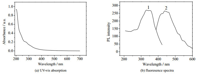
3 实验结果与讨论 3.1 碳点光学性能分析通过一步水热反应制备了N, S双掺杂的碳点,其具有良好的水溶性,且有一定的稳定性,在室温下放置数月不会聚沉。利用紫外可见光谱和荧光光谱分析碳量子点的光学性能。图 1(a)是制备的碳点的紫外可见吸收光谱。然而,当激发波长为270 nm时,并没有观察到制备的碳点的荧光发射峰。这可能是因为碳点内部的共轭结构并不是有效的荧光发射中心,制备的碳点的荧光来自其表面态而非核结构[30-31]。图 1(b)是碳点荧光激发光谱和荧光发射光谱。由图可知,在340 nm波长光激发下,碳点在443 nm处具有最强的荧光发射。
|
图 1 碳点的紫外可见吸收光谱和荧光光谱 Fig.1 UV-vis absorption and fluorescence spectra of the carbon dots |
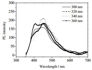
在短波激发的时候可以在405和445 nm处看到明显的双峰现象。随着激发波长红移,405 nm处的峰消失,445 nm处的峰显著增强。也就是说长波激发下,只有445 nm处的发射中心被激发出来;而这个发射峰随着激发光波长进一步红移,也发生相应的红移,且荧光强度随着激发波长红移先增大后减少,这种现象可能是由于碳点中不同发光中心决定的。
|
图 2 碳点在不同激发波长下的发射光谱 Fig.2 Fluorescence spectra of the carbon dots under excitation wavelengths from 300 nm to 360 nm |
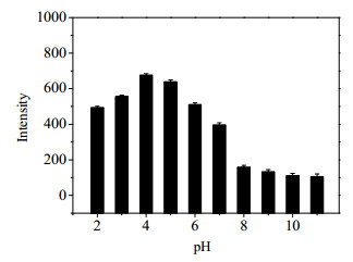
进一步考察不同pH值对碳量子点荧光性能的影响。由图 3可知,随着pH值的增加,碳点的荧光光谱强度先增加后减少,在pH值大于8后,碳点的荧光强度变化不大。
|
图 3 碳点在不同pH值下的荧光强度 Fig.3 Fluorescence intensity of the carbon dots in different pH solutions |
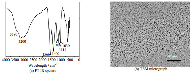
图 4(a)是制备的样品的红外谱图。碳量子点在3 380 cm-1处的宽峰属于─OH的伸缩振动,在3 200 cm-1处为─NH2的对称伸缩振动吸收峰,2 082 cm-1处无吸收峰,说明没有─SH基团,判断L-半胱氨酸中的─SH基本上反应完全;在1 566 cm-1处的峰是C=O的伸缩振动,1 400 cm-1处的峰是C─O的变形振动,在1 114和1 030 cm-1处的峰是S=O的伸缩振动。通过以上分析可知木质素磺酸钠和半胱氨酸作为前体经过脱水、聚合、炭化和生成了表面富含磺酸基、羰基、羟基等亲水基团的碳点。图 4(b)是碳量子点的透射电镜图。由图可知,制备的碳点大小均一,表面光滑,粒径分布在2~7 nm。
|
图 4 碳量子点的红外光谱和透射电镜图 Fig.4 FT-IR spectra and TEM micrograph of the carbon dots |
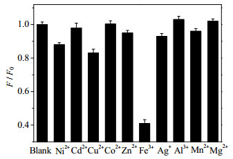
配制浓度为0.01 mol·L-1的各种不同的金属离子(Cu2+、Co2+、Ag+、Zn2+、Mg2+、Fe3+、Ni2+、Al3+、Cd2+、Mn2+)。将3 mL的碳点溶液加入比色皿中,然后,分别加入15 μL 0.01 mol·L-1的金属离子溶液,最终得到比色皿中的金属离子的溶液浓度为50 μmol·L-1,用荧光光谱仪分析所制备碳点的离子识别性能,结果如图 5。由图可知,加入Fe3+的溶液体系,荧光猝灭最强,而加入其他金属离子,荧光强度的变化均不大。结果表明制备的碳点可以实现对铁的检测。上述现象可能是由于Fe3+与碳点表面的羟基、羧基等通过分子间的螯合作用形成络合物的能力最强,这可能有利于电荷转移,荧光猝灭最明显。
|
图 5 碳点对不同金属离子的荧光响应 Fig.5 Quenching performance of the carbon dots with and without various metal ions |
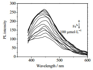
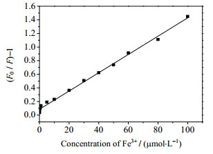
为分析所制备碳点用于检测Fe3+的灵敏度,配制不同浓度的Fe3+溶液,并测定荧光光谱,结果如图 6。由图可知,荧光分子探针的光谱位置基本没有改变,但不同浓度的Fe3+可以使制备的碳点发生不同程度的荧光猝灭。利用图 6中的数据,根据Stern-volmer方程,浓度对(F0/F-1)作图得图 7。由图可知,当铁离子的浓度由0增大到100 μmol·L-1时,对碳量子点的荧光淬灭呈线性关系,线性方程为:F0/F= 0.085 15+ 0.013 43 C(Fe3+),R2为0.995,检测限为0.2 μmol·L-1,检测线性范围为0.5~100 μmol·L-1。
|
图 6 铁离子浓度对碳点荧光强度的影响 Fig.6 PL emission spectra of the carbon dots under increasing Fe3+ concentrations |
|
图 7 铁离子浓度对碳点的Stern-volmer曲线 Fig.7 Stern–volmer curve of the carbon dots as a function of Fe3+ concentration |
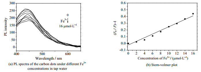
取自来水配制溶液来评价制备的碳点的性能,其结果如图 8所示,铁离子浓度从0逐渐增加到16 μmol·L-1时,不同浓度的铁离子可以使制备的碳点发生不同程度的荧光猝灭,且呈一定的线性,线性方程为:F0/F= 0.962 84 + 0.028 39 C(Fe3+),线性相关性系数为0.981 01(图 8(b))。综上,该碳点能成功应用于实际水样中铁离子的检测,可作为一种高灵敏度和高选择性的检测铁离子的荧光探针。
|
图 8 自来水中铁离子浓度对碳点荧光强度的影响及Stern-volmer曲线 Fig.8 PL spectra of the carbon dots under different Fe3+ concentrations in tap water and Stern-volmer curve of the carbon dots as a function of Fe3+ concentration |
本文以造纸废料木质素磺酸钠为碳源和硫源,半胱氨酸为氮源和硫源,通过一步水热反应制备得到具有荧光性能的N, S双掺杂的碳量子点。制备的碳点尺寸均一且具有良好的稳定性。该碳量子点可以作为荧光探针实现Fe3+的检测,在0.5~100 μmol·L-1,Fe3+浓度与碳点的荧光强度呈良好的线性关系,并在实际水样品的检测中获得了较好结果。该碳点的原料成本低廉,制备绿色简便,符合绿色可持续发展原则。设计并制备的新型生物基荧光传感器有望为木质素类天然可再生资源的高值化利用及生物质基功能材料的开发提供良好的理论与应用基础。
| [1] |
GILLET S, AGUEDO M, PETITJEAN L, et al. Lignin transformations for high value applications:Towards targeted modifications using green chemistry[J]. Green Chemistry, 2017, 19(18): 4200-4233. DOI:10.1039/C7GC01479A |
| [2] |
李伟, 佟国宾, 王梦茹, 等. 碱木质素基碳量子点/TiO2复合光催化剂的制备[J]. 林业工程学报, 2016, 1(5): 84-88. LI W, TONG G B, WANG M R, et al. Preparation of the alkaline lignin pyrolytic based carbon quantum dots/TiO2 composite photocatalyst[J]. Journal of Forestry Engineering, 2016, 1(5): 84-88. |
| [3] |
RAGAUSKAS A J, BECKHAM G T, BIDDY M J, et al. Lignin valorization:Improving lignin processing in the biorefinery[J]. Science, 2014, 344(6185): 1246843. DOI:10.1126/science.1246843 |
| [4] |
许利娜, 黄坤, 李守海, 等. 木质素磺酸钙-石墨烯复合量子点的制备及性能[J]. 化工进展, 2016, 35(11): 3595-3599. XU L N, HUANG K, LI S H, et al. Synthesis and properties of lignin/graphene quantum dots composites as fluorescent sensor[J]. Chemical Industry and Engineering Progress, 2016, 35(11): 3595-3599. |
| [5] |
RENDERS T, VAN DEN BOSCH S, KOELEWIJN S F, et al. Lignin-first biomass fractionation:The advent of active stabilisation strategies[J]. Energy & Environmental Science, 2017, 10(7): 1551-1557. |
| [6] |
SLUITER A, HAMES B, RUIZ R, et al. Determination of structural carbohydrates and lignin in biomass[J]. Laboratory Analytical Procedure, 2008, 1617: 1-16. |
| [7] |
ALBADARIN A B, COLLINS M N, NAUSHAD M, et al. Activated lignin-chitosan extruded blends for efficient adsorption of methylene blue[J]. Chemical Engineering Journal, 2017, 307: 264-272. DOI:10.1016/j.cej.2016.08.089 |
| [8] |
冯雪敏, 邱学青, 楼宏铭, 等. 氧化改性对木质素磺酸盐络合性能的影响[J]. 高校化学工程学报, 2015, 29(6): 1415-1421. FENG X M, QIU X Q, LOU H M, et al. Effects of oxidative modification on the chelating capacity of sodium lignosulfonate[J]. Journal of Chemical Engineering of Chinese Universities, 2015, 29(6): 1415-1421. DOI:10.3969/j.issn.1003-9015.2015.00.030 |
| [9] |
NAIR V, PANIGRAHY A, VINU R. Development of novel chitosan-lignin composites for adsorption of dyes and metal ions from wastewater[J]. Chemical Engineering Journal, 2014, 254: 491-502. DOI:10.1016/j.cej.2014.05.045 |
| [10] |
JIA K, HE X H, ZHOU X F, et al. Solid state effective luminescent probe based on CdSe@CdS/amphiphilic co-polyarylene ether nitrile core-shell superparticles for Ag+ detection and optical strain sensing[J]. Sensors and Actuators B:Chemical, 2018, 257: 442-450. DOI:10.1016/j.snb.2017.10.187 |
| [11] |
XU L N, FAN H, HUANG L X, et al. Eosinophilic nitrogen-doped carbon dots derived from tribute chrysanthemum for label-free detection of Fe3+ ions and hydrazine[J]. Journal of the Taiwan Institute of Chemical Engineers, 2017, 78: 247-253. DOI:10.1016/j.jtice.2017.06.011 |
| [12] |
JIANG Y L, WEI G, ZHANG W J, et al. Solid phase reaction method for preparation of carbon dots and multi-purpose applications[J]. Sensors and Actuators B:Chemical, 2016, 234: 15-20. DOI:10.1016/j.snb.2016.04.124 |
| [13] |
LI J J, ZUO G C, PAN X H, et al. Nitrogen-doped carbon dots as a fluorescent probe for the highly sensitive detection of Ag+ and cell imaging[J]. Luminescence, 2018, 33(1): 243-248. DOI:10.1002/bio.3407 |
| [14] |
XU L N, MAO W, HUANG J R, et al. Economical, green route to highly fluorescence intensity carbon materials based on ligninsulfonate/graphene quantum dots composites:Application as excellent fluorescent sensing platform for detection of Fe3+ ions[J]. Sensors and Actuators B:Chemical, 2016, 230: 54-60. DOI:10.1016/j.snb.2015.12.043 |
| [15] |
ZAN M H, RAO L, HUANG H M, et al. A strong green fluorescent nanoprobe for highly sensitive and selective detection of nitrite ions based on phosphorus and nitrogen co-doped carbon quantum dots[J]. Sensors and Actuators B:Chemical, 2018, 262: 555-561. DOI:10.1016/j.snb.2017.12.177 |
| [16] |
LI H T, KANG Z H, LIU Y, et al. Carbon nanodots:Synthesis, properties and applications[J]. Journal of Materials Chemistry, 2012, 22(46): 24230-24253. DOI:10.1039/c2jm34690g |
| [17] |
WANG F Y, LU Y X, CHEN Y, et al. Colorimetric nanosensor based on the aggregation of AuNP triggered by carbon quantum dots for detection of Ag+ ions[J]. ACS Sustainable Chemistry & Engineering, 2018, 6(3): 3706-3713. |
| [18] |
LIM S Y, SHEN W, GAO Z. Carbon quantum dots and their applications[J]. Chemical Society Reviews, 2015, 44(1): 362-381. |
| [19] |
ZHU S J, SONG Y B, ZHAO X H, et al. The photoluminescence mechanism in carbon dots (graphene quantum dots, carbon nanodots, and polymer dots):Current state and future perspective[J]. Nano Research, 2015, 8(2): 355-381. DOI:10.1007/s12274-014-0644-3 |
| [20] |
ZHENG X T, ANANTHANARAYANAN A, LUO K Q, et al. Glowing graphene quantum dots and carbon dots:Properties, syntheses, and biological applications[J]. Small, 2015, 11(14): 1620-1636. DOI:10.1002/smll.201402648 |
| [21] |
LI L L, JI J, FEI R, et al. A facile microwave avenue to electrochemiluminescent two-color graphene quantum dots[J]. Advanced Functional Materials, 2012, 22(14): 2971-2979. DOI:10.1002/adfm.201200166 |
| [22] |
WEI W L, XU C, WU L, et al. Non-enzymatic-browning-reaction:A versatile route for production of nitrogen-doped carbon dots with tunable multicolor luminescent display[J]. Scientific Reports, 2014, 4: 3564. |
| [23] |
ZHU A, QU Q, SHAO X, et al. Carbon-dot-based dual-emission nanohybrid produces a ratiometric fluorescent sensor for in vivo imaging of cellular copper ions[J]. Angewandte Chemie, 2012, 124(29): 7297-7301. DOI:10.1002/ange.201109089 |
| [24] |
FREIRE R M, LE N D B, JIANG Z, et al. NH2-rich carbon quantum dots:A protein-responsive probe for detection and identification[J]. Sensors and Actuators B:Chemical, 2018, 255: 2725-2732. DOI:10.1016/j.snb.2017.09.085 |
| [25] |
ZHENG X T, ANANTHANARAYANAN A, LUO K Q, et al. Glowing graphene quantum dots and carbon dots:Properties, syntheses, and biological applications[J]. Small, 2015, 11(14): 1620-1636. DOI:10.1002/smll.201402648 |
| [26] |
NIU W J, LI Y, ZHU R H, et al. Ethylenediamine-assisted hydrothermal synthesis of nitrogen-doped carbon quantum dots as fluorescent probes for sensitive biosensing and bioimaging[J]. Sensors and Actuators B:Chemical, 2015, 218: 229-236. DOI:10.1016/j.snb.2015.05.006 |
| [27] |
ZHANG R, CHEN W. Nitrogen-doped carbon quantum dots:Facile synthesis and application as a "turn-off" fluorescent probe for detection of Hg2+ ions[J]. Biosensors and Bioelectronics, 2014, 55: 83-90. DOI:10.1016/j.bios.2013.11.074 |
| [28] |
XU Q, PU P, ZHAO J, et al. Preparation of highly photoluminescent sulfur-doped carbon dots for Fe (Ⅲ) detection[J]. Journal of Materials Chemistry A, 2015, 3(2): 542-546. DOI:10.1039/C4TA05483K |
| [29] |
YANG Z, XU M, LIU Y, et al. Nitrogen-doped, carbon-rich, highly photoluminescent carbon dots from ammonium citrate[J]. Nanoscale, 2014, 6(3): 1890-1895. DOI:10.1039/C3NR05380F |
| [30] |
ZENG Y W, MA D K, WANG W, et al. N, S co-doped carbon dots with orange luminescence synthesized through polymerization and carbonization reaction of amino acids[J]. Applied Surface Science, 2015, 342: 136-143. DOI:10.1016/j.apsusc.2015.03.029 |
| [31] |
徐源, 陈艳华, 丁兰. 微波辅助法一步合成荧光碳点用于水中三价铁离子的检测[J]. 高等学校化学学报, 2018, 39(7): 1420-1426. XU Y, CHEN Y H, DING L. One-pot microwave-assisted synthesis of passivated fluorescent carbon dots for Fe(Ⅲ) detection[J]. Chemical Research in Chinese Universities, 2018, 39(7): 1420-1426. |


