 2023, Vol. 34


b College of Science and Technology, Hebei Agricultural University, Huanghua 061100, China;
c Department of Environmental Sciences, Kohsar University, Murree 47150, Pakistan
Graphene oxide (GO) is one of the most studied 2-dimensional carbon nanomaterials over the past decade [1]. As a dominant derivative of graphene, GO contains high surface area with numerous oxygen-containing functional groups, including carboxyl/carbonyl groups at the edges and epoxy/hydroxyl groups at the basal plane, which could maximize the benefit of the unique properties of this nanomaterial [2-4]. With unique structure and exceptional physicochemical characteristics, GO holds attractive promises in academic (research), environmental (i.e., desalination, filtration, adsorption), medical (i.e., bio-imaging, cancer therapy, and nanomedicine), and industrial (i.e., coating and sensors) applications [5-10]. It is estimated that production of graphene-based materials will shoot to 3800 tons annually in 2026, while the global graphene market will reach $656.9 million by 2027 [11]. GO comprises more than 60% of market share as compared to other graphene-based products [11]. The increase interest in GO application boomed its production, which certainly assure that it will eventually enter into the natural ecosystems, especially in aquatic environments [12, 13]. The concentrations of GO in wastewater from treatment plants or places near the discharge outlet of industries are higher than 10 mg/L [14]. Recent studies have shown that GO can be toxic toward bacteria and humans [15-17]. Thus, increasing research attention is being dedicated to determine its behavior and risks to the environment and ecological systems.
The stability of GO is one of the most important factors that ultimately control its environmental fate and ecological risks [18-20]. Aggregated GO particles have less available active sites and smaller surface-area-to-volume-ratio as compared to primary GO particles, thus decreasing the reactivity and bioavailability of aggregated GO particles [21]. GO has been announced as the most toxic particles among various graphene materials [22, 23], which depicts the importance of understanding the exposure of GO in environment for safe operation [24]. Due to many oxygen-containing groups, GO is dispersible in water, and can be transported in water through physical process or food chain. As a result, it might accumulate in the ecosystem, pose threat for aquatic organisms and eventually to human health [25]. The risks as well as toxicity of GO in aquatic environment might not be approached solely via its characteristics without taking into consideration the GO aggregation behavior in natural water bodies. Therefore, knowledge of the aggregation kinetics and stability of GO is critical to promote its industrial applications and evaluate its environmental risks.
After discharge into aquatic environments, GO particles come into contact with constituents in natural system that may include inorganic ions, natural organic matter, and colloidal particles [26]. These substances may influence the behavior of GO colloidal and further dictate the aggregation/stability of GO in natural water. Numerous findings have illustrated that the stability of GO in aqueous solutions is strongly affected by both solution chemistry (e.g., ionic strength and pH) [27, 28] and particle surface characteristics (e.g., surface oxygen content and particle sizes) [29]. Various inorganic ions including Ca2+, Mg2+, Na+, K+, SO42−, HCO3− and Cl−, have an obvious effect on the electro-kinetic behavior of GO colloids and consequently their aggregation/stability behavior [30]. For example, the aggregation of GO particles was enhanced by concentration increase of cations that suppress the electrostatic repulsive forces between GO particles [31]. Similarly, humic and fulvic acids, major constituents of NOM, have been considered to adsorb onto GO surface and significantly stabilize the GO particles [32].
The environmental conditions such as light, temperature, pH, and dissolved organic matter (DOM) may paly complex roles in the aggregation/stability of GO in natural water systems [33, 34]. The aggregation of GO was promoted when the temperature rose from 6 to 40 ℃ in the presence of Na+, K+ (monovalent) and Ca2+ (divalent) cations primarily because of the reduced electrostatic repulsion and enhanced cation dehydration [34]. The electron-hole pair generation under photoirradiation degrade GO into smaller fragments and low molecular weight species, which may influence the GO fate in natural waters [35, 36]. Therefore, it is necessary to shed light on various environmental factors that could influence the aggregation/stability behavior of GO in aquatic environment. This review will highlight various environmental factors that are responsible for the aggregation of GO in aquatic environment.
GO shows excellent dispersibility in various solvents, predominantly in water due to negatively charged carboxylic acid (−COO−) groups [37, 38]. These negatively charged groups create a repulsive electrostatic (EL) interaction between GO particles, which is higher than van der Waals (vdW) attractive force, thus promoting colloidal stability of GO [39, 40]. The colloidal stability of GO determines its application in industries, bioavailability, reactive oxygen species (ROS) generation, toxic effect, and adsorption/desorption capacity for various other contaminants (e.g., heavy metal ions and organic pollutants). Therefore, a comprehensive review of the aggregation kinetics of GO nanoparticle is required not only to promote their applications in industries but also for more accurate evaluation of their environmental fate, transport and prediction of their environmental influence and risks.
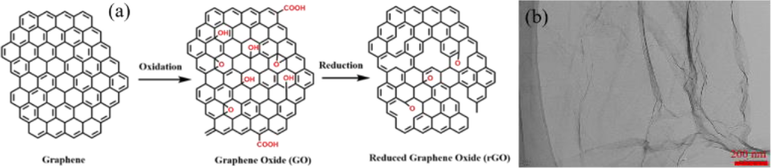
2. Structure of graphene and its derivativesGraphene-based materials comprise pristine graphene, GO, and reduced graphene oxide (rGO), where GO being an intermediate material formed during the formation of rGO (Fig. 1a). The most striking property of GO is that it may be reduced to material like graphene sheets by eliminating the oxygen-containing functional groups with the reformation of a conjugated structure [41, 42]. The reduction of GO to rGO enhance its electronic characteristics since the electrical conductivity of rGO is higher than that of GO [43].

|
Download:
|
| Fig. 1. Schematic diagram (a) showing the formation process of reduced GO (rGO) through reduction of graphene oxide (GO), and (b) TEM image of GO. (b) was copied with permission [32]. Copyright 2020, Elsevier. | |
GO structure is very hard to define and characterize largely due to its dependence on production methods and naturally nonstoichiometric structure [3]. According to the direct imaging evidence study by Dave et al., GO contains a long-chain arrangement in sp2 lattice [3]. The structure of GO is characterized as amorphous because of sp3 C−O bond distortions [44]. Macroscopically, GO sheet appears as wrinkled and clumped continuously via multiple samples [3]. However, single layer regions in GO sheet are visible as seen in Fig. 1b. The characterization of GO by oxygen-containing functional groups (hydroxyl and epoxy) on the basal plane, restraining its thermal and electrical transport or mobility, thus enhancing its applications in optical and electrical field [45, 46].
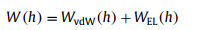
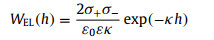
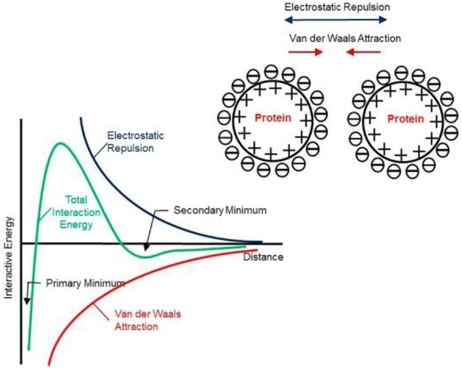
3. Aggregation behavior of GO in aquatic systemThe stability of charged particles is the consequences of the presence of various forces on surface of GO [47]. These forces of interaction (W(h)) have been measured by Derjaguin-Landau-Verwey-Overbeek (DLVO) theory, which indicates that the interfacial forces between two approaching colloidal particles are sum of electrostatic (WEL) repulsion and van der Waals' (WvdW) forces as shown in Fig. 2 [47-49]. Therefore, the aggregation/stability behavior of GO particles can be predicted by calculating the sum of WvdW(h) and WEL(h) according to the following equations (Eqs. 1–3) [50, 51].
|
(1) |
where WvdW(h) and WEL(h) can be calculated by Eqs. 2 and 3.

|
(2) |
|
(3) |
where H is Hamaker constant, h is separation distance, σ+ and σ− are the surface charge densities per unit area, ε0 is the permittivity of vacuum, ε is the dielectric constant of water, and κ is the inverse Debye length [51].

|
Download:
|
| Fig. 2. A schematic diagram depicting the balance between electrostatic repulsion and van Der Waals attraction for colloids (DLVO theory). Copied with permission [49]. Copyright 2019, Elsevier. | |
The decrease in surface charge density due to reduction of GO causes obvious decrease in the content of charged functional group, thereby noticeably decreasing the EL repulsion strength. Also, the restoration of the conjugated structure of graphene strengthens the attraction of vdW forces [52]. Hence, these above-mentioned driving forces (EL and vdW) are frequently calculated to explain the stability/instability of GO. The stability of GO is higher than that of rGO in aqueous solutions because of the following two reasons. First, the reduction of GO decreases the surface charge density due to an obvious decrease in the number of charged groups, thus expressively decreasing the strength of electrostatic repulsion [50, 53]. Second, the restoration of the conjugated structure of graphene strengthens the van Der Waals attraction [50, 53]. There are two different situations that may exist for the aggregation of particles; i.e., symmetrical situation or homoaggregation (deals with two identical particles) and asymmetrical situation or heteroaggregation (deals with two different particles).
3.1. GO homoaggregationHomoaggregation involves the interaction of two particles with the same type. Although the presence of the O-containing functional groups on GO surface make it extremely hydrophilic [6], the presence of ionized functional groups on surface of GO due to electrolytes in natural waters generates an electrical double layer (EDL) [54]. The electrolytes have capacity to compress the EDL which screen the surface charge of GO and thus promoting the irreversible homoaggregation [55]. Generally, the attachment efficiency (α) is commonly used to investigate the aggregation of GO, where, the reaction-limited regime (α < 1) and diffusion-limited regime (α = 1) determine the GO homoaggregation in the presence of various electrolytes [56]. Also, the values of zeta potential provide magnitude and sign of the effective surface charge density related to EDL around the colloid particle [57]. Usually, negatively charged surface colloids with a zeta potential lower than −30 mV or positively charged surface colloids with a zeta potential higher than 30 mV is assumed to be electrostatically stable [28]. The divalent cations (Ca2+, Mg2+) showed more aggressive compression for EDL as compared to monovalent cations (Na+, K+), following the Schulze−Hardy rule [27, 30]. During homoaggregation, multivalent ions showed high propensity to bind hydroxyl and carboxyl groups on GO sheets, enhancing the bridging between GO sheets [58].
3.2. GO heteroaggregationIn natural waters, the interaction probability of NPs with each other is much less than collision with other colloids [59, 60]. NPs interaction with solid-phase constituents, result in limitation of their mobility [61]. In aquatic system, the natural colloid concentration is likely to be several folds of magnitude higher than the concentration of GO particles [62]. Therefore, the GO heteroaggregation with other natural colloidal particles (e.g., clay particles, metal oxides, and macromolecular NOM), rather than the homoaggregation of GO, is supposed to be the governing process that controls the mobility of GO [63, 64]. Some previous studies indicated that the aggregation of GO might be affected by other NPs such as clays (kaolinite and montmorillonite), metal oxides (ZnO, MGO, and TiO2), minerals (layered double hydroxides, goethite, and hematite), and metal–organic frameworks [56, 65, 66]. For example, GO aggregation and deposition in aquatic environment was noticed due to the formation of multilayered structure with goethite [67]. The flocculation behavior of Al2O3 decreased the mobility of GO, thus enhancing its aggregation and deposition in aquatic systems [68]. Moreover, the aggregation behavior of GO was enhanced significantly when treated with kaolinite at high ionic strength (> 50 mmol/L NaCl) due to the compression of EL layer [69].
4. Effect of GO properties on its aggregationVarious physicochemical characteristics (e.g., specific surface area, size, morphology, surface charge, functional groups, and polarity) of GO are responsible for different interactions of graphene substrates with themselves or aqueous media and eventually change the GO aggregation behavior.
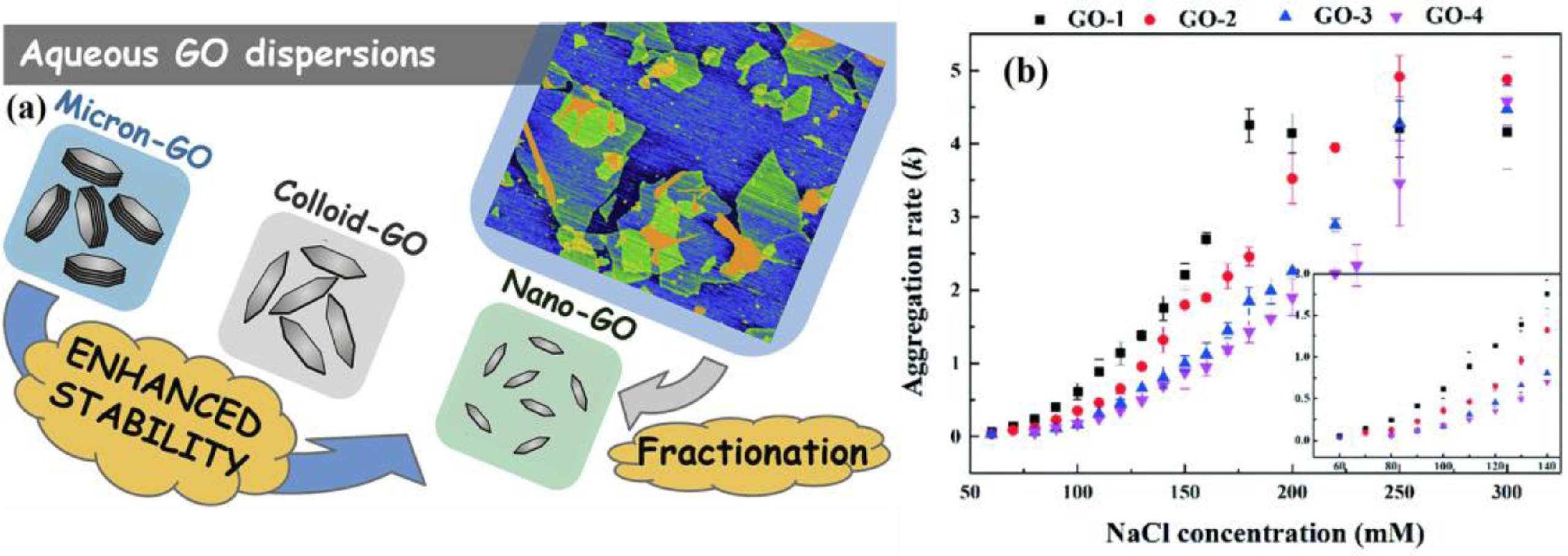
4.1. SizeThe size may act as an important and intrinsic factor affecting GO aggregation [70]. The smaller the size of GO nanosheets, the more intense the Brownian motion would be and vice versa. The intensity of Brownian motion not only enhanced the possibility of collision among GO nanosheets but also enhanced the possibility of collision among ions and GO nanosheets in dispersion system [71]. Fig. 3a illustrates an obvious dispersity of GO with decreasing size and vice versa. A comparative study was conducted to explore the stability/aggregation behavior of nano-sized GO along with colloid and micron-sized GO. They have noticed the higher stability with critical coagulation concertation (CCC) value (360 mmol/L) for nano-sized (150 nm) GO as compared to colloid and micron-sized particles [72]. Our previous research also found that small-sized GO particle showed higher stability as compared to large sized GO. Their CCC values in NaCl solution were 150 mmol/L and 49 mmol/L, respectively [32]. A study by Ding et al. detected the strong interaction between large-sized GO particles, indicating their faster aggregation due to slow Brownian motion [71]. When the collision between GO nanosheets and ions in the dispersion system happened, cross-linking occurred between them, and the formation of aggregate with strong force of interaction among GO nanosheets and surrounding ions was noticed [73, 74]. A size dependent study found the higher aggregation rates for smaller-sized GO as compared to larger-sized GO (Fig. 3b). This study summarized the CCC values were 278.4, 249.2, 225.3, and 173.9 mmol/L NaCl for GO with particle sizes of 93 (GO-4), 128 (GO-3), 170 (GO-2), and 240 (GO-1) nm, respectively [75]. The results from these studies suggest that the smaller the size of GO particles, the more likely the exposure to organisms and long-term transport in the aquatic environment.

|
Download:
|
| Fig. 3. (a) Schematic diagram showing effect of various sizes (micron, colloid, and nano) of GO on aggregation behavior. Copied with permission [72]. Copyright 2020, Elsevier. (b) Effect of various sizes (GO-1:240, GO-2:170, GO-3:128, and GO-4:93 nm) of GO on aggregation rates in NaCl solutions. Copied with permission [75]. Copyright 2020, Royal Society of Chemistry. | |
GO particles usually show variation in morphology, have random functional groups for each sheet/layer and show differences in physical structure (such as shape, molecular weight, and defects) [76]. GO with 2D structure can be physically modified into 3D structures, such as wrinkled (corrugated) and paper ball-like spheres (crumpled) [77]. The stability of GO derivatives (like GO with different morphological transformation and reduction degrees) may alter from pristine GO under the same characteristic of aqueous solution. A previous study by Jiang et al. observed the higher CCC values (81.7 mmol/L NaCl) for crumpled GO as compared to pristine GO (68.7 mmol/L NaCl) [78]. This can be assigned to the modification (crumpling) of GO structures, which can minimize the π–π stacking between discrete sheets, resulting in inhibition of aggregation [53].
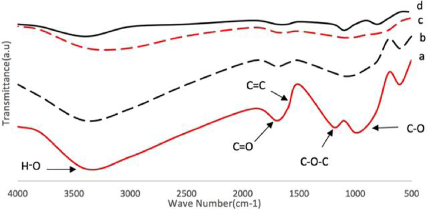
4.3. Functional groupsGO contains an extensive variety of hydrophilic oxygenated functional groups such as carbonyl, phenol, carboxyl, and hydroxyl [79]. The different ionization strength for the acidic oxygen-containing functional groups on the surface of GO may ultimately influence the stability of GO in water [80]. GO disperse well in water due to the electrostatic repulsion (EL) between the negatively charged ionized edges attributed to the ionization of carboxylate groups [81]. Because of the sufficient hydrophilic functional groups, such as carboxylic (-COOH) groups on the periphery of the planes, and epoxide and hydroxyl (-OH) on the basal planes, GO may disperse uniformly in aqueous solutions [82]. A study by Shams et al. revealed that removal of hydrophilic functional groups via photodegradation could result in conversion of GO into rGO, thus promoting hydrophobicity and aggregation [29]. Fig. 4 describes an obvious reduction via sonication of GO into rGO after sufficient removal of oxygen-containing functional groups from GO surface. The pristine GO contained 49% oxygen groups, while after reduction rGO-31, rGO-19, and rGO-9 contained 31%, 19%, and 9% oxygen groups, respectively [2]. The results show an obvious removal in oxygen functional groups from GO surface indicating the formation of rGO with hydrophobic nature that can promote GO aggregation.

|
Download:
|
| Fig. 4. FTIR spectra of (a) GO, (b) rGO-31, (c) rGO-19, (d) rGO-9. Copied with permission [16]. Copyright 2018, Springer Nature. | |
Complex environmental conditions may alter the surface physicochemical properties, including hydrophobicity, surface charge, and surface area. Therefore, the environmental factors, such as light, pH, dissolved organic matter (DOM), ionic strength, and ion types of water, may affect the stability and aggregation of GO in natural waters, which will be comprehensively summarized as follows.
5.1. Salt type and ionic strengthGround and surface water ecosystems contain different inorganic ions including Ca2+, Mg2+, Na+, K+, SO42−, HCO3−, Cl− and other ions, which varies with geographical location and may have an obvious effect on the surface charge of nanoparticles and consequently their colloidal characteristics [30]. Usually, GO aggregation behavior in presence of different electrolytes can be defined as a balance between the interfacial concentration of cation (Na+, Ca2+ and Mg2+) and anion (Cl–, SO42– and H2PO4–). On one hand, GO becomes more negative due to the adsorption of anions, which enhance the stabilization of GO in aqueous solutions [68]. On the other hand, GO particles in aqueous solutions might easily be destabilized by introducing salts, acid, or base (HCl, LiOH, LiCl, LiBr, NaCl, KCl, or KBr) [83]. The electrolyte type has significant influence on destabilization of GO, where GO aggregated more significantly in the presence of divalent cations than monovalent [34, 53]. Wang et al. compared the aggregation behavior of GO in aqueous solution containing 24 and 2.5 mmol/L of NaCl and MgCl2, respectively [84]. Mg2+ showed higher screening or charge neutralization effect than Na+ (Figs. 5a and b).

|
Download:
|
| Fig. 5. Aggregation rates of GO dispersions in different concentrations of (a) NaCl, (b) MgCl2 (0.02, 0.05, 0.25, 0.50, 2.50, 5.00, 24.00 and 91.00 mmol/L from left to right). Copied with permission [84]. 2016, Royal Society of Chemistry. (c) Cross linking by divalent cations. Copied with permission [73]. Copyright 2013, American Chemical Society. (d) Effect of ionic strength on stability ratio of nano, colloid, and micron GO. Copied with permission [72]. Copyright 2020, Elsevier. | |
The strength of charge neutralization depends on the concentration of electrolyte, counter-ions valency, and interaction between electrolyte and colloids [83, 85, 86]. As the concentration of counter-ion approaches and exceeds the CCC value, GO dispersion enters a favorable aggregation condition [87]. The ionic strength of solution plays an essential role in controlling the GO stability in suspension [30, 73]. A study compared the CCC values of GO and found they were 44 mmol/L for NaCl, 0.9 mmol/L for CaCl2, and 1.3 mmol/L for MgCl2 [88]. Our research group also detected CCC values as 150 mmol/L and 49 mmol/L NaCl for small (200 nm) and large (589 nm) GO particles, respectively [32]. Ca2+ and Mg2+ (divalent cations) were found to be more significantly effective than Na+ (monovalent cations) in destabilization of GO suspensions [73]. The CCC value of GO in NaCl (188 mmol/L) was much smaller than those in CaCl2 (2.6 mmol/L) and MgCl2 (3.9 mmol/L) solutions [73], representing that the divalent cations would be much effective to destabilize GO suspensions. This might be due to the strong cross-linkage between divalent cations and GO sheets occurred via functional groups on surface of GO, primarily at the edges (Fig. 5c). A comparative study found that ionic strength affected the stability of three sized (nano, colloid, and macron) GO, and the fastest aggregation rate was noticed at high ionic strength for all sized GO as shown in Fig. 5d [72].
5.2. Effect of pHThe pH of solution is a main parameter for determination the stability of graphene suspension [89]. The surface charges and pH dependent aggregation/stability of GO particles in solutions usually depend on the extent of ionization degree of the ionizable groups (carboxylic and/or hydroxyl group) that is influenced by protonation and deprotonation conditions [54, 57, 90]. At low pH, the ionized functional groups (mostly hydroxyl/carboxylic) attract proton and gain more positive charge that cause aggregation of GO due to compression of EL. At high pH, functional groups (hydroxyl/carboxylic) on surface of GO dissociate and gain more negative charge, indicting higher stability due to strong electrostatic repulsion (Fig. 6a). The density of surface charge is directly proportional to the concentration of available ionized functional groups at various pH conditions [57]. Overall, the protonation of ionizable functional groups occurred at low pH. While the shifting of pH to alkaline promoted deprotonation and the surface charge of GO becomes sufficiently negative, which inhibited GO aggregation as seen in Fig. 6b.

|
Download:
|
| Fig. 6. (a) Schematic illustration shows the surface charge of GO under various pH conditions. The shadowed sections indicate the basal plane of GO. And the circle with subtraction sign depicts the negative charge of GO. Copied with permission [57]. Copyright 2013, Elsevier. (b) Aggregation mechanism of GO in solutions with different pH: deprotonation of the carboxyl groups at the edges at alkaline solutions and protonation in acidic solutions. Copied with permission [73]. Copyright 2013, American Chemical Society. | |
Shih et al. performed a series of comparative and molecular dynamics studies under the desired pH conditions to elucidate the influence of pH (1 to 14) in the behavior of GO in aqueous solutions [91]. They revealed that GO exhibited high dispersion at pH 14, while aggregated easily at low pH 1. Also, the reported point of zero charge (PZC) values range between 3.5–4 for GO [92, 93]. The PZC is the pH at which the net charge on the particle surface is found to be zero. The surface charge of GO is positive at pH < pHpzc and negative at pH ˃ pHpzc. When pH of solution is higher than pHpzc, the surface of GO is negatively charged with high dispersity, while providing favorable conditions for the adsorption of cationic species [92]. The aggregation of GO at low pH is due to the effective acceleration of π–π interaction, as the restoration of aromatic structure may decrease the steric hindrance mediated by water [94]. Some other studies also confirmed that pH sensitive surface of GO tends to induce variations in surface charges of GO nanoparticles, and thus affect the stability and dispersity of GO [88, 95].
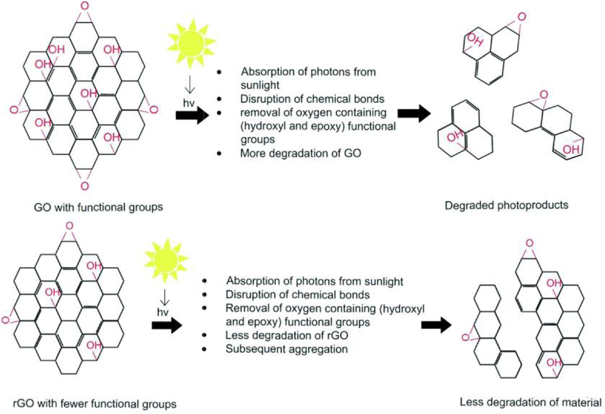
5.3. Effect of lightAggregation of various graphene nanomaterials in natural aquatic systems have been altered upon exposure to sunlight [96, 97]. Fig. 7 shows the schematic illustration of how photodegradation promote aggregation for GO and rGO. In direct irradiation by light, the functional groups of GO act as chromophores [98]. Epoxy (C–O) and hydroxyl (C–OH) functional groups are hypothesized to break in initial period of irradiation as they are single bonded groups. Various functional groups on the edges are usually double bonded groups, which need more energy to break as compared to single bonds, and are thus less likely to react initially [99, 100]. The strong electron donating epoxy and hydroxyl functional groups, absorb more photons that excite electrons from the ground state to the excited state, generating excited holes and electrons [101]. Finally, these holes and electrons destruct the chemical bonds of the functional groups and break the covalent bonds, thus contributing to the physical disruption of the GO material [102, 103]. Due to the loss of oxygen groups, GO is converted to rGO with significantly fewer O-containing functional groups than pristine GO. The reduction in functional groups causes the enhancement of hydrophobicity for rGO particles, thus enhancing its aggregation more easily. A study by Hou et al. recently reduced GO under sunlight for 25 days and found that GO was completely reduced to low molecular weight species in 8 days like rGO that are comparatively smaller in molecular weight than pristine material [35].

|
Download:
|
| Fig. 7. Schematics of the proposed photodegradation pathway for graphene oxide nanomaterial. (Top) GO is shown with oxygen-containing (hydroxyl and epoxy) functional groups. Due to absorption of photons from sunlight, the functional groups are removed, accelerating the physical degradation. (Bottom) rGO with fewer functional groups undergoes slower degradation than GO under similar irradiation conditions. Copied with permission [29]. Copyright 2019, Royal Society of Chemistry. | |
While, the hydrodynamic diameter of GO was found to increase abruptly after exposure to UV light for 30 min, probably because of the elimination of surface functional groups and the hydrogen bonds breakup among GO particles. After 30 min, the hydrodynamic diameter decreased slowly due to the reverse folding or crumpling of the partially reduced GO sheets [104]. Indirect irradiation by UV light also induced the variation in GO physico-chemical properties. With the increasement in cation concentration, more obvious aggregation could be observed for UV-transformed GO. Similarly, different types of cation (monovalent/divalent) has different capability to destabilize the UV-irradiated GO (i.e., Ca2+ > Mg2+ > Na+) [105]. It should be noted that once the reduced photoproducts formed via degradation of GO, they are more resistant to further photodegradation but favorable for aggregation [29]. Moreover, a study described that 120 h UV irradiation exposure at 28–74 µW/cm2 might bring important alteration in GO structure via elimination of C=O and -OH functional groups. This reduction decreased the negative zeta potential from −41 mV to −37 mV and enhanced the aggregation of GO in water with prolonging the exposure time [106]. Du et al. observed that UV irradiation showed remarkable variations in the content of oxygen-containing functional groups on GO surface [107]. One must keep in mind that aggregation degree mainly depends on the kind of light source; i.e. intensity and range of radiation [108]. GO showed an increase of 31% and 41% in C/O ratio after irradiation with UV and simulated solar light, respectively. The loss of oxygen-containing functional groups converted GO to rGO, which is more prone to aggregation rather than degradation via photoirradiation.
5.4. Effect of dissolved organic matterDOM in natural aquatic system plays a significant role in GO stability/aggregation, even in the presence of cationic salts (Na+, K+, Ca2+, and Mg2+) [30, 109]. It has been noticed that DOM could be adsorbed by GO via its Lewis acid-base, hydrogen bonds, and π-π interactions [110, 111]. A long-term study showed that occurrence of DOM in natural waters prominently enhanced the GO stability via steric repulsion [88]. Shen et al. compared the effect of DOM molecular weight on aggregation behavior of GO in presence of NaCl and CaCl2 [31]. The results from the study noted an obvious inhibition of aggregation with high molecular weight DOM as compared to low molecular weight in presence of both NaCl and CaCl2 electrolyte. Also, the effect of DOM on GO stability was more obvious in NaCl and CaCl2 solutions with low concentrations as compared to high concentrations (Fig. 8).

|
Download:
|
| Fig. 8. Aggregation profiles of GO in the absence or presence of Suwannee River DOM in different concentrations of (a–c) NaCl and (d–f) CaCl2 solutions. Copied with permission [31]. Copyright 2019, Elsevier. | |
The intrinsic physical and chemical properties of DOM might be responsible for its adsorption on most of the natural particle's surfaces in natural aquatic systems [112, 113]. Humic acid (HA) and fulvic acid (FA) are main constituents of DOM [114, 115]. It has been proved that HA showed most effectiveness for stabilization of GO in NaCl than in CaCl2, due to the formation of bridging between Ca2+ and carboxyl functional groups on GO [88, 116]. Generally, HA with complex structure has higher molecular weight, longer chain length of hydrophobic groups, and less polarity than FA [117]. Thus, HA inhibited GO aggregation more obviously than FA because of the higher steric repulsion power of HA than FA [118]. Also, the aggregation of GO nanoparticles is concentration dependent, indicating higher stability with enhancing the concentrations of HA (0.1–10 mg/L) due to thicker coating of HA on GO surface [30]. Our research group also found that the stability of GO is concentration and DOM's component (HA or FA) dependent, where HA/FA with higher concentration and HA component of DOM collected from various climate zones of China showed more stability for GO [32]. Another study by Shen et al. also found that GO aggregation was altered significantly by large molecular weight and highly aromatic content of DOM in NaCl [31]. The study calculated the CCC values for GO in NaCl as a function of molecular weights of Suwannee River DOM. They observed higher CCC values (326 mmol/L NaCl) with less aggregation for GO in the presence of DOM with higher molecular weight (> 100 kD). The exact mechanism of interaction for GO-DOM depends on both DOM physicochemical properties (size, source, structure, aromaticity, polarity, and charge density) as well as GO functional derivatization and properties.
6. Environmental implicationsAs the rapid increase in GO production due to its potential for numerous applications, it is certain that a sufficient amount will be released into the aquatic environment during its synthesis, transportation, application, and disposal. The fate and transport of GO was primarily governed by its aggregation/stability in natural aquatic environments [88]. Moreover, previous studies have observed that GO may be hazardous for various organisms including human, animals, and bacteria [119, 120]. After disposal, GO may also interact with various constituents in natural waters, that can alter its stability leading to adverse impact on ecosystems [53].
6.1. Effect of GO aggregation on its transportationGO aggregation affects its subsequent transportation through sedimentation, adsorption, redox reaction, photochemical and biologically mediated reactions in natural waters [21, 30]. Sedimentation is one of the most crucial processes affected by GO aggregation in aquatic environments [121]. To assess the GO transportation and mobility, the concentrations of GO and natural colloids need to be accounted. The sedimentation of GO via aggregation with other particles are concentration dependent with higher aggregation rates found near the point of GO release [56]. Also, aggregation of GO in ground water due to low concentration of DOM and high concentration of salts may reduce its mobility in ground water [30]. Usually, large size of aggregated GO stops it to enter the sediments, while dispersed GO with small size easily enters the sediments and travels far away [122]. Also, the reduction of GO to rGO decreases the available reactive sites, thus increasing the hydrophobicity that ultimately decrease the transportation and enhance the sedimentation of GO in aquatic environment [24]. In natural waters, natural colloids are likely to be several orders of magnitude higher than concentration of GO [56, 62]. Therefore, GO heteroaggregation with natural colloids such as clay particles, metal oxides, and macromolecular organic matter, rather than GO homoaggregation, is supposed to be the dominant process that governs GO transport [63]. Feng et al. concluded that the hetero aggregation of hematite with GO nanoparticles in natural waters reduced the mobility of GO [56].
6.2. Effect of GO aggregation on its toxicityIn natural waters, the investigation of GO aggregation behavior is of key importance to highlight and manage its potential toxicity. The potential effect of GO in natural waters is the consequences of its aggregation behavior that eventually alters its hazardous impacts to aquatic biota [17]. Substantial risks related to aggregation of GO are usually dependent on their synthesis methods, sizes, morphology, and functional groups as well as the complex chemistry of natural waters including pH, slat types, ionic strength, light, and DOM presence. The aggregation can alter the GO physical and chemical properties like particle size and surface area, which may control the toxicity to biota in aquatic systems [26]. Both physical and chemical toxic effects via cell membrane damage and reactive oxygen species generation were noticed for GO [53]. GO can be toxic toward a variety of organisms including bacteria, animals, and humans [15, 17]. A close contact of GO nanoparticle with cytoplasmic membrane or cell wall mostly destructs the cell membrane or produce obvious membrane stress, leaking the substances present in cell and ultimately cause the cell death [123]. A study by Liu et al. stated that the aggregated large-sized graphene sheets showed lower cytotoxicity as compared to dispersed GO with smaller size due to lack of active sites responsible for interaction [124]. Also, the reduction of GO to rGO may form sharpness on its nanowall edges, which result in more severe damage of cell membrane and higher toxicity of rGO toward bacteria [22]. Therefore, understanding the aggregation behavior of GO is crucial to evaluate, monitor, and manage its fate, transport/mobility and toxicity behavior in natural waters.
7. Conclusions and future prospectiveGO, as an important graphene derivative, has gained a huge attraction in recent years due to its potential for academic, environmental and industrial applications. As the production of GO increases, serious concerns due to its expected exposure to the natural aquatic ecosystem prevail. After being released into natural waters, main transformation process of GO in waters are interrelated, including aggregation/stability, sedimentation, adsorption, oxidation/reduction, and photo-degradation. For example, high pH deprotonates the ionizable functional groups of GO particles, which might be adsorbed to naturally occurring colloids, combine with ligands, aggregated and finally deposited.
Knowledge regarding the aggregation of GO in natural water ecosystem is essential for predicting its adverse impacts. We have summarized several critical parameters controlling the stability of GO in aquatic ecosystem. It is a difficult task to highlight the action mechanisms of GO because of the combined effects in environmental relevant conditions. In general, cations (monovalent and bivalent) and DOM have been proved as the main controlling factors for GO aggregation, whereas light (visible and UV) plays a key role in the transformation of GO. In addition to above mentioned environmental factors, the aggregation of GO also depends on its own physicochemical characteristics like morphology, size, concentration, and presence of various surface functional groups.
There are still numerous difficulties in determining the environmental behavior of GO in aquatic system. For example, the challenge in understanding the transport of GO was due to confusion in synthesis techniques, complicated parameters in aquatic ecosystem, and short-term evaluation. Since there are many methods for GO synthesis, GO composition and functional groups on the surface may vary, that can make a huge difference in results for the estimation of GO stability. In addition, once GO was exposed to natural aquatic ecosystem, long-term assessment of GO aggregation was required since the current research were mostly performed for short term. The regions and time could affect the components in natural waters. This alteration could affect the environmental transport of GO, which has not been investigated yet.
The production and applications of various nanomaterials are increasing day by day. Water bodies would be the sink for other nanomaterials. Still, how the existence of other nanomaterials in waters influences the stability of GO remains poorly understood. Hence, new research regarding practical dynamic changes in complicated water systems is required to evaluate the complex role of different nanomaterials on GO transport. Tracking the transformation and mobility of GO within and among the environmental ecosystems (especially in water and soil) is also essential to manage the adverse impacts of GO.
Declaration of competing interestThe authors declare that they have no known competing financial interests or personal relationships that could have appeared to influence the work reported in this paper.
AcknowledgmentsThe study was financially supported by the National Key R & D Program of China (No. 2021YFC3200401), the National Natural Science Foundation of China (Nos. 21677015, 52170024 and 22006031), and the Natural Science Foundation of Hebei Province (No. B2019204315).
| [1] |
T. Malina, E. Maršálková, K. Holá, R. Zbořil, B. Maršálek, J. Hazard. Mater. 399 (2020) 123027. DOI:10.1016/j.jhazmat.2020.123027 |
| [2] |
F. Mouhat, F.X. Coudert, M.L. Bocquet, Nat. Commun. 11 (2020) 1566. DOI:10.1038/s41467-020-15381-y |
| [3] |
S.H. Dave, C. Gong, A.W. Robertson, J.H. Warner, J.C. Grossman, ACS Nano 10 (2016) 7515-7522. DOI:10.1021/acsnano.6b02391 |
| [4] |
L.C. Chen, S. Lei, M.Z. Wang, J. Yang, X.W. Ge, Chin. Chem. Lett. 27 (2016) 511-517. DOI:10.1016/j.cclet.2016.01.057 |
| [5] |
G. Reina, J.M. González-Domínguez, A. Criado, et al., Chem. Soc. Rev. 46 (2017) 4400-4416. DOI:10.1039/C7CS00363C |
| [6] |
Y. Zhao, Y. Liu, X. Zhang, W. Liao, Chemosphere 262 (2021) 127885. DOI:10.1016/j.chemosphere.2020.127885 |
| [7] |
G. Eda, M. Chhowalla, Adv. Mater. 22 (2010) 2392-2415. DOI:10.1002/adma.200903689 |
| [8] |
A.T. Smith, A.M. LaChance, S. Zeng, B. Liu, L. Sun, Nano Mater. Sci. 1 (2019) 31-47. DOI:10.1016/j.nanoms.2019.02.004 |
| [9] |
L. Saghatforoush, M. Hasanzadeh, N. Shadjou, Chin. Chem. Lett. 25 (2014) 655-658. DOI:10.1016/j.cclet.2014.01.014 |
| [10] |
X. Hu, S. You, F. Li, Y. Liu, Front. Environ. Sci. Eng. 16 (2022) 48. DOI:10.1007/s11783-021-1482-7 |
| [11] |
Y. Gao, X. Zeng, W. Zhang, et al., Sci. Total Environ. 806 (2022) 150942. DOI:10.1016/j.scitotenv.2021.150942 |
| [12] |
Y. Zhu, H. Ji, H.M. Cheng, R.S. Ruoff, Natl. Sci. Rev. 5 (2018) 90-101. DOI:10.1093/nsr/nwx055 |
| [13] |
Y. Yang, Z. Yu, T. Nosaka, et al., Front. Environ. Sci. Eng. 9 (2015) 823-831. DOI:10.1007/s11783-015-0787-9 |
| [14] |
K. Ko, M.J. Kim, J.Y. Lee, W. Kim, H.J. Chung, Sci. Total Environ. 651 (2019) 1087-1095. DOI:10.1016/j.scitotenv.2018.09.124 |
| [15] |
P. Kumar, P. Huo, R. Zhang, B. Liu, Nanomaterials 9 (2019) 737. DOI:10.3390/nano9050737 |
| [16] |
S. Azizighannad, S. Mitra, Sci. Rep. 8 (2018) 10083. DOI:10.1038/s41598-018-28353-6 |
| [17] |
J. Zhao, Z. Wang, J.C. White, B. Xing, Environ. Sci. Technol. 48 (2014) 9995-10009. DOI:10.1021/es5022679 |
| [18] |
B. Sun, Y. Zhang, R. Li, et al., Water Res. 200 (2021) 117213. DOI:10.1016/j.watres.2021.117213 |
| [19] |
Y. Gao, X. Zeng, W. Zhang, et al., Sci. Total Environ. 806 (2022) 150942. DOI:10.1016/j.scitotenv.2021.150942 |
| [20] |
J.L. Suter, R.C. Sinclair, P.V. Coveney, Adv. Mater. 32 (2020) 2003213. |
| [21] |
Q. Abbas, B. Yousaf, Amina, et al., Environ. Int. 138 (2020) 105646. DOI:10.1016/j.envint.2020.105646 |
| [22] |
O. Akhavan, E. Ghaderi, ACS Nano 4 (2010) 5731-5736. DOI:10.1021/nn101390x |
| [23] |
M.C. Duch, G.S. Budinger, Y.T. Liang, et al., Nano Lett. 11 (2011) 5201-5207. DOI:10.1021/nl202515a |
| [24] |
X. Hu, Q. Zhou, Chem. Rev. 113 (2013) 3815-3835. DOI:10.1021/cr300045n |
| [25] |
S. Ouyang, X. Hu, Q. Zhou, ACS Appl. Mater. Interfaces 7 (2015) 18104-18112. DOI:10.1021/acsami.5b05328 |
| [26] |
N. Malhotra, O.B. Villaflores, G. Audira, et al., Molecules 25 (2020) 3618. DOI:10.3390/molecules25163618 |
| [27] |
S. Yu, X. Wang, R. Zhang, et al., Sci. Rep. 7 (2017) 39625. DOI:10.1038/srep39625 |
| [28] |
M. Bayati, M. Fidalgo de Cortalezzi, J. Environ. Eng. 145 (2019) 04019050. DOI:10.1061/(ASCE)EE.1943-7870.0001561 |
| [29] |
M. Shams, L.M. Guiney, L. Huang, et al., Environ.Sci. Nano 6 (2019) 2203-2214. DOI:10.1039/C9EN00355J |
| [30] |
J.D. Lanphere, B. Rogers, C. Luth, C.H. Bolster, S.L. Walker, Environ. Eng. Sci. 31 (2014) 350-359. DOI:10.1089/ees.2013.0392 |
| [31] |
M. Shen, X. Hai, Y. Shang, et al., Sci. Total Environ. 656 (2019) 843-851. DOI:10.1016/j.scitotenv.2018.11.387 |
| [32] |
J. Ali, Y. Li, X. Wang, et al., Sci. Total Environ. 721 (2020) 137682. DOI:10.1016/j.scitotenv.2020.137682 |
| [33] |
X. Huangfu, Y. Xu, C. Liu, et al., Chemosphere 219 (2019) 766-783. DOI:10.1016/j.chemosphere.2018.12.044 |
| [34] |
M. Wang, B. Gao, D. Tang, et al., Colloid Surf. A Physicochem. Eng. Asp. 538 (2018) 63-72. DOI:10.1016/j.colsurfa.2017.10.061 |
| [35] |
W.C. Hou, I. Chowdhury, D.G. Goodwin, et al., Environ. Sci. Technol. 49 (2015) 3435-3443. DOI:10.1021/es5047155 |
| [36] |
A. Ramos-Corona, R. Rangel, J. Espino, et al., Catal. Today 392- 393 (2022) 81-92. |
| [37] |
R. Ma, Y. Zhou, H. Bi, et al., Prog. Mater. Sci. 113 (2020) 100665. DOI:10.1016/j.pmatsci.2020.100665 |
| [38] |
Z. Teng, B. Wang, Y. Hu, D. Xu, Chin. Chem. Lett. 30 (2019) 717-720. DOI:10.1016/j.cclet.2018.08.017 |
| [39] |
P. Sun, K. Wang, H. Zhu, Adv. Mater. 28 (2016) 2287-2310. DOI:10.1002/adma.201502595 |
| [40] |
K.Y. Yoon, S.J. An, Y. Chen, et al., J. Colloid Interface Sci. 403 (2013) 1-6. DOI:10.1016/j.jcis.2013.03.012 |
| [41] |
B.W. Pratama, W.S.B. Dwandaru, Nano Express 1 (2020) 010023. DOI:10.1088/2632-959X/ab8685 |
| [42] |
F.T. Johra, W.G. Jung, Appl. Surf. Sci. 357 (2015) 1911-1914. DOI:10.1016/j.apsusc.2015.09.128 |
| [43] |
L. Silipigni, G. Salvato, B. Fazio, et al., J. Mater. Sci. Mater. Electron. 31 (2020) 11847-11854. DOI:10.1007/s10854-020-03738-4 |
| [44] |
K.A. Mkhoyan, A.W. Contryman, J. Silcox, et al., Nano Lett. 9 (2009) 1058-1063. DOI:10.1021/nl8034256 |
| [45] |
K. Erickson, R. Erni, Z. Lee, et al., Adv. Mater. 22 (2010) 4467-4472. DOI:10.1002/adma.201000732 |
| [46] |
K.P. Loh, Q. Bao, G. Eda, M. Chhowalla, Nat. Chem. 2 (2010) 1015-1024. DOI:10.1038/nchem.907 |
| [47] |
S. Ghazizadeh, P. Duffour, N.T. Skipper, M. Billing, Y. Bai, Cem. Concr. Res. 99 (2017) 116-128. DOI:10.1016/j.cemconres.2017.05.011 |
| [48] |
F. Rubbi, L. Das, K. Habib, et al., J. Mol. Liq. 338 (2021) 116771. DOI:10.1016/j.molliq.2021.116771 |
| [49] |
J. Li, Y. Cheng, X. Chen, S. Zheng, Int. J. Pharm. X 1 (2019) 100002. |
| [50] |
M.M. Gudarzi, Langmuir 32 (2016) 5058-5068. DOI:10.1021/acs.langmuir.6b01012 |
| [51] |
G. Trefalt, S.H. Behrens, M. Borkovec, Langmuir 32 (2016) 380-400. DOI:10.1021/acs.langmuir.5b03611 |
| [52] |
Q. Pan, E. Shim, B. Pourdeyhimi, W. Gao, Langmuir 33 (2017) 7452-7458. DOI:10.1021/acs.langmuir.7b01508 |
| [53] |
K. He, G. Chen, G. Zeng, et al., Nanoscale 9 (2017) 5370-5388. DOI:10.1039/C6NR09931A |
| [54] |
J. Luo, L.J. Cote, V.C. Tung, et al., J. Am. Chem. Soc. 132 (2010) 17667-17669. DOI:10.1021/ja1078943 |
| [55] |
B.J. Hong, O.C. Compton, Z. An, I. Eryazici, S.T. Nguyen, ACS Nano 6 (2012) 63-73. DOI:10.1021/nn202355p |
| [56] |
Y. Feng, X. Liu, K.A. Huynh, et al., Environ. Sci. Technol. 51 (2017) 6821-6828. DOI:10.1021/acs.est.7b00132 |
| [57] |
X. Hu, Y. Yu, W. Hou, J. Zhou, L. Song, Appl. Surf. Sci. 273 (2013) 118-121. DOI:10.1016/j.apsusc.2013.01.201 |
| [58] |
Z. Zeng, Y. Wang, Q. Zhou, K. Yang, D. Lin, Environ. Pollut. 250 (2019) 366-374. DOI:10.1016/j.envpol.2019.03.112 |
| [59] |
D. Shevlin, N. O'Brien, E. Cummins, Sci. Total Environ. 621 (2018) 1033-1046. DOI:10.1016/j.scitotenv.2017.10.123 |
| [60] |
T.Y. Sun, F. Gottschalk, K. Hungerbühler, B. Nowack, Environ. Pollut. 185 (2014) 69-76. DOI:10.1016/j.envpol.2013.10.004 |
| [61] |
B. Zhu, X. Xia, S. Zhang, Y. Tang, Environ. Pollut. 234 (2018) 581-589. DOI:10.1016/j.envpol.2017.11.086 |
| [62] |
S. Wagner, A. Gondikas, E. Neubauer, T. Hofmann, F. von der Kammer, Angew. Chem. Int. Ed. 53 (2014) 12398-12419. |
| [63] |
B.M. Smith, D.J. Pike, M.O. Kelly, J.A. Nason, Environ. Sci. Technol. 49 (2015) 12789-12797. DOI:10.1021/acs.est.5b03486 |
| [64] |
L. Jiang, Y. Liu, G. Zeng, et al., Chem. Eng. J. 343 (2018) 371-378. DOI:10.1016/j.cej.2018.03.026 |
| [65] |
N.P. Sotirelis, C.V. Chrysikopoulos, Environ. Sci. Technol. 49 (2015) 13413-13421. DOI:10.1021/acs.est.5b03496 |
| [66] |
J. Wang, S. Yu, Y. Zhao, et al., Sep. Purif. Technol. 184 (2017) 88-96. DOI:10.1016/j.seppur.2017.03.058 |
| [67] |
J. Zhao, F. Liu, Z. Wang, X. Cao, B. Xing, Environ. Sci. Technol. 49 (2015) 2849-2857. DOI:10.1021/es505605w |
| [68] |
X. Ren, J. Li, X. Tan, et al., Environ. Sci. Technol. 48 (2014) 5493-5500. DOI:10.1021/es404996b |
| [69] |
N.P. Sotirelis, C.V. Chrysikopoulos, Sci. Total Environ. 579 (2017) 736-744. DOI:10.1016/j.scitotenv.2016.11.034 |
| [70] |
J. Amaro-Gahete, A. Benítez, R. Otero, et al., Nanomaterials 9 (2019) 152. DOI:10.3390/nano9020152 |
| [71] |
G. Ding, N. Zhang, C. Wang, et al., J. Nanopart. Res. 20 (2018) 313. DOI:10.1007/s11051-018-4421-1 |
| [72] |
T. Szabo, P. Maroni, I. Szilagyi, Carbon 160 (2020) 145-155. DOI:10.1016/j.carbon.2020.01.022 |
| [73] |
L. Wu, L. Liu, B. Gao, et al., Langmuir 29 (2013) 15174-15181. DOI:10.1021/la404134x |
| [74] |
Y. He, Y. Liu, F. Guo, et al., Chin. Chem. Lett. 31 (2020) 1625-1629. DOI:10.1016/j.cclet.2019.10.010 |
| [75] |
B. Sun, Y. Zhang, Q. Liu, et al., Environ. Sci. Nano 7 (2020) 634-644. DOI:10.1039/C9EN01040H |
| [76] |
A. Bagri, C. Mattevi, M. Acik, et al., Nat. Chem. 2 (2010) 581-587. DOI:10.1038/nchem.686 |
| [77] |
J. Luo, H.D. Jang, T. Sun, et al., ACS Nano 5 (2011) 8943-8949. DOI:10.1021/nn203115u |
| [78] |
Y. Jiang, Q. Zeng, P. Biswas, J.D. Fortner, J. Membr. Sci. 581 (2019) 453-461. DOI:10.1016/j.memsci.2019.03.056 |
| [79] |
C. Liao, X.R. Zhao, X.Y. Jiang, J. Teng, J.G. Yu, Microchem. J. 152 (2020) 104288. DOI:10.1016/j.microc.2019.104288 |
| [80] |
X. Ren, J. Li, C. Chen, et al., Environ. Sci. Nano 5 (2018) 1298-1340. DOI:10.1039/C7EN01258F |
| [81] |
Y. Tang, H. Liu, X. Wang, et al., J. Mol. Struct. 1224 (2021) 129196. DOI:10.1016/j.molstruc.2020.129196 |
| [82] |
Y. Si, E.T. Samulski, Nano Lett. 8 (2008) 1679-1682. DOI:10.1021/nl080604h |
| [83] |
H. Wang, Y.H. Hu, J. Colloid Interface Sci. 391 (2013) 21-27. DOI:10.1016/j.jcis.2012.09.056 |
| [84] |
M. Wang, Y. Niu, J. Zhou, et al., Nanoscale 8 (2016) 14587-14592. DOI:10.1039/C6NR03503E |
| [85] |
M. Li, M. Kobayashi, Colloid Surf, A Physicochem. Eng. Asp. 626 (2021) 127021. DOI:10.1016/j.colsurfa.2021.127021 |
| [86] |
J. Zhao, Y. Li, X. Wang, et al., Environ. Pollut. 279 (2021) 116926. DOI:10.1016/j.envpol.2021.116926 |
| [87] |
K.L. Chen, M. Elimelech, J. Colloid Interface Sci. 309 (2007) 126-134. DOI:10.1016/j.jcis.2007.01.074 |
| [88] |
I. Chowdhury, M.C. Duch, N.D. Mansukhani, M.C. Hersam, D. Bouchard, Environ. Sci. Technol. 47 (2013) 6288-6296. DOI:10.1021/es400483k |
| [89] |
A. Griffith, S.M. Notley, J. Colloid Interface Sci. 369 (2012) 210-215. DOI:10.1016/j.jcis.2011.11.081 |
| [90] |
T. Szabó, E. Tombácz, E. Illés, I. Dékány, Carbon 44 (2006) 537-545. DOI:10.1016/j.carbon.2005.08.005 |
| [91] |
C.J. Shih, S. Lin, R. Sharma, M.S. Strano, D. Blankschtein, Langmuir 28 (2012) 235-241. DOI:10.1021/la203607w |
| [92] |
X. Li, X. Tang, Y. Fang, J. Mol. Liq. 199 (2014) 237-243. DOI:10.1016/j.molliq.2014.09.020 |
| [93] |
V. Sabna, S.G. Thampi, S. Chandrakaran, Water Sci. Technol. 78 (2018) 732-742. DOI:10.2166/wst.2018.311 |
| [94] |
H. Tang, S. Zhang, T. Huang, F. Cui, B. Xing, Environ. Sci. Nano 7 (2020) 984-995. DOI:10.1039/C9EN01365B |
| [95] |
W. Wu, Y. Hu, Q. Guo, et al., J. Hazard. Mater. 297 (2015) 59-65. DOI:10.1016/j.jhazmat.2015.04.078 |
| [96] |
T.P.D. Shareena, D. McShan, A.K. Dasmahapatra, P.B. Tchounwou, Nano Micro Lett. 10 (2018) 53. DOI:10.1007/s40820-018-0206-4 |
| [97] |
I. Chowdhury, W.C. Hou, D. Goodwin, et al., Water Res. 78 (2015) 37-46. DOI:10.1016/j.watres.2015.04.001 |
| [98] |
H. Bai, W. Jiang, G.P. Kotchey, et al., J. Phys. Chem. C 118 (2014) 10519-10529. DOI:10.1021/jp503413s |
| [99] |
G. Gündüz, Chemistry, Materials, and Properties of Surface Coatings: Traditional and Evolving Technologies, DEStech Publications, Inc, 2015.
|
| [100] |
R.C. Neuman, Organic Chemistry, in Organic Molecules and Chemical Bonding, California (US): University of California, 1999, pp. 1–55.
|
| [101] |
M. Mohandoss, S.S. Gupta, A. Nelleri, T. Pradeep, S.M. Maliyekkal, RSC Adv. 7 (2017) 957-963. DOI:10.1039/C6RA24696F |
| [102] |
M.P. Fasnacht, N.V. Blough, Environ. Sci. Technol. 36 (2002) 4364-4369. DOI:10.1021/es025603k |
| [103] |
T. Mill, W. Mabey, B. Lan, A. Baraze, Chemosphere 10 (1981) 1281-1290. DOI:10.1016/0045-6535(81)90045-X |
| [104] |
N.S. Andryushina, O.L. Stroyuk, I.B. Yanchuk, A.V. Yefanov, Colloid Polym. Sci. 292 (2014) 539-546. DOI:10.1007/s00396-013-3134-3 |
| [105] |
Y. Gao, X. Ren, G. Song, et al., J. Hazard. Mater. 382 (2020) 121097. DOI:10.1016/j.jhazmat.2019.121097 |
| [106] |
W.R. Gallegos-Pérez, A.C. Reynosa-Martínez, C. Soto-Ortiz, et al., Chemosphere 249 (2020) 126160. DOI:10.1016/j.chemosphere.2020.126160 |
| [107] |
T. Du, A.S. Adeleye, T. Zhang, et al., Environ. Sci. Nano 5 (2018) 2590-2603. DOI:10.1039/C8EN00593A |
| [108] |
K. Spilarewicz-Stanek, A. Jakimińska, A. Kisielewska, M. Dudek, I. Piwoński, Mater. Sci. Semicond. Process 123 (2021) 105525. DOI:10.1016/j.mssp.2020.105525 |
| [109] |
Z. Qi, T. Du, P. Ma, F. Liu, W. Chen, Sci. Total Environ. 657 (2019) 1450-1459. DOI:10.1016/j.scitotenv.2018.12.143 |
| [110] |
S. Bele, V. Samanidou, E. Deliyanni, Chem. Eng. Res. Des. 109 (2016) 573-585. DOI:10.1016/j.cherd.2016.03.002 |
| [111] |
N. Cai, D. Peak, P. Larese-Casanova, Chem. Eng. J. 273 (2015) 568-579. DOI:10.1016/j.cej.2015.03.108 |
| [112] |
I. Chowdhury, M.C. Duch, N.D. Mansukhani, M.C. Hersam, D. Bouchard, Environ. Sci. Technol. 48 (2014) 9382-9390. DOI:10.1021/es5020828 |
| [113] |
W. Chen, J. Song, S. Jiang, et al., Front. Environ. Sci. Eng. 16 (2022) 16. |
| [114] |
M.A. Islam, D.W. Morton, B.B. Johnson, M.J. Angove, Sep. Purif. Technol. 247 (2020) 116949. DOI:10.1016/j.seppur.2020.116949 |
| [115] |
Y. Luo, Y. Zhang, M. Lang, et al., Front. Environ. Sci. Eng. 15 (2021) 96. DOI:10.1007/s11783-020-1340-z |
| [116] |
M. Pham, E.A. Mintz, T.H. Nguyen, J. Colloid Interface Sci. 338 (2009) 1-9. DOI:10.1016/j.jcis.2009.06.025 |
| [117] |
Y. Li, J. Niu, E. Shang, J.C. Crittenden, Environ. Sci. Technol. 49 (2015) 965-973. DOI:10.1021/es505089e |
| [118] |
E. Shang, Y. Li, J. Niu, et al., Water Res. 124 (2017) 595-604. DOI:10.1016/j.watres.2017.08.001 |
| [119] |
Y. Sun, B. Gao, S.A. Bradford, et al., Water Res. 68 (2015) 24-33. DOI:10.1016/j.watres.2014.09.025 |
| [120] |
S. Gurunathan, J. Han, J.H. Park, J.H. Kim, Int. J. Nanomed. 9 (2014) 1783-1797. |
| [121] |
J.T.K. Quik, I. Velzeboer, M. Wouterse, A.A. Koelmans, D. van de Meent, Water Res. 48 (2014) 269-279. DOI:10.1016/j.watres.2013.09.036 |
| [122] |
A. Beryani, M.R. Alavi Moghaddam, T. Tosco, et al., Sci. Total Environ. 698 (2020) 134224. DOI:10.1016/j.scitotenv.2019.134224 |
| [123] |
F. Zou, H. Zhou, D.Y. Jeong, et al., ACS Appl. Mater. Interfaces 9 (2017) 1343-1351. DOI:10.1021/acsami.6b15085 |
| [124] |
S. Liu, T.H. Zeng, M. Hofmann, et al., ACS Nano 5 (2011) 6971-6980. DOI:10.1021/nn202451x |