 2023, Vol. 34


Diabetes mellitus (mainly type 2) and related complications are leading causes of morbidity and mortality worldwide. The global market of hypoglycemic drugs expands rapidly, and multiple of hypoglycemic agents such as biguanides, dipeptidyl peptidase-4 (DPP-4) inhibitors, glucagon-likepeptide-1 (GLP-1) receptor agonists, and sodium-glucose co-transporter type 2 (SGLT2) inhibitors are used to treat type 2 diabetes in clinical [1-3]. However, the unignored side effects, such as pancreatitis caused by GLP-1 receptor agonists [4], and acidosis or urinary tract infection induced by SGLT2 inhibitors [5, 6] limited their clinical application. Therefore, development of anti-diabetic drugs with high safety profile and novel mechanism is of scientific importance.
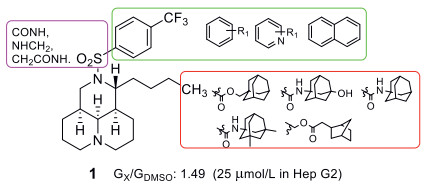
Recently, we found that 12N-p-trifluoromethylbenzenesulfonyl matrinane (1, Fig 1), derived from natural product matrine (2), displayed promising anti-diabetic effects through suppression of glucose aerobic oxidation and induction of glycolysis with the advantages of high safety profile and ameliorating diabetic nephropathy [7]. In order to develop a new type of hypoglycemic candidates, a series of tricyclic matrinic analogues was continuously synthesized to expand the glucose-lowering structure-activity relationship and in-depth mechanism research of its kind.

|
Download:
|
| Fig. 1. Chemical structure and modification of lead 1. | |
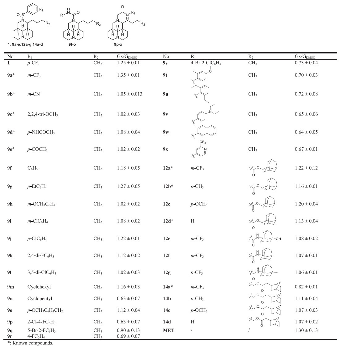
According to the primary structure-activity relationship (SAR) analysis, 11-butane side chain was beneficial for activity [7]. SAR investigation was mainly focused on the substitution on the 12N atom in this study while 11-side chain was set as lipophilic butane or bulky ester and amide moiety, by which a panel of tricyclic matrinic analogues (Table 1) were prepared. All the target compounds were tested in vitro for their ability to stimulate glucose consumption in skeletal muscle L6 cell, and the druglike property, in vivo hypoglycemic effect as well as mode of action of the key compound were illustrated in this study.
|
|
Table 1 The structures and glucose consumptions of target compounds in L6 myotubes. |
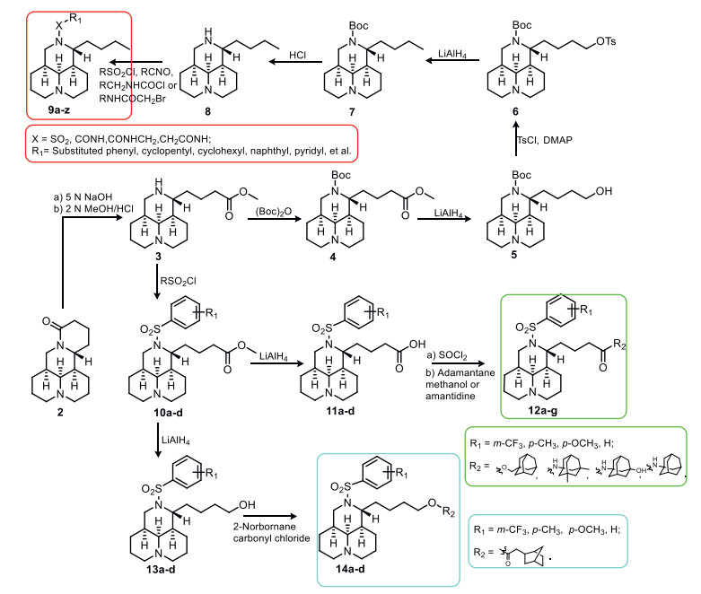
A total of 35 tricyclic matrinic analogues, of which 26 were new, were prepared from 2, as depicted in Scheme 1. Compounds 9a–e, 12a, 12b, 12d and 14a were prepared as we described [8-12]. The key intermediate matrinane 8 was acquired from 2 through a seven-step procedure in a good yield as reported previously [8], and the aimed 12N-substituted matrinane derivatives 9f–x were obtained from an electrophilic substitution reaction of 8 in 33%–73% yields [8-10, 13]. Target compounds 12a–g and 14a–d were gained from a two-step sequence reaction from intermediates 10a–d, respectively [11, 12].

|
Download:
|
| Scheme 1. The synthesis of all target compounds. | |
All target compounds were evaluated for their stimulatory efficacy on glucose consumption at the concentration of 20 µmol/L in L6 rat skeletal muscle cells, taking 1 (20 µmol/L) and metformin (MET, 5 mmol/L) as the positive controls. The activity was identified as the ratio of Gx /GDMSO. The structures and activities of all target compounds were listed in Table 1.
Our previous SAR study disclosed that the p-CF3 on the benzene ring (1) owned higher activity than o-CF3 [8], then compound 9a with m-CF3 was generated and gave a superior activity to the lead 1 and MET (5 mmol/L), with the Gx/GDMSO value of 1.35. However, compounds 9b–e with various of other substitutions on the benzene ring gave significantly declined activities, indicating CF3 might be beneficial for the activity.
Then, the sulfonyl linker was replaced by aminocarbonyl linker, and compounds 9f–o with a benzene ring were synthesized and evaluated. Except for compounds p-Et (9g) and p-chloro (9j) gave a comparable activity to 1, all other compounds gave decreased activities to varying degrees. The alternation of benzene ring into cyclohexyl (9m) or cyclopentyl (9n) ring led to comparable or declined activities. The alternation of aminocarbonyl into metheneaminocarbonyl or aminocarbonylmethene also brought descended activities, as seen in 9o or 9p–x.
In addition, compounds 12a–g with a lipophilic bulky 11-side chain were obtained. Compounds 12a–d with an adamantly ester moiety gave comparable or slightly inferior activities to the lead 1. The alternation of ester bond into amide bond gave decreased activities, as seen in compounds 12e–g. Meanwhile, another series of matrinyl 2-norbornane acetates 14a–c displayed inferior activities too, indicating the replacement of 11-butane into the bulky ester or amide moiety was unfavorable for activity.
Then the glucose consumption-stimulating activities of 9a at different concentrations (10, 20 and 40 µmol/L) were verified in L6 myotubes, HepG2 cells and 3T3-L1 cells, taking 1 as the positive control. It dose-dependently promoted glucose consumption in all three cells (Fig. S1 in Supporting information), and the efficacies were higher than those of 1 when administered at a same concentration.
Then the druglike property of 9a was evaluated. In acute toxicity test, it gave the median lethal dose (LD50) values of over 1000 mg/kg via oral route, comparable to that of lead 1 [8], as well as the LD50 values of 400 mg/kg and over 40 mg/kg via intraperitoneal and intravenous routes respectively. And it gave the LD50 value between 50–60 µmol/L in the zebrafish embryonic developmental toxicity assay (Fig. S2 in Supporting information). The PK profiles of compound 9a was investigated in SD rat model at the dosage of 25 mg/kg via oral route. As depicted in Table S1 and Fig. S3 (Supporting information), it gave the T1/2 value of 4.03 h, maximum concentration (Cmax) of 1.04 µmol/L, area under curve (AUC) of 7.36 µmol h/L, and an oral bioavailability of 16.7%. These results suggested that 9a owned a high druglike profile.
The beneficial effects of 9a on glucose metabolism were evaluated in a high-fat diet (HFD) + STZ-induced diabetic mouse model. The research committee of the Institute of Medicinal Biotechnology reviewed and approved the protocols of the animal experiments. Animals were treated humanely and cared for according to the guidelines of CAMS & PUMC. As shown in Table S2 (Supporting information), the diabetic model was developed successfully before treatment. After 8 weeks of administration, the fasting blood glucose (FBG) values of the diabetic mice were lowered by 9a dose-dependently. It lowered FBG at 100 mg/kg to an extent similar to that of MET at 300 mg/kg, comparable to 1's efficacy in KK-Ay mice [8]. The decreased serum insulin level was greatly restored by 9a, and 9a at 50 mg/kg gave a comparable efficacy to 1 at 100 mg/kg in KK-Ay mice [8]. It also restored the liver and kidney functions by reducing serum creatinine (Scr), alanine aminotransaminase (ALT), aspartate aminotransferase (AST) and total bile acid (TBA) levels, similar to those of MET.
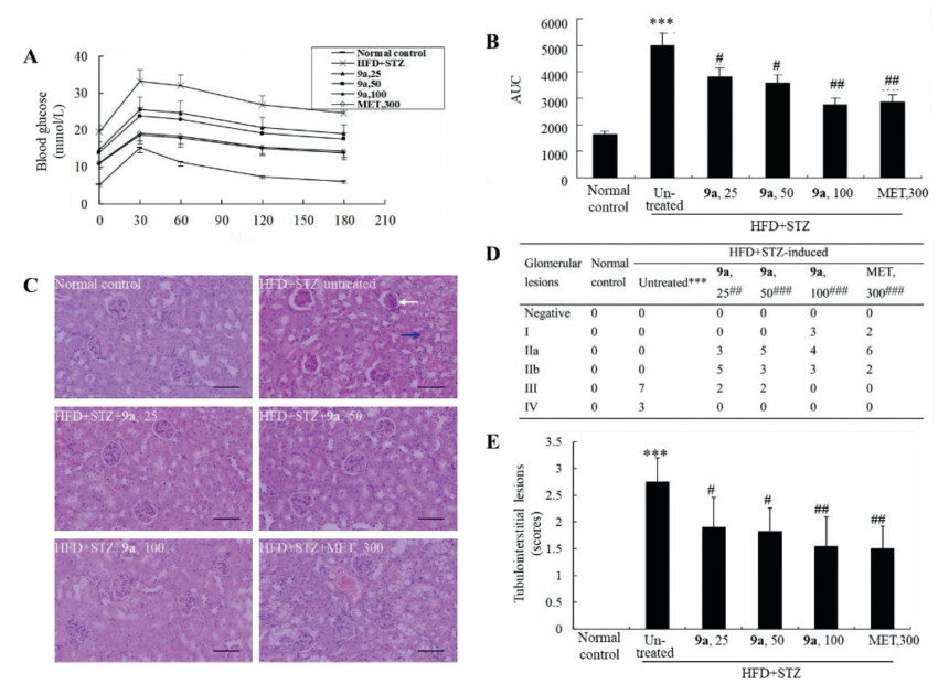
In oral glucose tolerance test (OGTT), the HFD + STZ induced diabetic mice had impaired glucose tolerance as compared to normal control. Compound 9a improved glucose tolerance of the diabetic mice significantly and dose-dependently (Figs. 2A and B), the activities of 9a at 50 mg/kg were comparable to those of 1 at 100 mg/kg in KK-Ay mice [8], and the activities of 9a at 100 mg/kg were comparable to those of MET at 300 mg/kg. Significant abnormalities appeared in the kidney of diabetic mice (Fig. 2C), which included glomerular sclerosis and tubulointerstitial lesions (Figs. 2D and E), and these pathological changes were greatly ameliorated by 9a administration (Figs. 2C–E), in a comparable efficacy to 1 in KK-Ay mice [8].

|
Download:
|
| Fig. 2. (A, B) Effects of 9a on OGTT in HFD + STZ-induced diabetic mice. (C-E) Effects of 9a on pathological changes of kidney in HFD + STZ-induced diabetic mice (200×, scale bar = 100 µm). The white arrow indicates glomerular lesions and the blue arrow indicates tubulointerstitial lesions. Values are mean ± SD of 10 mice of each group in panel C. ***P < 0.001 vs. that of normal control group; # P < 0.05, ## P < 0.01, ### P < 0.01 vs. that of untreated diabetic mice. | |
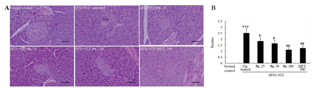
Meanwhile, the islets of the diabetic mice largely atrophied accompanied with structure disorder due to STZ injection (Fig. 3). Compound 9a significantly improved the abnormalities of islets, in a comparable efficacy to 1 in KK-Ay mice (Fig. 3) [8]. As the positive control, the efficacies of MET at 300 mg/kg were similar to those of 9a at 100 mg/kg.

|
Download:
|
| Fig. 3. Effects of 9a on pathological changes of pancreas in HFD + STZ-induced diabetic mice (200×, scale bar = 100 µm). Typical images are presented panel A, in which a blue arrow indicates the atrophy of islet. The scores of pathological changes are presented in panel B, in which values are mean ± SD of 10 mice in each group. ***P < 0.001 vs. normal control group; # P < 0.05, ## P < 0.01 vs. untreated diabetic mice. | |
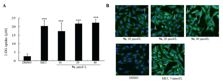
Next, the detailed anti-diabetic mechanism of 9a was explored. First, the influence of 9a on glucose uptake was evaluated in L6 myotubes. As shown in Fig. 4A, at concentrations of 10, 20, and 40 µmol/L, 9a greatly stimulated the uptake of 2-DG, a glucose analogue, to about 6.73 to 8.64-fold of DMSO-treated cells. Then, cellular localization of glucose transporter 4 (GLUT4) was assayed by immunofluorescence staining. As shown in Fig. 4B, GLUT4 protein (green) was mainly localized in cytoplasm in DMSO-treated cells. After 9a treatment, the surface level of GLUT4 increased in a concentration-dependent manner, indicating that it promoted GLUT4 membrane translocation in L6 myotubes, similar to those of MET. These results indicate that 9a increases glucose uptake and GLUT4 membrane translocation in L6 myotubes.

|
Download:
|
| Fig. 4. Effects of 9a on 2-DG uptake (A) and GLUT4 translocation (B). Values are ± SD of 4 repeated experiments, ***P < 0.001 vs. DMSO-treated cells. GLUT4 membrane translocation was determined by immunofluorescence assay (green) and DAPI was used to stain nucleus (blue). The white arrows indicate membrane localization of GLUT4 protein. One of 3 repeated experiments (×100) is presented in panel B. | |
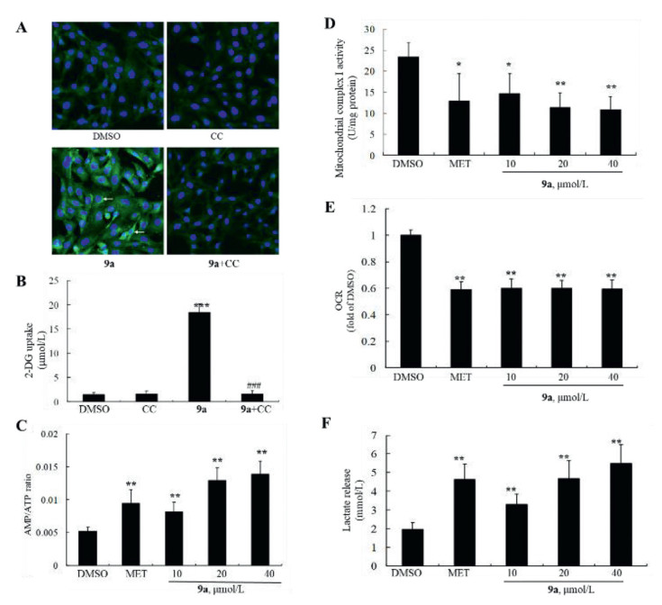
In light of the crucial role of AMP-activated protein kinase (AMPK) in GLUT4 membrane translocation [14, 15], the stimulatory effect of 9a upon AMPK was investigated in L6 myotubes, taking MET as the positive control. As shown in Fig. S4A (Supporting information), compound 9a stimulated AMPKα phosphorylation in a dose-dependent manner, as indicated by the significant increase of p-AMPKα (Thr172) level after treatment, and the stimulatory efficacy of 9a at 40 µmol/L was comparable to that of MET at 5 mmol/L. In addition, the stimulatory effects of 9a on AMPKα phosphorylation were completely blocked by compound C, a selective AMPK inhibitor, as shown in Fig. S4B (Supporting information). Accordingly, the promoting activities of 9a on GLUT4 membrane translocation (Fig. 5A) and 2-DG uptake (Fig. 5B) were also abolished in the presence of compound C in L6 myotubes, similar to that of MET. These results suggest that AMPKα is indispensable for the stimulatory effects of 9a on glucose metabolism.

|
Download:
|
| Fig. 5. (A, B) Blocking effects of CC on GLUT4 translocation (×100) and 2-DG uptake. Values are mean ± SD of 4 repeated experiments, ***P < 0.001 vs. DMSO-treated cells, ###P < 0.001 vs. cells treated with 9a alone. (C) Effects of 9a on AMP/ATP ratio. (D) Effects of 9a on mitochondria complex I activity. (E) Effects of 9a on OCR. (F) Effects of 9a on lactate release. For C-F, values are mean ± SD 3-4 repeated experiments. P < 0.05, **P < 0.01 vs. DMSO-treated cells. | |
AMPK could be activated by the increase of AMP/ATP ratio or upstream AMPK kinases (AMPKKs) [16, 17]. First, the influence of 9a on AMP/ATP ratio was evaluated in L6 myotubes. It increased AMP/ATP ratio in a dose-dependent manner, as disclosed in Fig. 5C. Mitochondrial complex I plays a crucial role in ATP production [18, 19], as anticipated, 9a suppressed mitochondrial complex I activity dose-dependently (Fig. 5D). Accordingly, as shown in Figs. 5E and F, it also decreased cell oxygen consumption rate (OCR), and promoted cellular lactate release, in agreement of 1's effects in our earlier study [8].
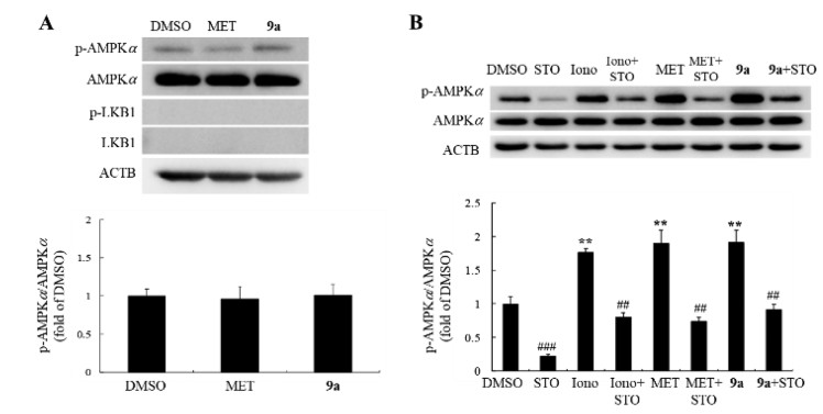
Next, its influences on upstream AMPKKs, namely liver kinase B1 (LKB1) and calcium/calmodulin-dependent protein kinase (CaMKK) [17], were investigated. It greatly increased the phosphorylation level of LKB1 at Ser334 (Fig. S4A). In HeLa cells with LKB1-deficency [20], its stimulation on AMPKα phosphorylation were abolished (Fig. 6A). STO-609 (a CaMKK inhibitor) was used to evaluate the role of CaMKK in the AMPK-stimulating activity of 9a. As shown in Fig. 6B, the stimulating activities of 9a, MET, as well as ionomycin, an ionophore for calcium, on AMPKα phosphorylation were totally blocked in the presence of STO-609. Therefore, 9a activates AMPKα in a AMPKKs-dependent manner.

|
Download:
|
| Fig. 6. (A) Effects of 9a on the phosphorylation of AMPKα in HeLa cells. (B) Blocking effect of STO-609 (STO) on AMPKα phosphorylation. Values are mean ± SD of 3 repeated experiments. **P < 0.01 vs. DMSO-treated cells, ##P < 0.01, ###P < 0.001 vs. same-treated cells without STO. | |
In summary, 35 tricyclic matrinic derivatives, of which 26 are new, are synthesized and determined for their stimulation effects on glucose consumption in L6 myotubes, taking 1 as the lead. The most potent compound 9a, with a highly druglike feature, effectively lowers blood glucose and ameliorates diabetic nephropathy and islet damage in HFD and STZ induced diabetic mice. Compared to 1, it exerts comparable potencies on restoring serum insulin level and improving glucose tolerance at a lower dosage. The mechanism study indicates that compound 9a increases AMP/ATP ratio through inhibiting mitochondrial complex I and affects LKB1 and CaMKK, which synergically induces AMPKα activation and successively stimulates GLUT4 membrane translocation and 2-DG uptake to accelerate glucose consumption. Our results suggest that compound 9a may be a potential novel anti-diabetic candidate with the advantage of multiple-target mechanism, worthy of further investigation.
Declaration of competing interestThe authors declare that they have no known competing financial interests or personal relationships that could have appeared to influence the work reported in this paper.
AcknowledgmentsThis work was supported by CAMS Innovation Fund for Medical Sciences (No. 2021-12M-1-030), the Natural Science Foundation of Beijing Municipality (No. 7202131) and Chinese Pharmaceutical Association-Yiling Pharmaceutical Innovation Fund for Biomedicine (No. GL-1-B04-20190397).
Supplementary materialsSupplementary material associated with this article can be found, in the online version, at doi:10.1016/j.cclet.2022.05.075.
| [1] |
E.M. Vaughan, J.J. Rueda, S.L. Samson, et al., Curr. Diabetes Rev. 16 (2020) 851-858. DOI:10.2174/1573399816666200206112318 |
| [2] |
Z.G. Sun, Z.N. Li, H.L. Zhu, Mini Rev. Med. Chem. 20 (2020) 1709-1718. DOI:10.2174/1389557520666200628032507 |
| [3] |
S. Cornell, J. Clin. Pharm. Ther. 45 (2020) 17-27. DOI:10.1111/jcpt.13230 |
| [4] |
E. Brown, H.J.L. Heerspink, D.J. Cuthbertson, et al., Lancet 398 (2021) 262-276. DOI:10.1016/S0140-6736(21)00536-5 |
| [5] |
G. Savarese, B. Schrage, F. Cosentino, et al., ESC Heart Fail. 7 (2020) 3438-3451. DOI:10.1002/ehf2.12937 |
| [6] |
M. Singh, A. Kumar, Curr. Drug Saf. 13 (2018) 84-91. DOI:10.2174/1574886313666180226103408 |
| [7] |
S. Tang, C. Wang, Y.H. Li, et al., Eur. J. Med. Chem. 201 (2020) 112315. DOI:10.1016/j.ejmech.2020.112315 |
| [8] |
S. Tang, Y.H. Li, X.Y. Cheng, et al., Future Med. Chem. 8 (2016) 495-508. DOI:10.4155/fmc-2015-0019 |
| [9] |
X.Y. Cheng, Y.H. Li, S. Tang, et al., Eur. J. Med. Chem. 126 (2017) 133-142. DOI:10.1016/j.ejmech.2016.09.097 |
| [10] |
S.G. Wang, L.Y. Kong, Y.H. Li, et al., Bioorg. Med. Chem. Lett. 25 (2015) 3690-3693. DOI:10.1016/j.bmcl.2015.06.043 |
| [11] |
T. Niu, X. Zhao, J. Jiang, et al., Molecules 24 (2019) 921. DOI:10.3390/molecules24050921 |
| [12] |
J. Jiang, X. Zhao, T. Niu, et al., Pharmazie 74 (2019) 265-269. DOI:10.1001/jamainternmed.2018.5295 |
| [13] |
S. Tang, Z.G. Peng, X. Zhang, et al., Chin. Chem. Lett. 27 (2016) 1052-1057. DOI:10.1016/j.cclet.2016.03.006 |
| [14] |
F. Dehghan, F. Hajiaghaalipour, A. Yusof, et al., Sci. Rep. 6 (2016) 25139. DOI:10.1038/srep25139 |
| [15] |
J.O. Lee, S.K. Lee, J.H. Kim, et al., J. Biol. Chem. 287 (2012) 44121-44129. DOI:10.1074/jbc.M112.361386 |
| [16] |
S. Herzig, R.J. Shaw, Cell Biol. 19 (2018) 121-135. DOI:10.1038/nrm.2017.95 |
| [17] |
A. Gormand, E. Henriksson, K. Ström, et al., J. Cell Biochem. 112 (2011) 1364-1375. DOI:10.1002/jcb.23053 |
| [18] |
K. Fiedorczuk, L.A. Sazanov, Trends Cell Biol. 28 (2018) 835-867. DOI:10.1016/j.tcb.2018.06.006 |
| [19] |
H. Zhu, B. Zhang, N. Zhu, et al., Chin. Chem. Lett. 32 (2021) 1220-1223. DOI:10.1016/j.cclet.2020.09.003 |
| [20] |
J. Han, J. Yi, F. Liang, et al., Mol. Cell Endocrinol. 405 (2015) 63-73. DOI:10.1016/j.mce.2015.02.008 |