 2021, Vol. 32


b College of Material Science and Art Design, Inner Mongolia Agricultural University, Hohhot 010018, China
Genetic engineering is a process of introducing nucleic acids to modulate specific protein expression, which has fueled tremendous interest in the biomedical fields [1, 2]. To date, although some non-viral gene delivery systems based on polymer nanoparticles, dendrimers, mesoporous silica nanoparticles, liposomes, and core-shell nanoparticles have presented promising alternatives to viral vehicles [3-10], there still exist several challenges in constructing these systems, such as complicated synthetic procedure, poor aqueous solubility, high toxicity, lack of specificity, and insufficient gene release [11-15]. Therefore, far more efficient gene vehicles are still in imperative demand. In this regard, supramolecular chemistry exhibits great potential [16-21]. Among the representative macrocyclic molecules, such as cyclodextrins (CDs), are a popular class of biocompatible artificial receptors with a hydrophilic external periphery and a well-defined hydrophobic hollow cavity that can selectively bind a variety of guest species [22-29]. On the other hand, hyaluronic acid (HA), a sort of water-soluble biocompatible and biodegradable polysaccharide with specific recognition ability toward cancer cells, which could be used as the targeting agent and building block for the construction of targeted drug/gene delivery system [30-34]. Previously, we developed a pH-responsive and targeted polysaccharide conjugate based on the boronate linkage of gluconamide-grafted HA with anticancer drug bortezomib, which could exhibit targeted drug release behaviors at acidic pH and possess lower cytotoxicity and a higher inhibition effect toward cancer cells [35]. Chen et al. reported a platform for siRNA delivery by ZnⅡ−dipicolylamine complexes functionalized HA nanoconjugate, which has a high affinity for siRNA and targeted delivery it into cancer cells, demonstrating a safe and efficient vector for targeted gene therapy [36].
Recently, grafting CDs/adamantane onto HA backbone has attracted much attention from supramolecular chemists. For instance, Liu et al. developed a photocontrolled supramolecular nanoassembly for targeted delivery of siRNA, which was successfully constructed from α-cyclodextrin modified HA and a cationic azobenzene-modified diphenylalanine derivative, demonstrating this strategy has great potential to be an attractive one for targeted, controlled gene delivery [37]. The incorporation of supramolecular host-guest interactions with cell-specific polysaccharide building blocks confer several practical superiorities [38, 39]. Firstly, the grafting of HA could be achieved by facile modification, and the supramolecular host-guest interactions provide a facile and easy strategy to construct assembly, which could avoid complicated synthesis and purification process [40, 41]. Besides, the targeting ligands could selectively deliver the system to tumor cells, which could improve therapeutic efficiency and decrease side effects [42, 43]. Also, owing to the biocompatible polysaccharide skeleton in HA, the release of drug/gene could be readily achieved by the enzymatic degradation with hyaluronidase (HAase) [44-46]. Thus, this strategy may have potential application in gene therapy.
In this work, the adamantane-grafted HA (HA-ADA) was intermolecularly connected with the mono-(6-(tetraethylenepentamine)-6-deoxy)-β-cyclodextrin (TEPA-CD) to construct an enzyme-responsive polysaccharide nanoassembly HA-ADA/TEPA-CD, which could be utilized as a functional nanocarrier to encapsulate plasmid DNA (pDNA) (Scheme 1). Subsequently, benefiting from the biocompatible HA skeleton, the gene could be released through the enzymatic degradation. The constructed nanoassembly possesses some inherent characteristics as (1) the introduction of HA and β-CD could efficiently increase the water solubility and biocompatibility, (2) the nanoassembly could enrich the polyamine chains together and facilitate the interaction with DNA, thus leading to the improvement of DNA encapsulation, (3) HA–HAase pair were successfully applied in the construction of enzyme-responsive nanoassembly, controllable pDNA binding and release could be realized [47-49]. Consequently, the enzyme-responsive polysaccharide supramolecular nanoassembly could be utilized as a convenient platform for controlled gene delivery.

|
Download:
|
| Scheme 1. Construction of the enzyme-responsive HA-ADA/TEPA-CD polysaccharide nanoassembly. | |
All chemicals were reagent grade unless noted otherwise. Sodium hyaluronate (Mw = 550 kDa) were purchased from commercial sources. Hyaluronic acid was prepared by treating sodium hyaluronate with cation exchange resin. Adamantane-grafted hyaluronic acid (HA-ADA) was synthesized according to the reported method [50], mono-(6-(tetraethylenepentamine)-6-deoxy)-β-cyclodextrin (TEPA-CD) was synthesized according to the reported method [51]. 293 T human embryonic kidney cell line was obtained from China Infrastructure of Cell Line Resource.
The absorbance spectra were recorded in a conventional quartz cell (light path 10 mm) by using UV–vis spectrophotometer (U-2900, Hitachi). TEM images were acquired by a FEI Tecnai G2 F20 transmission electron microscope. The samples were prepared by placing a drop of solution onto a carbon-coated copper grid and air-dried. The SEM images were recorded on a JSM-6700 F scanning electronic microscope. The DLS and zeta potential were recorded on Malvern Zetasizer Nano ZS90 (Malvern Instruments Ltd., Worcestershire, UK). The zeta potential were examined on NanoBrook 173 Plus at 25 ℃. Gel electrophoresis experiments were measured on a 1% (w/v) agarose gel at 60 V for 45 min and photographed using a UV transilluminator and WD-9415 B gel documentation system (Beijing Liuyi Instrument Factory, China). The confocal laser scanning microscopy was performed on a fluorescence inverted microscope (Zeiss LSM 800).
HA-ADA (9.75 mg), TEPA-CD (6.53 mg) were dissolved in 5 mL deionized water, then the solution was ultrasonicated for 10 min. The resulting solution was stored at 4 ℃.
The controlled condensation ability of polysaccharide nanoassembly to pDNA was measured by evaluating the electrophoretic mobility on agarose gel at different N/P ratios. The gel electrophoresis experiments were performed in TAE (0.04 mol/L Tris, 0.02 mol/L acetic acid, and 2.0 mmol/L EDTA) buffer at 25 ℃. After samples loading and electrophoresis process, pDNA bands were stained in Gen Green solution and were photographed using UV light at 302 nm.
293 T human embryonic kidney cell line was cultured in Dulbecco's modified Eagle's medium (DMEM) supplemented with 10% fetal bovine serum (FBS). 293 T cells were seeded in 96-well plates (5×10⁴ cells/mL, 100 μL per well) for 24 h, then the cells were incubated with HA-ADA, TEPA-CD, HA-ADA/TEPA-CD and PEI25k at different concentrations for 48 h, respectively. The relative cellular viability was determined by MTT assay. All data were presented as the mean±standard deviation.
293 T cells were cultured in 6-well plates (5×10⁴ cells/mL, 2 mL per well) for 24 h at 37 ℃ in 5% CO2. The cells incubated with TEPA-CD@pDNA, HA-ADA/TEPA-CD@pDNA and PEI25k@pDNA for 6 h, and then the medium in each well was replaced with fresh medium. The cells were further incubated for 42 h, and then the cells were washed with PBS buffer for three times. After that the cells were fixed with 4% paraformaldehyde for 15 min, then observed by a confocal laser scanning microscope (λex = 480 nm).
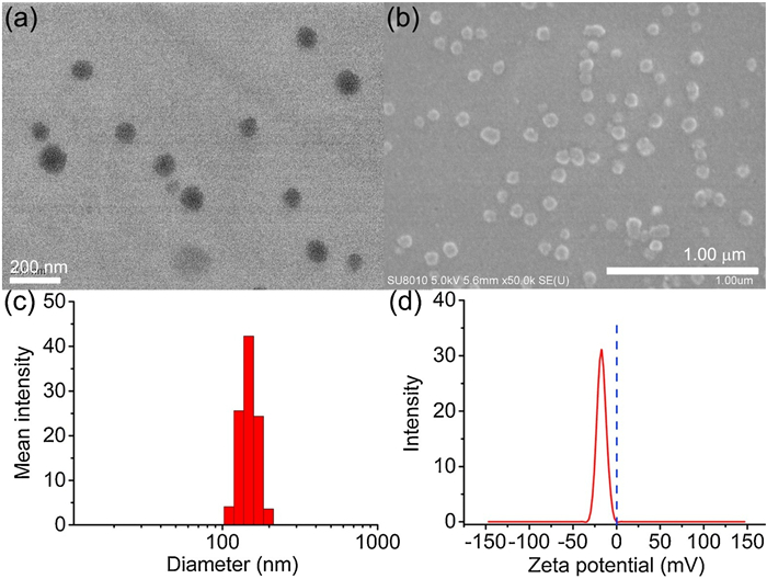
Owing to the large association constant and high reliability of the interactions between adamantane and β-cyclodextrin, the enzyme-responsive polysaccharide supramolecular nanoassembly (HA-ADA/TEPA-CD) was constructed by simply mixing the aqueous solutions of HA-ADA and TEPA-CD together. The critical aggregation concentration of the nanoassembly was measured by monitoring the dependence of optical transmittance at 400 nm upon increasing nanoassembly concentration from 2 μmol/L to 35 μmol/L, an inflection point at 6 μmol/L was observed due to the formation of a large-sized nanoassembly (Fig. S1 in Supporting information). The size and morphology of the enzyme-responsive polysaccharide nanoassembly comes from the transmission electron microscopy (TEM), scanning electron microscopy (SEM), dynamic light scattering (DLS), and zeta potential experiments. As shown inFig. 1a, the nanoassembly existed as homogeneous spherical nanoparticles with an average diameter of ca. 110 nm was observed in TEM image, and the SEM image (Fig. 1b) also showed about 110 nm spherical nanoparticles with homogeneous dispersity, which was also suitable for passive tumor targeting via the enhanced permeability and retention (EPR) effect [52]. Moreover, the results of DLS experiments (Fig. 1c) gave a hydrodynamic diameter of HA-ADA/TEPA-CD nanoassembly as ca. 140 nm with a narrow distribution, which was in accordance with the results in electron microscopy. Furthermore, the zeta potential of HA-ADA/TEPA-CD nanoassembly was measured as ca. −17.5 mV (Fig. 1d) due to the negative charged hydrophilic HA surface of the nanoassembly, which would facilitate the stability, dispersibility, and biocompatibility of HA-ADA/TEPA-CD nanoassembly in biological environments and prolong the circulation time in vivo. In addition, HA-ADA/TEPA-CD nanoassembly also exhibited an excellent stability in aqueous solution for months (Fig. S2 in Supporting information).

|
Download:
|
| Fig. 1. Typical (a) TEM and (b) SEM images of HA-ADA/TEPA-CD polysaccharide nanoassembly. (c) DLS and (d) zeta potential results of polysaccharide nanoassembly in deionized aqueous solution. | |
Binding with DNA is one of the crucial prerequisites for polysaccharide nanoassembly to achieve efficient loading of the therapeutic gene and then transfer it into target cells. Considering that the polyamine chain of TEPA-CD was positively charged in aqueous solution, it is believed that the HA-ADA/TEPA-CD polysaccharide nanoassembly may impart good condensation abilities to DNA. Therefore, agarose gel electrophoresis was firstly performed to examine the pDNA condensation ability of nanoassembly at different N/P ratios. As shown in Fig. 2, the nanoassembly completely retarded the pDNA while TEPA-CD partially retarded at N/P ratio of 80, indicating the stronger ability of the nanoassembly to condense pDNA than that of TEPA-CD. This phenomenon revealed that the nanoassembly could enrich the polyamine chains together to increase the charge density, thus leading to the enhanced DNA encapsulation. Simultaneously, other information about the pDNA condensation by HA-ADA/TEPA-CD polysaccharide nanoassembly was further confirmed by TEM, SEM, DLS and zeta potential experiments. The TEM image (Fig. S3a in Supporting information) revealed that pDNA was condensed into even tighter spherical nanoparticles with an average diameter of 90 nm at the N/P ratio of 80, similar phenomenon was also observed by SEM (Fig. S3b in Supporting information). The DLS result and zeta potential further indicated that the HA-ADA/TEPA-CD polysaccharide nanoassembly could effectively condense pDNA into nanoparticles with a hydrodynamic diameter of 126 nm (Fig. S3c in Supporting information) as well as zeta potential of ca.−30 mV (Fig. S3d in Supporting information).

|
Download:
|
| Fig. 2. Agarose gel electrophoresis retardation of pDNA by (a) TEPA-CD, (b) HA-ADA/TEPA-CD polysaccharide nanoassembly at N/P ratios of 5, 10, 30, 60, 70, 80 and 90 from lane 1 to lane 7, respectively, (c) HA-ADA/TEPA-CD polysaccharide nanoassembly with pDNA before (lane 1) and after (lane 2) treated with HAase at the N/P ratio of 80. | |
After confirming the pDNA condensation ability of the obtained nanoassembly, we further investigated the controlled pDNA release behavior of supramolecular nanoassembly after enzymatic hydrolysis of HA. As shown in Fig. 2c, pDNA could be released from HA-ADA/TEPA-CD polysaccharide nanoassembly after treating with HAase, in agreement with the decrease of the Tyndall effect (Fig. S4 in Supporting information). These results jointly indicated that the obtained polysaccharide nanoassembly could be specifically degraded as soon as the polysaccharide backbone of HA was hydrolysed to low-molecular-weight oligomers upon exposure to HAase, which realize the controlled binding and release of pDNA.
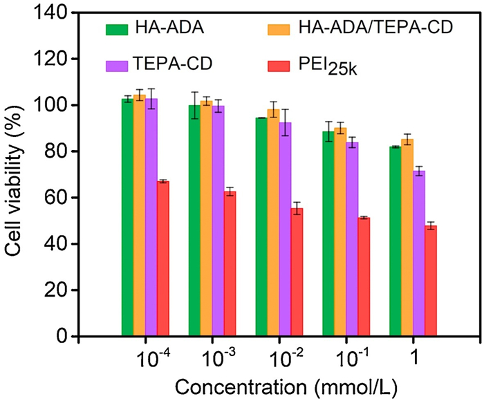
Subsequently, the cytotoxicity of the supramolecular nanoassembly was investigated by measuring the cellular viability of 293 T human embryonic kidney cells treated by HA-ADA, HA-ADA/TEPA-CD, TEPA-CD by means of MTT assay, with molecular weight of 25kDa polyethylenimine (PEI25k), the golden standard of gene transfection among the polymeric gene carriers as the control. Owing to the satisfactory biocompatibility, HA-ADA/TEPA-CD nanoassembly exhibited no cytotoxicity even when the concentration was up to 1 mmol/L after 48 h of incubation (Fig. 3). Moreover, the cellular viabilities of 293 T cells treated with HA-ADA/TEPA-CD nanoassembly was higher than 80% after 48 h even when the concentration was up to 1 mmol/L, which was much higher than that of treated with PEI25k, further collaborating the low cytotoxicity of the polysaccharide supramolecular nanoassembly. In addition, the toxicity of the HA-ADA/TEPA-CD nanoassembly was always lower than that of TEPA-CD at any concentration, implying that the introduction of HA exactly improve biocompatibility to some extent.

|
Download:
|
| Fig. 3. Cellular viability of 293 T cells after the treatment with HA-ADA, HA-ADA/TEPA-CD, TEPA-CD, PEI25k at different concentrations in 48 h. | |
After verifying the low cytotoxicity of the HA-ADA/TEPA-CD nanoassembly, we continued to investigate its ability to transport plasmid DNA into 293 T cells by the confocal laser scanning microscope with EGFP gene as a reporter gene. As shown inFig. 4, 293 T cells exhibited bright green fluorescence in cytoplasm after transfected with HA-ADA/TEPA-CD nanoassembly (Fig. 4b) and 25 kDa PEI (Fig. 4c), but in contrast, slight green fluorescence was observed in TEPA-CD group (Fig. 4a). These phenomena suggested that the supramolecular nanoassembly was able to act as a gene carrier to transfer the EGFP information into cells, which might have potential application value in genetic engineering.

|
Download:
|
| Fig. 4. Confocal fluorescence images of 293 T cells transfected with (a) TEPA-CD, (b) HA-ADA/TEPA-CD at N/P ratio of 80, and (c) PEI25k at N/P ratio of 10 for 48 h. | |
In conclusion, an enzyme-responsive pDNA encapsulation polysaccharide nanoassembly HA-ADA/TEPA-CD was successfully constructed by the strong supramolecular interaction between HA-ADA and positively charged TEPA-CD, thereby displaying a satisfactory enhanced DNA encapsulation ability along with good biocompatibility. The release of pDNA could be readily achieved by the enzymatic degradation of polysaccharide nanoassembly with HAase due to the biocompatible HA skeleton in nanoassembly. Moreover, as investigated using confocal laser scanning microscope, it can be seen that the transfection efficiency of TEPA-CD was dramatically improved by introducing HA-ADA. Considering the facile construction of polysaccharide nanoassembly and its specific targeting ability, we can envision that the obtained nanoassembly has great potential to be an attractive one for targeted, controlled gene delivery.
Declaration of competing interestThe authors report no declarations of interest.
AcknowledgmentsWe thank the Programs of Higher-level Talents of Inner Mongolia Agricultural University (No. NDGCC2016-21), the Program for Young Talents of Science and Technology in Universities of Inner Mongolia Autonomous Region (No. NJYT-19-B26), the Grassland Talents of Inner Mongolia Autonomous Region (No. DC2000000745) and the Inner Mongolia Autonomous Region Natural Science Fund Project (No. 2017 BS0206) for financial support.
Appendix A. Supplementary dataSupplementary material related to this article can be found, in the online version, at doi: https://doi.org/10.1016/j.cclet.2021.01.032.
| [1] |
C.O. Mellet, J.G. Fernández, J.M. Benito, Chem. Soc. Rev. 40 (2011) 1586-1608. DOI:10.1039/C0CS00019A |
| [2] |
W.C. Geng, Q. Huang, Z. Xu, R. Wang, D.S. Guo, Theranostics 9 (2019) 3094-3106. DOI:10.7150/thno.31914 |
| [3] |
S. Roy, D.C. Zhu, W.J. Parak, N. Feliu, ACS Nano 14 (2020) 8012-8023. DOI:10.1021/acsnano.9b10219 |
| [4] |
H. Cabral, K. Miyata, K. Osada, K. Kataoka, Chem. Rev. 118 (2020) 6844-6892. |
| [5] |
A.B. Cook, S. Perrier, Adv. Funct. Mater. 30 (2020) 1901001. DOI:10.1002/adfm.201901001 |
| [6] |
Y.Y. Cheng, X.Y. Jiao, W.P. Fan, et al., Biomaterials 256 (2020) 120191. DOI:10.1016/j.biomaterials.2020.120191 |
| [7] |
Y. Lin, J.H. Wu, W.H. Gu, et al., Adv. Sci. 5 (2018) 1700611. DOI:10.1002/advs.201700611 |
| [8] |
B.D. Rodrigues, A. Banerjee, T. Kanekiyo, J. Singh, Int. J. Pharm. 566 (2019) 717-730. DOI:10.1016/j.ijpharm.2019.06.026 |
| [9] |
M. Liu, Y. Peng, Y.B. Nie, et al., Acta Biomater. 110 (2020) 242-253. DOI:10.1016/j.actbio.2020.04.041 |
| [10] |
X.S. Li, J.Y. Han, X. Wang, et al., Mater. Chem. Front. 3 (2019) 103-110. DOI:10.1039/C8QM00509E |
| [11] |
Z.X. Zhou, X.R. Liu, D.C. Zhu, et al., Adv. Drug Delivery Rev. 115 (2017) 115-154. DOI:10.1016/j.addr.2017.07.021 |
| [12] |
T.V. Mashel, Y.V. Tarakanchikova, A.R. Muslimov, et al., Biomaterials 258 (2020) 120282. DOI:10.1016/j.biomaterials.2020.120282 |
| [13] |
R. Mohammadinejad, A. Dehshahri, V.S. Madamsetty, et al., J. Control. Release 325 (2020) 249-275. DOI:10.1016/j.jconrel.2020.06.038 |
| [14] |
K. Chatterjee, S. Sarkar, K.J. Rao, S. Paria, Adv. Colloid Interface Sci. 209 (2014) 8-39. DOI:10.1016/j.cis.2013.12.008 |
| [15] |
T.X. Xiao, W.W. Zhong, L.X. Xu, et al., Org. Biomol. Chem. 17 (2019) 1336-1350. DOI:10.1039/C8OB03095B |
| [16] |
C.P. Jiang, Z.T. Qi, H.B. Jia, et al., Biomacromolecules 20 (2019) 478-489. DOI:10.1021/acs.biomac.8b01395 |
| [17] |
X. Jin, L.J. Zhu, B. Xue, X.Y. Zhu, D.Y. Yan, Natl. Sci. Rev. 6 (2019) 1128-1137. DOI:10.1093/nsr/nwz018 |
| [18] |
J.Y. Chen, Y.D. Zhang, Z. Meng, et al., Chem. Sci. 11 (2020) 6275-6282. DOI:10.1039/D0SC01756F |
| [19] |
Q. Zhang, C.Y. Shi, D.H. Qu, et al., Sci. Adv. 4 (2018) eaat8192. DOI:10.1126/sciadv.aat8192 |
| [20] |
M.X. Wu, H.J. Yan, J. Gao, et al., ACS Appl. Mater. Interfaces 10 (2018) 34655-34663. DOI:10.1021/acsami.8b13758 |
| [21] |
P.Y. Li, Y. Chen, Y. Liu, Chin. Chem. Lett. 30 (2019) 1190-1197. DOI:10.1016/j.cclet.2019.03.035 |
| [22] |
Y.M. Zhang, Y.H. Liu, Y. Liu, Adv. Mater. 32 (2020) 1806158. DOI:10.1002/adma.201806158 |
| [23] |
Q.D. Hu, G.P. Tang, P.K. Chu, Acc. Chem. Res. 47 (2014) 2017-2025. DOI:10.1021/ar500055s |
| [24] |
Y.T. Wen, H.Z. Bai, J.L. Zhu, et al., Sci. Adv. 6 (2020) eabc2148.. DOI:10.1126/sciadv.abc2148 |
| [25] |
M.A. Raquel, G.S. Laura, W. Verboom, J. Huskens, J.Mater.Chem.B 5 (2017) 36-52. DOI:10.1039/C6TB02776H |
| [26] |
K.I. Assaf, J. Holub, E. Bernhardt, et al., ChemPhysChem 21 (2020) 971-976. DOI:10.1002/cphc.201901225 |
| [27] |
L. Liang, Y. Chen, X.M. Chen, Y. Zhang, Y. Liu, Chin. Chem. Lett. 29 (2018) 989-991. DOI:10.1016/j.cclet.2017.12.022 |
| [28] |
L. Zhang, Y.M. Zhang, G.X. Liu, Y. Liu, Chin. Chem. Lett. 30 (2019) 120-122. DOI:10.1016/j.cclet.2018.04.028 |
| [29] |
Q. Zhao, Y. Chen, Y. Liu, Chin. Chem. Lett. 29 (2018) 84-86. DOI:10.1016/j.cclet.2017.07.024 |
| [30] |
Y.J. Zhang, T. Sun, C. Jiang, Acta Pharm. Sin. B 8 (2018) 34-50. DOI:10.1016/j.apsb.2017.11.005 |
| [31] |
W. Zhong, L. Pang, H.H. Feng, et al., Carbohydr. Polym 238 (2020) 116204. DOI:10.1016/j.carbpol.2020.116204 |
| [32] |
N. Zheng, D. Xie, C.S. Wang, et al., ACS Appl. Mater. Interfaces 11 (2019) 44007-44017. DOI:10.1021/acsami.9b19546 |
| [33] |
X. Wang, M.H. Li, Y.H. Hou, et al., Adv. Funct. Mater. 30 (2020) 2000229. DOI:10.1002/adfm.202000229 |
| [34] |
T.L. Nascimento, H. Hillaireau, J. Vergnaud, E. Fattal, Nanomedicine 11 (2016) 1865-1887. DOI:10.2217/nnm-2016-5000 |
| [35] |
Y.H. Zhang, Y.M. Zhang, J. Yu, J. Wang, Y. Liu, Chem. Commun. 55 (2019) 1164-1167. DOI:10.1039/C8CC09956A |
| [36] |
K.Y. Choi, O.F. Silvestre, X.L. Huang, et al., Nat. Protoc. 9 (2014) 1900-1915. DOI:10.1038/nprot.2014.128 |
| [37] |
F.Q. Li, Q.L. Yu, Y.H. Liu, et al., Chem. Commun. 56 (2020) 3907-3910. DOI:10.1039/D0CC00629G |
| [38] |
X. Ma, Y.L. Zhao, Chem. Rev. 115 (2015) 7794-7839. DOI:10.1021/cr500392w |
| [39] |
H.Q. Song, W.T. Pan, R.Q. Li, et al., Small 14 (2018) 1703152. DOI:10.1002/smll.201703152 |
| [40] |
Y. Yang, Y.M. Zhang, Y. Chen, J.T. Chen, Y. Liu, Sci. Rep. 6 (2016) 19212. DOI:10.1038/srep19212 |
| [41] |
Y. Bai, C.P. Liu, D. Chen, et al., Carbohydr. Polym. 246 (2020) 116654. DOI:10.1016/j.carbpol.2020.116654 |
| [42] |
H.P. Zhang, J. Liu, Q.X. Chen, P. Mi, J. Control. Release 320 (2020) 314-327. DOI:10.1016/j.jconrel.2020.01.026 |
| [43] |
X.Y. Xu, Z.S. Zeng, J. Chen, et al., Chem. Eng. J. 390 (2020) 124628. DOI:10.1016/j.cej.2020.124628 |
| [44] |
K.Y. Choi, H.S. Han, E.S. Lee, et al., Adv. Mater. 31 (2019) 1803549. DOI:10.1002/adma.201803549 |
| [45] |
L. Hou, Y.L. Zhang, X.M. Yang, et al., ACS Appl. Mater. Interfaces 11 (2019) 255-268. DOI:10.1021/acsami.8b17750 |
| [46] |
Y.H. Zhang, Y.M. Zhang, X.L. Sheng, J. Wang, Y. Liu, Chem. Commun. 56 (2020) 1042-1045. DOI:10.1039/C9CC08491F |
| [47] |
P. Singh, L. Wu, X.H. Ren, et al., Int. J. Pharm. 586 (2020) 119542. DOI:10.1016/j.ijpharm.2020.119542 |
| [48] |
Z. Tan, Y. Jiang, W. Zhang, et al., J. Am. Chem. Soc. 141 (2019) 15804-15817. DOI:10.1021/jacs.9b06218 |
| [49] |
P. Ayaz, B.J. Xu, X.S. Zhang, et al., Appl. Surf. Sci. 527 (2020) 146806. DOI:10.1016/j.apsusc.2020.146806 |
| [50] |
Y.M. Zhang, Y. Cao, Y. Yang, J.T. Chen, Y. Liu, Chem. Commun. 50 (2014) 13066-13069. DOI:10.1039/C4CC04533E |
| [51] |
J.C. Beeson, A.W. Czarnik, Bioorg. Med. Chem. 2 (1994) 297-303. DOI:10.1016/S0968-0896(00)82172-0 |
| [52] |
C. Kinnear, T.L. Moore, R.L. Laura, R.R. Barbara, P.F. Alke, Chem. Rev. 117 (2017) 11476-11521. DOI:10.1021/acs.chemrev.7b00194 |