 2021, Vol. 32


b Department of Clinical Laboratory, the Affiliated Drum Tower Hospital of Nanjing University Medical School, Nanjing 210008, China;
c Economical Forest Cultivation and Utilization of 2011 Collaborative Innovation Center in Hunan Province, Hunan Key Laboratory of Biomedical Nanomaterials and Devices, Hunan University of Technology, Zhuzhou 412007, China
Electrochemical biosensing principles can be classified into four categories including amperometry, potentionetry, conductometry and impedance [1, 2]. Biosensors using electrochemical biosensing principles such as electrochemical biosensors, electrochemiluminescence (ECL) biosensors and photochemical biosensors have been widely reported for decades and a series of novel biosensors consisting of antibodies and aptamers as sensing components have been developed [3-7]. Aptamers are single-strand DNA or RNA ligands obtained through an in vitro process named systematic evolution of ligands by exponential enrichment (SELEX) which is aimed at selecting nucleotide sequences specific to different targets ranging from metal irons, organic molecules to proteins and cells from a random library [8-18]. Aptamers exhibit several unique advantages compared with antibodies. They are chemically synthesized and can be modified with various functional groups to produce physical signals, to improve their stability or to achieve efficient immobilization [19-24]. And unlike antibodies, the refolding ability of aptamers is not influenced by extreme change of temperature or pH, which makes them easy to handle or store. Due to those unique properties mentioned above, aptamers have been widely applied in different analytical equipment [25-34]. A typical aptamer-based electrochemical biosensor which employs aptamer as the biological recognition element and utilizes transducer converting the biologic interaction into electrical signals for the quantitative measurement of targets is shown in Fig. 1 [35-39].

|
Download:
|
| Fig. 1. Basic analytical principle of a typical aptamer-based electrochemical biosensor. μM: μmol/L. Copied with permission [39]. Copyright 2008, American Chemical Society. | |
Although aptamer-based biosensors using electrochemical biosensing principles hold many advantages such as high selectivity, low cost and rapid response, it has been noted that efficient detection is a bottleneck as some of the detection targets are either with low abundance or have difficulty to isolate from biological samples [40-43]. Therefore, exploring electrochemical biosensors with high sensitivity and selectivity is in urgent need and a large amount of signal amplification strategies have been developed. Enzymes, dyes perform strong chemical signals and biomaterials such as polymeric nanocomposites, carbon-based nanomaterials, metallic nanoparticles and nanowires have been ultrasensitive biosensors [44-46]. These materials are employed to assist constructing electrodes which can transfer the biological reactions more efficiently, or attempt to obtain enhanced signal through multi-labeled carriers, or catalyze redox cycling to produce amplified electrical signals. Some nanomaterials can also serve as tracers themselves based on their direct electrochemical characteristics. Here we introduced five categories of materials employed in the electrochemical aptasensors including gold materials, carbon nanomaterials, enzymes, nucleic acids and polymers. We hope this review could inspire researchers to overcome the drawbacks of previous methods and to design more simple, practical and repeatable biosensors for clinical applications.
2. Amplification strategies based on different signal amplification materials 2.1. Gold and other metallic nanomaterialsGold nanomaterials possess extraordinary properties such as high specific surface area and being easy to modify, making them the excellent choice to either be the carriers for labeling or serve as electroactive tracers themselves. The good biocompatibility, chemical inertness and rapid response gold nanomaterials have been taken advantage of to amplify signal of biorecognition event [47-50]. Gold nanomaterials can be employed as vehicles to carry aptamer probes [51-56]. For example, carcinoembryonic antigen (CEA) is a highly glycosylated protein over-expressed on a large variety of cancer cells. Shu and co-workers constructed a sandwich structured by means of the CEA-specific aptamer and 6-(ferrocenyl)hexanethiol (Fc) modified gold nanoparticles (AuNPs), CEA and Au electrode gold. They performed differential pulse (DPV) to quantify the concentration of CEA. Human epidermal growth factor receptor (HER2) is also an effective biomarker overexpressed in several cancer cells especially breast cancer cells [57]. Zhu and co-workers developed a ultrasensitive sensor based on hydrazine-AuNP-aptamer structure to detect both HER2 protein and HER2-positive cells, wherein AuNP was used to immobilize aptamers and to achieve signal amplification through AuNP-promoted silver enhancement (Fig. 2). This novel aptasensor offered a detection limit of 26 cells/mL and could be applied to a variety of biomarkers and cells [58].

|
Download:
|
| Fig. 2. Gold nanoparticle-assisted electrochemical aptasensor for the detection of HER2 protein and HER2-overexpressed cancer cells. Copied with permission [58]. Copyright 2013, American Chemical Society. | |
Gold nanomaterials can also be immobilized with tags to achieve signal amplification of one single reaction. Silver particles have been reported to enhance assay sensitivity through a technology named silver staining in which silver particles are precipitated on the gold nucleus [59-62]. For example, mitochondria released-cytochrome c can induce a series of biochemical reactions including apoptosis. The research on cytochrome c is of significant importance to reveal the mechanism and regulation of the apoptosis pathways and their potential therapeutic values [63]. Ocaña and co-workers developed an aptamer-antibody interaction-based biosensor for cytochrome c detection. The addition of cytochrome c would lead to the formation of an aptamer/target/antibody sandwich structure on electrode surface, which caused the change in interfacial charge transfer resistance (Rct). Then streptavidin gold nanoparticles with silver enhancement treatment were introduced to further increase the Rct and reached a detection limit of 12 pmol/L [59].
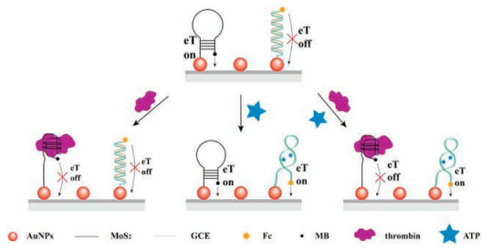
Furthermore, gold nanoparticles or hybrid nanomaterials consisted of gold nanoparticles [64] and other metallic nanomaterials including copper sulfide nanosheets [65], Zinc oxide nanopencils [66], and platinum nanoparticles [67] have been reported as nanoscale electrodes to electrically communicate between bulk electrode materials and reaction mixture [68, 69]. Su and co-workers constructed a dual-target aptasensor based on gold nanoparticles-decorated MoS2 coupled dual signaling detection strategy. The presence of adenosine triphosphate (ATP) and thrombin (a multifunctional serine protease generats at site of vascular injury converting circulating fibrinogen to fibrin monomer and has a host of direct actions on cells [70]) could induce structure switching of the aptasensor and lead to the ferrocene close or the methylene blue far from the electrode surface (Fig. 3) [68].

|
Download:
|
| Fig. 3. Gold nanoparticles decorated MoS2 nanocomposites served as carriers for labeling applied in dual-target electrochemical aptasensor. Copied with permission [68]. Copyright 2016, American Chemical Society. | |
Besides gold nanomaterials, iron and iron-oxide magnetic nanoparticles [71, 72] and nanoporous PtTi [73] have also emerged as potential candidates as carries for immobilizing aptamers or signal groups. For example, Fang and co-workers reported an electrochemical aptasensor to detect proteins in whole blood using plasma-polymerized 4 vinyl pyridine (PP4VP) modified ferriferous oxide (Fe3O4) and three-dimensional graphene nanocomposite. In this aptasensor, the immobilization of aptamer was promoted by amino groups contained in the three-component nanocomposite [71].
As several metallic nanoparticles (Au, Ag, etc.), especially bimetallic core-shell nanoparticles, exhibit an extraordinary catalytic behavior in the meantime and yield large surface area, nanomaterials are also applied as efficient catalysts to avoid instability at finite temperature and pH caused by using biological materials like enzymes (Fig. 4) [74-78]. For example, Mazloum-Ardakani and co-workers reported a tumor necrosis factor-alpha aptasensor employing graphene oxide nanosheets supported Ag@Pt nanoparticles as the nanocatalyst. The detection limit of this aptasensor was 2.07 pg/mL and the practicability was verified using human serum samples [76].

|
Download:
|
| Fig. 4. Gold nanoparticles participated electrocatalysis applied in electrochemical aptasensor. Copied with permission [78]. Copyright 2016, American Chemical Society. | |
Although gold and other metallic nanomaterials such as nanosheets, nanorods, core-shell nanoparticles, and metal-organic frameworks (MOFs), a type of zeolite-like crystalline porous material with metal ions as nodes and organic ligands as linkers to form 3-D frameworks [79-81], have many advantages over traditional approaches, there still remains the problem of electrical instability as metallic nanoparticles are susceptible to salt concentrations. Thus, proper surface modifications on nanoparticles are indispensable to resist salt induced aggregation. Second, when gold and other metallic nanomaterials are used as carriers, further studies are required to ensure the diffusional limitation inside the nanostructures would not affect the biosensor. And the uniformity, distribution and shape of these nanomaterials need to be well established to increase the efficiency [45]. Moreover, it is important to minimize the non-specific adsorption to lower the background.
2.2. Carbon nanomaterialsCarbon nanomaterials including carbon nanotubes (CNTs), graphene and graphene oxide (GO) are frequently used as supporting electrode matrices in electrochemical biosensors owning to their large surface area, remarkable mechanical, chemical and electrical properties [82-86]. Carbon nanomaterials modified electrodes then provide solid supports for the immobilization of biorecognition units such as antibodies and aptamers [87-91]. The immobilization strategies could be divided into covalent and non-covalent functionalization. In the former method, carbon nanomaterials are oxidized to introduce carboxylic groups to bind aptamers via amine bond formation. While physical adsorption or bifunctional linker groups are employed in the latter method [92-94]. Moreover, the large surface-to-bulk ratio nanomaterials provided to capture biomolecules including enzyme and aptamer were also utilized to fabricate protein biosensors [95, 96]. Chen and co-workers developed a sandwich-type aptasensor for the determination of mucin 1 (MUC1), which is a high molecular weight and cell surface glycoprotein with the ability to identify breast cancer in the early stages [95]. They used poly(o-phenylenediamine)-AuNPs hybrid film as the carrier for the immobilization of the primary aptamer and AuNPs/SiO2@MWCNTs (multi-walled carbon nanotubes) to enhance the surface area for stabilizing the secondary aptamer assembly. The aptasensor was proved to be of good stability and reproducibility with a detection limit of 1 pmol/L. Jing Li and co-workers established an aptasensor for thrombin detection. Carbon nanotubes (CNTs) labeled with aptamer and horseradish peroxidase (HRP) were used as a probe to combine thrombin. In the presence of H2O2, postelectropolymerization of the precipitates was produced by the enzymatically biocatalyzed reaction between HRP and 3, 3-diaminobenzidine (DAB) producing amplified signal. The detection limit of thrombin was 0.05 pmol/L [96] (Fig. 5).

|
Download:
|
| Fig. 5. Carbon nanomaterials based-amplification applied in applied in electrochemical aptasensor. Copied with permission [96]. Copyright 2015, American Chemical Society. | |
Recently, carbon nanomaterials have been reported to support metallic nanoparticles with catalytic activity [97-101]. For example, Shokoh and co-workers fabricated a dopamine aptasensor using functionalized carbon nanomaterials to support silver nanoparticles (AgNPs) to amplify signals [97]. Aptamer specific to dopamine (a neuroactive chemical released by a unique set of amacrine cells with either inhibitory or excitatory activity [102]) was immobilized based on the attachment between the amino groups of the chitosan trapped in the AgNPs/CNTs/GO nanocomposite and the phosphate groups of the aptamer. The formation of target-aptamer complexes would lead to the inhibition of electrocatalytic activity of AgNPs, leading to the decrease in the peak currents. This aptasensor was fast, simple and sensitive with the detection limit of 700±19.23 pmol/L.
Other novel biosensors such as single-walled carbon nanotubes (SWCNTs) based cocaine aptasensor which utilized the characteristic of SWCNTs possessed stronger interaction with ssDNA than that with dsDNA [103], chloramphenicol aptasensor which was developed by combining MWCNTs, mesoporous carbon and three-dimensional porous graphene [104], etc. were also reported lately.
It is clear that carbon nanomaterials-assisted aptasensors offer numerous advantages owning to their excellent physical and electrical properties, low costs and easy operation. However, the performances of these aptasensors are discrepant from each other due to the difficulty to well control the chirality, diameter, level of agglomeration of CNTs, and/or the impurities and substrate of graphenethe [82]. And the interaction between aptamers and carbon nanomaterials should also be investigated to make full use of their signal amplification ability [105, 106]. In brief, carbon nanomaterials-based biosensors are still in the preliminary stages and efforts including increasing the reproducibility, biocompatibility and nanotoxicity are necessary to drive the development of these detection platforms towards broader application areas [107].
2.3. Quantum dotsQuantum dots (QDs) are colloidal nanocrystalline semiconductors with 1~10 nm in size mainly formed by cationic groups Ⅱ-Ⅵ, Ⅲ-Ⅴ, and Ⅳ-Ⅵ. QDs contain three major kinds including homogeneous structures, core-shell structures and ternary structures [108]. Due to their prominent electrochemical properties and other advantages such as miniaturization, good biocompatibility, controllable morphology, low power requirements and low cost, QDs have been served as transduction layers or labels for signal amplification in aptamer-based biosensing [109-112].
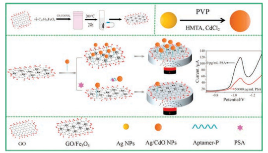
There are four primary methods to immobilize aptamers onto QDs. The first one is through self-assembly between aptamers and QDs. The second is based on specific interactions such as biotin-streptavidin interaction [113, 114]. Aptamers can also be attached to QDs via hybridization to complementary sequences modified on the surface of the nanoparticles [115]. For example, Yuan Zhao and co-workers designed a simple magnetic electrochemical aptasensor for the detection of prostatic specific antigen (PSA) in human serums using Ag/CdO NPs as electroactive labels. As shown in Fig. 6, they assembled aptamer-modified Ag/CdO NPs on the surface of superparamagnetic Fe3O4/graphene oxide nanosheets (GO/Fe3O4 NSs) through the hydrophobic and π-π stacking interaction of aptamers and GO NSs firstly. With the concentrations of PSA increasing, the high affinity of aptamers to PSA enabled the separation of Ag/CdO NPs from GO/Fe3O4 NSs, making the change of electrochemical signal. Ag/CdO possessed superior electroactive properties and efficient electron transfer, which might amplify the detecting signal [116]. Last but not least is covalently binding [117]. Liu and co-workers fabricated an ultrasensitive cancer cell biosensor based on aptamer-DNA concatamer-QDs probe. Aptamers were first immobilized on QDs through amidation reaction to form the cancer detection probe. Signal amplification was accomplished through adding two designed nucleotide sequences to hybridize with the probe to construct aptamer-DNA concatamer-QDs formation. The aptasensor had a low detection limit of 50 cell/mL. Moreover, this aptasensor could distinguish cancer cells from normal cells so it held a great potential for cancer diagnosis [118].

|
Download:
|
| Fig. 6. Ag/CdO NP assisted-amplification applied in magnetic electrochemical aptasensor for PSA detection. Copied with permission [116]. Copyright 2019, American Chemical Society. | |
Nowadays, the applied research of QDs in aptamer-based electrochemical detection has been developed rapidly [119-122]. However, the selectivity and reproducibility of these aptasensors towards real samples still need to be verified. And non-specific adsorption on the electrode surface may affect some of the aptasensors which measure targets according to the suppression of the voltammetric signal of QD ions. Another limitation of QDs-assisted aptasensors is the facility request as the synthesis of QDs is usually performed at high temperatures [123].
2.4. Nucleic acidIn nucleic acid-based signal amplification strategies, molecular biological technologies such as rolling circle amplification (RCA), strand-displacement amplification (SDA) [124], hybridization chain reaction (HCR) [125], PCR and DNA origami are involved to generate repetitive oligonucleotide sequences which are able to incorporate detectable elements [126-130]. Rolling circle amplification (RCA) uses a circular DNA template and special polymerases to synthesize long single stranded DNA or RNA [131]. The RCA production gives rise to tens to hundreds of tandem repeats complementary to the template. The most common approach of RCA-assisted methodologies is using RCA produces as carriers to load numerous electrochemical tracers to amplify the electrical signals [132, 133]. For example, Fan and co-workers reported a label-free electrochemical aptasensor for the detection of thrombin. The RCA produce contains tens to hundreds of tandem repeats complementary to the template. The present of thrombin could trigger RCA process to generate poly adenine production for the adsorption of AuNPs. Then the AuNPs electrocatalytic reduction toward H2O2 was measured. The aptasensor showed a detection limit of 35 fmol/L and had a great potential for clinical diagnosis applications [134]. Moreover, as RCA generates long DNA with numerous tandem copies, Yang and co-workers fabricated an aptamer network-modified electrode interface to achieve efficient capturing and detection of CTCs. The network interface produced via RCA contained repeated aptamer segments and could capture CTCs in a multivalent cooperative manner, which demonstrated it potential application in monitoring CTCs in blood [135].
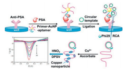
Moreover, copper nanoparticles (CuNPs) [136], CdS nanoparticles [137, 138], and electrochemical active compounds are also employed as signal-reporting units for signal amplification [139]. Zhu and co-workers combined RCA with CuNPs to develop an ultrasensitive aptasensor for the detection of prostate specific antigen (PSA). Different pulse stripping voltammetry was used to detect the copper ions released from poly(thymine)-templated CuNPs. Besides, large amounts of oligonucleotide sequences generated through nucleic acid amplification technologies can also be used as signal units (Fig. 7). The aptasensor achieved a remarkable detection limit of 0.020 fg/mL and had potential for clinical practice [136].

|
Download:
|
| Fig. 7. Rolling circle amplification-based aptasensor for the detection of prostate specific antigen. Copied with permission [136]. Copyright 2016, American Chemical Society. | |
Hybridization chain reaction (HCR) mimics molecular chain-growth polymerization to generate long double-strand DNA [140]. HCR opens up avenues for meeting the requirement of efficient signal amplification for biosensor due to its kinetics-controlled reaction, high sensitivity and selectivity [141]. Similar with RCA, HCR productions can also be used to load electrochemical tracers. Methyl blue (MB), enzymes like HRPs and alkaline phosphatase (ALP) [125, 142, 143], artificial mimicking enzymes such as hemin [144], metal nanoparticles with catalytic behavior, and electroactive cation like [Ru(NH3)6]3+ [145-147], are widely employed as signal-reporting units for signal amplification. An and co-workers reported an ultrasensitive aptasensor for the determination of exosomes, which were lipid bilayer membrane vesicles secreted by various kinds of cells able to guide pathological and physiological processes. By combining the ability of HRP molecules to catalyze the reaction of o-phenylenediamine and H2O2 and HCR to achieve signal amplification, the aptasensor allowed the sensitive detection of targets ranging from 102 exosomes/mL to 108 exosomes/mL.
Despite the fact that molecular biological technologies offer remarkable advantages including efficiency, programmability, biocompatibility, without toxicity and immunogenicity, it still possessed several problems. For example, The limitation of HCR is low sensitivity and low catalytic rate for the formation of DNA concatemers [44]. And the steps to prepare circular templates for RCA is also expensive and time-consuming [40].
2.5. EnzymesEnzymes have been widely employed in electrochemical detection due to their catalytic activity and substrate specificity. Various nucleases have been used for target recycling to increase the signal/target ratio [148]. Endonuclease [149, 150], exonuclease [151-154] and deoxyribonuclease [155-157] are three representative kinds of nucleases applied in aptasensors. For example, Zhao and co-workers achieved ultrasensitive detection of thrombin combining DNA ligase-catalyzed ligation of thrombin-specific aptamer with an extension strand and exonuclease Ⅲ-catalyzed selective degradation of probe DNA (Fig. 8). The electrochemical biosensor they described exhibit excellent performance with a detection limit of 33 fmol/L. Moreover, the biosensor had the ability to distinguish thrombin in serum samples and might hold the potential in clinical applications [158].

|
Download:
|
| Fig. 8. Exonuclease based aptasensor for the detection of thrombin. Copied with permission [158]. Copyright 2014, American Chemical Society. | |
Besides target recycling method, polymeric enzyme formed by a number of enzyme molecules is also used to enhance analytical sensitivity. For example, Xiong and co-workers presented an AFP (alfa-fetoprotein) aptasensor based on HRP-functionalized Envision antibody complex. The immunocomplex formed on the electrode surface allowed large amounts of HRP to amplify electrocatalytic signal [159].
Furthermore, artificial mimicking enzymes such as hemin, [160, 161], catalytic/electroactive nanoparticles [162-164] have been explored for biosensing due to their high stability and are easier to syntheses and modify than natural enzymes. Hemin is the catalytic center of many artificial mimicking enzymes. Hemin conjugated G-quadruplex [165-169], graphene sheet [170], and other nanomaterials [81] have been widely used to achieve ultrasensitive assays. Meanwhile, researchers have also draw attention to metallic alloy and semiconductor nanomaterials exhibit enzyme-like activities [171-173].
Nuclease-based amplification holds several advantages including easy to design, simple in construction, rapid reaction and suitable for homogenous assay. However, this kind of aptasensor is susceptible to sample matrix. It is strictly to establish optimal buffer systems as the enzyme reaction during detection process may be influenced by impurities in the sample.
2.6. PolymersOwing to the rapid development of nanotechnologies, researchers are now able to design complicated and optimized soft structures with synergistic properties [174]. Polymers such as poly(ethylene glycol) exhibit remarkable properties such as responsiveness to electronic, optical, mechanical, magnetic and thermal stimulation have been widely applied to the clinical and biochemical analysis [175-179]. For example, Min You and co-workers developed a novel sandwich assay electrochemical biosensor to detect AβO using molecularly imprinted polymers (MIPs) and aptamer as the recognition element. In the work, the AβO was captured by the film of MIPs and the AβO-specific aptamer, forming a MIPs/target/aptamer sandwich structure. Small amount of AβO could trigger the electrochemical reaction and amplified electrochemical signal could be generate by the sandwich structure [180].
Jifan Li and co-workers also developed a dual binding site and dual signal amplifying electrochemical aptasensor to detect carcinoembryonic antigen (CEA). In their experiment, self-polymerized dopamine (PDA)-decorated Au coordinated with Fe-MOF (Au@PDA@Fe-MOF) and aptamer were employed as electrochemical labels. This aptasensor offered excellent electrochemical performance because of a detection limit of 0.33 fg/mL (Fig. 9) [181]. Ali R. Jalalvand established a novel and efficient aptasensor to detect prostate specific antigen (PSA). Aptamer against PSA was immobilized onto Au nanoparticles/fullerene C60-chitosan-ionic liquid/multiwalled carbon nanotubes/screen printed carbon electrode which was used as aptasensor to detect PSA. The aptasensor has high surface-to-volume ratio and good in-plane conductivity at the existence of MWCNTs and C60, and the amount of immobilized AuNPs was increased by the CS-IL which made effective signal amplification [182]. Hui-Min Wang and co-workers built a novel label-free aptasensor to detect thrombin (TB). The aptasensor was constructed based on palladium nanocones (Pd NCs)/amine-terminated polyamidoamine (PAMAM)/glassy carbon electrode (GCE) as platform coupled with tripropylamine (TPA) system, and aptamer against TB was modified on Pd NCs as capture probe. In the aptasensor, Pd NCs were functionalized with mercaptoethanol (MCH) and attached onto the PAMAM/GCE. PAMAM could amplify electrochemical signal and the electrochemical intensity of the Pd NCs/TPA system showed about 2.5 times enhancement than that of the bare GCE [183].

|
Download:
|
| Fig. 9. Au@PDA@Fe-MOF applied in electrochemical aptasensor for CEA detection. Copied with permission [181]. Copyright 2020, American Chemical Society. | |
Polymers are often used together with other materials usually, such as metallic nanomaterials, carbon nanomaterials, quantum dots. Polymers can provide large specific surface area and have favorable biocompatibility. More materials can bind on polymers by the help of these properties which may make other materials using their own strengths and may produce lots of signals. Also, polymers can accelerate electron conductivity and enhance electrochemical intensity further. Therefore, polymers will provide a promising approach to develop efficient biosensors associated with other materials for researchers.
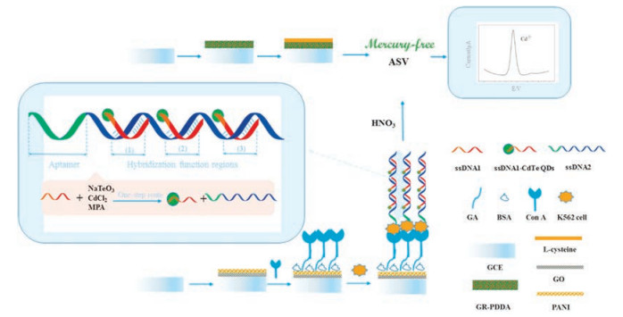
2.7. Combined application of several materialsRecently, in order to acquire the amplified signal, each material cannot be avoided in combination with other materials [71, 95, 102, 181, 182]. Each material plays its own properties in the same detecting method. For example, Yue Zheng and co-workers designed a green and ultrasensitive electrochemical biosensing strategy for detecting K562 cells. First, new electrode modification composite, graphene (GR)-poly diallyldimethylammonium chloride (PDDA)/L-Cysteine (L-Cys), was fabricated as the capture electrode. Second, aptamer-DNA concatamer-CdTe QDs was used as signal amplification probes. In the strategy, HCR could produce tens to hundreds of repetitive sequence that acts as carriers to load numerous CdTe QDs to amplify the electrical signals. CdTe QDs had been served as labels for signal amplification in this biosensing. L-Cys contained nitrogen and oxygen elementsin which could adsorpt heavymetal ions such as Cd2+ and Pb2+ in the solution strongly. And, PDDA could increase the specific surface area of the electrode by capturing much more signal recognition material. The schematic was showed in Fig. 10 [184].

|
Download:
|
| Fig. 10. A novel supersandwich cytosensor using aptamer-DNA concatamer-QDs and GR-PDDA/l-Cys for detecting K562 cells. Copied with permission [184]. Copyright 2018, Elsevier. | |
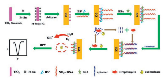
Lingling Xu and co-workers constructed a sandwich-type electrochemical aptasensor for the detection of carcinoembryonic antigen (CEA) [185]. The aptasensor contained two kinds of nanomaterials. Firstly, graphene oxide was combined with hemin and modified gold nanoparticles (Hemin-rGO-AuNPs) as the sensing nanomatrix, and the CEAapt1 was immobilized on Hemin-rGO-AuNPs for capturing CEA. The redox reaction of hemin on the electrode surface could produce powerful signal. Secondly, horseradish peroxidase-modified organic-inorganic hybrid nanoflowers linked to gold nanoparticles (HRP-Cu3(PO4)2-HNF-AuNP-CEAapt2) were used as nanocarriers. At the existence of 4-chloro-1-naphthol (4-CN) and H2O2, both HRP-Cu3(PO4)2HNF and Hemin-rGO-AuNPs were catalyzed to start precipitation reaction to produce a layer of precipitate film. The precipitate film would impede the electron transmission and decrease current signal obviously. LeLe Li and co-workers designed a highly sensitive and selective electrochemical aptasensor for the detection of streptomycin based on a dual signal amplification system [186]. Initially, Pt and Sn nanoparticles were modified on TiO2 nanorods to form Pt-Sn@TiO2. Then, Pt-Sn@TiO2 was immobilized on glassy carbon electrode and a DNA with a complementary sequence to a streptomycin aptamer (cDNA) was captured on the electrode. Next, the streptomycin aptamer would be hybridized with cDNA. Followed, with the adding of streptomycin and RecJf exonuclease, both the streptomycin-aptamer complex and the cDNA were cleaved from the electrode, producing the electrochemical signal. RecJf exonuclease would make streptomycin release from the streptomycin-aptamer complex and the streptomycin would combine with aptamers on the electrode again. In this system, the Pt-Sn@TiO2 composite could enhance the oxygen reduction current because of the large specific surface loaded with biomolecules and excellent electrocatalytic activity which amplified the detecting signal. The cyclic amplification mechanism initiated by streptomycin and RecJf would lead to a 0.85-fold enhancement of the oxygen reduction peak current. These two factors made the detecting signal amplified (Fig. 11).

|
Download:
|
| Fig. 11. A electrochemical aptasensor for the detection of streptomycin based on dual signal amplification system. Copied with permission [186]. Copyright 2019, Elsevier. | |
Jiaxin Zheng and co-workers built a triple-amplified electrochemical aptasensor for the detection of MUC1. The aptasensor was established based on exonuclease Ⅲ (Exo Ⅲ)-assisted with strand displacement reaction and enzyme catalytic strategy. First, Exo Ⅲ could make MUC1 and aptamer recycled during the cycle I, produce the single stranded DNA-1 (S-1). S-1 would be introduced to the hybride reaction on the electrode. During the cycle II, strand displacement reaction was triggered by hairpin DNA-2 (H-2) on the electrode. Then, the AuNPs-DNA-enzyme conjugates as signal probe could be combined on the electrode conjugating with H-2 which produced signal. In this strategy, two factors amplified the signal. On one hand, AuNPs-DNA-enzyme conjugates amplified signal by catalyzing the H2O2-TMB system; on the other hand, the target triggered the Exo Ⅲ-assisted cycling and strand displacement reaction which made vast of S-1 produced resulting in signal amplification (Fig. 12) [187].

|
Download:
|
| Fig. 12. A triple-amplified electrochemical aptasensor for the detection of MUC1. Copied with permission [187]. Copyright 2019, Elsevier. | |
In these examples described above, all materials gave their advantages to amplify electrochemical signal. Metallic nanoparticles and metal oxide nanomaterials have high biocompatibility, high conductivity, distinctive electronic and electrocatalytic properties, and sensor activities which can be used as carriers of biomolecules or be used to amplify the signal. The carbon materials have their unique properties such as high electrical conductivity, large surface area, good mechanical and chemical stabilities and can be modified with functional groups easily for detecting targets. Polymer nanomaterials can be combined with biomolecules through covalent bond, increase electrochemical signal transduction, and have high recognition in bioanalyses. The collaboration of these materials with enzymes or DNA can improve the analytical performances of the designed methods by the synergistic effect.
3. Current challenges and future outlookAlthough tremendous developments and routine applications have been made, aptamer-based electrochemical biosensing are confronted with several challenges. Although most of the aptasensors mentioned above exhibit excellent performance in laboratory, it is difficult to detect target molecules in real sample [127]. The reasons of failure in the practical application include cost, not viable to generalize due to their complicated design and high requirement for equipment, and most importantly, the sensor-to-sensor variability of their properties and ultimately their analytical performance [188]. For instance, although nanomaterials like metallic nanoparticles and carbon nanomaterials have been proved to be excellent materials in various areas, it still requires large amounts of investment to transform academic research to cost-effective products. And the sensor-to-sensor variability caused by the structure and physicochemical properties of nanomaterials and enzymes are inevitable. Moreover, real biological samples are far more comprehensive compared with the samples used in laboratory, which makes the remarkable stability, sensitivity and reproducibility achieved by reported electrochemical aptasensors obtained under optimal conditions difficult to a transcribe.
The development of products fit for universal popularization and application demands the establishment of standard for effective and low-cost molecular biological technologies and the synthesis methods for nanomaterials. It is still a long way to go for the aptamer-based electrochemical biosensing to overcome accuracy, stability and other problems before applied in practice. There is no doubt that with the solution of the above challenges, aptamer-based electrochemical biosensing will make distinguished contributions in clinical research.
4. ConclusionIn this review, we have summarized various kinds of amplification strategies to improve the sensitivity of electrochemical aptasensors (Table 1). Novel nanomaterials such as gold nanomaterials and carbon nanotube, molecular biological technologies and enzymes are commonly applied as high-performance electrode-supporting materials, or label carriers, or electroactive tracers, or to achieve target recycling. The use of these strategies has led to the unprecedented success in improving the performance of aptamer-based electrochemical biosensing. And the application of these ultrasensitive aptasensors to real samples will undoubtedly show great potential in clinical diagnosis.
|
|
Table 1 Summary of different signal amplification strategies. |

The authors declare that they have no known competing financial interests or personal relationships that could have appeared to influence the work reported in this paper.
AcknowledgmentsThis work was financially supported by the National Key Research and Development Program of China (No. 2017YFA0205301), the National Natural Science Foundation of China (Nos. 61527806, 81902153 and 61871180), the Clinical Advanced Technology of Social Development Projects in Jiangsu Province (No. BE2018695) and the Natural Science Foundation of Hunan Province (No. 2017JJ2069).
| [1] |
N.J. Ronkainen, H.B. Halsall, W.R. Heineman, Chem. Soc. Rev. 39 (2010) 1747-1763. DOI:10.1039/b714449k |
| [2] |
Z.X. Shi, G.K. Li, Y.F. Hu, Chin. Chem. Lett. 30 (2019) 1600-1606. DOI:10.1016/j.cclet.2019.04.066 |
| [3] |
Y.M. Nie, X.D. Yuan, P. Zhang, Y.Q. Chai, R. Yuan, Anal. Chem. 91 (2019) 3452-3458. DOI:10.1021/acs.analchem.8b05001 |
| [4] |
A. Devadoss, P. Sudhagar, C. Terashima, K. Nakata, A. Fujishima, J. Photoch. Photobio. C 24 (2015) 43-63. DOI:10.1016/j.jphotochemrev.2015.06.002 |
| [5] |
Y.H. Pang, L.L. Guo, X.F. Shen, N.C. Yang, C. Yang, Electrochim. Acta 341 (2020) 136055. DOI:10.1016/j.electacta.2020.136055 |
| [6] |
M. Liu, A. Khan, Z.F. Wang, et al., Biosens. Bioelectron. 130 (2019) 174-184. DOI:10.1016/j.bios.2019.01.006 |
| [7] |
R.R. Huang, L. He, Y.Y. Xia, et al., Small 15 (2019) e1900735. DOI:10.1002/smll.201900735 |
| [8] |
A.D. Ellington, J.W. Szostak, Nature 346 (1990) 818-822. DOI:10.1038/346818a0 |
| [9] |
R.R. Huang, Z.J. Xi, N.Y. He, Sci. China Chem. 58 (2015) 1122-1130. DOI:10.1007/s11426-015-5344-7 |
| [10] |
Z.Y. Zhang, A. Runa, J. Wu, H. Zhang, X. Li, Chin. Chem. Lett. 30 (2019) 779-782. DOI:10.1016/j.cclet.2018.10.019 |
| [11] |
C. Wang, L.L. Sun, Q. Zhao, Chin. Chem. Lett. 30 (2019) 1017-1020. DOI:10.1016/j.cclet.2019.01.029 |
| [12] |
D.L. Liu, Y. Li, R. Sun, et al., J. Nanosci. Nanotechnol. 20 (2020) 2114-2121. DOI:10.1166/jnn.2020.17358 |
| [13] |
Z.S. Chen, Z. Ali, S. Li, B. Liu, N.Y. He, J. Nanosci. Nanotechnol. 16 (2016) 9346-9358. DOI:10.1166/jnn.2016.12641 |
| [14] |
R.R. Huang, Z.J. Xi, Y. Deng, N.Y. He, Sci. Rep. 6 (2016) 31103. DOI:10.1038/srep31103 |
| [15] |
Z.J. Xi, R.R. Huang, Y. Deng, N.Y. He, J. Biomed. Nanotechnol. 10 (2014) 3043-3062. DOI:10.1166/jbn.2014.1979 |
| [16] |
M.Q. Gu, J.L. Liu, D.L. Li, et al., Nanosci. Nanotech. Lett. 11 (2019) 1139-1144. DOI:10.1166/nnl.2019.2981 |
| [17] |
Z.J. Xi, B. Zheng, Nanosci. Nanotech. Lett. 10 (2018) 309-319. DOI:10.1166/nnl.2018.2624 |
| [18] |
Z.K. Guo, C. Wang, S. Li, et al., J. Nanosci. Nanotechnol. 20 (2020) 3373-3377. DOI:10.1166/jnn.2020.17424 |
| [19] |
M.P. Landry, H. Ando, A.Y. Chen, et al., Nat. Nanotechnol. 12 (2017) 368-377. DOI:10.1038/nnano.2016.284 |
| [20] |
S. Yu, X.J. Bi, L. Yang, et al., J. Biomed. Nanotechnol. 15 (2019) 1135-1148. DOI:10.1166/jbn.2019.2751 |
| [21] |
D.X. Liu, T.T.T. Tien, D.T. Bao, et al., J. Biomed. Nanotechnol. 15 (2019) 204-211. DOI:10.1166/jbn.2019.2667 |
| [22] |
W.R. Shin, S.S. Sekhon, S.G. Kim, et al., J. Biomed. Nanotechnol. 14 (2018) 1992-2002. DOI:10.1166/jbn.2018.2634 |
| [23] |
L.Y. Jiang, H. Wang, S.Z. Chen, J. Nanosci. Nanotechnol. 20 (2020) 2025-2031. DOI:10.1166/jnn.2020.17301 |
| [24] |
M. Liu, X.C. Yu, Z. Chen, et al., J. Nanobiotechnology 15 (2017) 81. DOI:10.1186/s12951-017-0311-4 |
| [25] |
A.T.V. Nguyen, T.T.T. Trinh, V.T. Hoang, et al., J. Biomed. Nanotechnol. 15 (2019) 1185-1200. DOI:10.1166/jbn.2019.2772 |
| [26] |
L.P. Zhong, H. Zou, Y. Huang, et al., J. Biomed. Nanotechnol. 15 (2019) 352-362. DOI:10.1166/jbn.2019.2688 |
| [27] |
Z.X. Hu, J. He, W.L. Gong, et al., J. Biomed. Nanotechnol. 14 (2018) 1645-1653. DOI:10.1166/jbn.2018.2619 |
| [28] |
Z.Y. He, Z. Chen, M.D. Tan, et al., Cell Prolif. 53 (2020) e12822. |
| [29] |
X.C. Yu, L. He, M. Pentok, et al., Nanoscale 11 (2019) 15589-15595. DOI:10.1039/C9NR04050A |
| [30] |
R.R. Huang, L. He, S. Li, et al., Nanoscale 12 (2020) 2445-2451. DOI:10.1039/C9NR08747H |
| [31] |
M. Liu, T. Yang, Z.S. Chen, Z.F. Wang, N.Y. He, Biomater. Sci. 6 (2018) 3152-3159. DOI:10.1039/C8BM00787J |
| [32] |
M. Liu, Z.F. Wang, T. Tan, et al., Theranostics 8 (2018) 5772-5783. DOI:10.7150/thno.28949 |
| [33] |
R.R. Huang, Z.S. Chen, M. Liu, et al., Sci. China Chem. 60 (2017) 786-792. |
| [34] |
T.T. Li, J.J. Yang, Z. Ali, et al., Sci. China Chem. 60 (2017) 370-376. DOI:10.1007/s11426-016-0159-2 |
| [35] |
G.K. Mishra, V. Sharma, R.K. Mishra, Biosensors 8 (2018) 28. DOI:10.3390/bios8020028 |
| [36] |
L. Hong, F. Zhou, D.M. Shi, X.J. Zhang, G.F. Wang, Biosens. Bioelectron. 95 (2017) 152-159. DOI:10.1016/j.bios.2017.04.023 |
| [37] |
N. Saraf, E.R. Woods, M. Peppler, S. Seal, Biosens. Bioelectron. 117 (2018) 40-46. DOI:10.1016/j.bios.2018.05.031 |
| [38] |
W. Chen, C. Yan, L. Cheng, et al., Biosens. Bioelectron. 117 (2018) 845-851. DOI:10.1016/j.bios.2018.07.012 |
| [39] |
E.E. Ferapontova, E.M. Olsen, K.V. Gothelf, J. Am. Chem. Soc. 130 (2008) 4256-4258. DOI:10.1021/ja711326b |
| [40] |
H.X. Ju, J. Anal. Test. 1 (2017) 7. DOI:10.1007/s41664-017-0008-6 |
| [41] |
V. Plaks, C.D. Koopman, Z. Werb, Science 341 (2013) 1186-1188. DOI:10.1126/science.1235226 |
| [42] |
A.P. Catherine, P. Klaus, Cancer Discov. 6 (2016) 479-491. DOI:10.1158/2159-8290.CD-15-1483 |
| [43] |
N. Bushati, S.M. Cohen, Annu. Rev. Cell Dev. Biol. 23 (2007) 175-205. DOI:10.1146/annurev.cellbio.23.090506.123406 |
| [44] |
T. Fozooni, H. Ravan, H. Sasan, Appl. Biochem. Biotech. 183 (2017) 1224-1253. DOI:10.1007/s12010-017-2494-4 |
| [45] |
I.H. Cho, J. Lee, J. Kim, et al., Sensors 18 (2018) 207. DOI:10.3390/s18010207 |
| [46] |
H.X. Ju, Appl. Mater. Today 10 (2018) 51-71. DOI:10.1016/j.apmt.2017.11.001 |
| [47] |
X.D. Cao, Y.K. Ye, S.Q. Liu, Anal. Biochem. 417 (2011) 1-16. DOI:10.1016/j.ab.2011.05.027 |
| [48] |
D.S. Zhen, F.F. Zhong, D. Yang, Q.Y. Cai, Y. Liu, Mater. Express 9 (2019) 319-327. DOI:10.1166/mex.2019.1499 |
| [49] |
C.L. Tang, Z.Y. He, H.M. Liu, et al., J. Nanobiotechnol 18 (2020) 62. DOI:10.1186/s12951-020-00613-6 |
| [50] |
Z.J. Xi, R.R. Huang, Y. Deng, E.B. Su, N.Y. He, Sci. Adv. Mater. 8 (2016) 1678-1682. DOI:10.1166/sam.2016.2447 |
| [51] |
S. Ranjbar, S. Shahrokhian, F. Nurmohammadi, Sens. Actuators B: Chem. 255 (2018) 1536-1544. DOI:10.1016/j.snb.2017.08.160 |
| [52] |
G.Y. Li, S.S. Li, Z.H. Wang, et al., Anal. Biochem. 547 (2018) 37-44. DOI:10.1016/j.ab.2018.02.012 |
| [53] |
J.K. Bhattarai, D. Neupane, B. Nepal, et al., Nanomaterials 8 (2018) 171. DOI:10.3390/nano8030171 |
| [54] |
G.J. Yang, Z.Q. Xiao, C.L. Tang, et al., Biosens. Bioelectron. 141 (2019) 111416. DOI:10.1016/j.bios.2019.111416 |
| [55] |
Y.X. Lai, H. Huang, Z.H. Xia, et al., Mater. Express 9 (2019) 444-450. DOI:10.1166/mex.2019.1524 |
| [56] |
Y.X. Lai, C.X. Zhang, Y. Deng, et al., Chin. Chem. Lett. 30 (2019) 160-162. DOI:10.1016/j.cclet.2018.07.011 |
| [57] |
M.A. Owens, B.C. Horten, M.M.D. Silva, Clin. Breast Cancer 5 (2004) 63-69. DOI:10.3816/CBC.2004.n.011 |
| [58] |
Y. Zhu, P. Chandra, Y.B. Shim, Anal. Chem. 85 (2013) 1058-1064. DOI:10.1021/ac302923k |
| [59] |
C. Ocaña, S. Lukic, M.D. Valle, Microchim. Acta 182 (2015) 2045-2053. DOI:10.1007/s00604-015-1540-6 |
| [60] |
J.J. Zhang, F.F. Cheng, T.T. Zheng, J.J. Zhu, Biosens. Bioelectron. 89 (2017) 937-945. DOI:10.1016/j.bios.2016.09.087 |
| [61] |
R. Liu, Y. Zhang, S.Y. Zhang, W. Qiu, Y. Gao, Appl. Spectrosc. Rev. 49 (2014) 121-138. DOI:10.1080/05704928.2013.807817 |
| [62] |
G.J. Yang, Y.X. Lai, Z.Q. Xiao, C.L. Tang, Y. Deng, Chin. Chem. Lett. 29 (2018) 1857-1860. DOI:10.1016/j.cclet.2018.11.030 |
| [63] |
X.J. Jiang, X.D. Wang, Annu. Rev. Biochem. 73 (2004) 87-106. DOI:10.1146/annurev.biochem.73.011303.073706 |
| [64] |
M. Baghayeri, R. Ansari, M. Nodehi, I. Razavipanah, H. Veisi, Electroanalysis 30 (2018) 2160-2166. DOI:10.1002/elan.201800158 |
| [65] |
K.J. Huang, Y.J. Liu, J.Z. Zhang, Microchim. Acta 182 (2015) 409-417. DOI:10.1007/s00604-014-1352-0 |
| [66] |
Y.F. Qiao, J. Li, H.B. Li, et al., Biosens. Bioelectron. 86 (2016) 315-320. DOI:10.1016/j.bios.2016.06.062 |
| [67] |
Z.S. Beiranvand, A.R. Abbasi, S. Dehdashtian, Z. Karimi, A. Azadbakht, Anal. Biochem. 518 (2017) 35-45. DOI:10.1016/j.ab.2016.10.001 |
| [68] |
S. Su, H.F. Sun, W.F. Cao, et al., ACS Appl. Mater. Interfaces 8 (2016) 6826-6833. DOI:10.1021/acsami.5b12833 |
| [69] |
B. Posha, S.R. Nambiar, N. Sandhyarani, Biosens. Bioelectron. 101 (2018) 199-205. DOI:10.1016/j.bios.2017.10.030 |
| [70] |
S.R. Coughlin, Nature 407 (2000) 258-264. DOI:10.1038/35025229 |
| [71] |
M. Hasanzadeh, N. Shadjou, M.D.L. Guardia, Trends Anal. Chem. 72 (2015) 1-9. DOI:10.1016/j.trac.2015.03.016 |
| [72] |
S.M. Fang, X.D. Dong, S.L. Liu, et al., Electrochim. Acta 212 (2016) 1-9. DOI:10.1016/j.electacta.2016.06.128 |
| [73] |
W.J. Guo, N. Sun, X.L. Qin, M.S. Pei, L.Y. Wang, Biosens. Bioelectron. 74 (2015) 691-697. DOI:10.1016/j.bios.2015.06.081 |
| [74] |
R. Tian, X.J. Chen, Q.W. Li, C. Yao, Anal. Biochem. 500 (2016) 73-79. DOI:10.1016/j.ab.2016.01.021 |
| [75] |
C.C. Zhang, J. Tang, L.L. Huang, Y.P. Li, D.P. Tang, Microchim. Acta 184 (2017) 2445-2453. DOI:10.1007/s00604-017-2223-2 |
| [76] |
M.A. Mohammad, H. Laleh, T. Zahra, Biosens. Bioelectron. 74 (2015) 30-36. DOI:10.1016/j.bios.2015.05.072 |
| [77] |
Y.H. Luo, X.H. Zhang, D.M. Yao, et al., RSC Adv. 4 (2014) 28052-28055. DOI:10.1039/c4ra02857k |
| [78] |
C.L. Bentley, M. Kang, P.R. Unwin, J. Am. Chem. Soc. 138 (2016) 12755-12758. DOI:10.1021/jacs.6b08124 |
| [79] |
B. Panella, M. Hirscher, H. Pütter, U. Müller, Adv. Funct. Mater. 16 (2006) 520-524. DOI:10.1002/adfm.200500561 |
| [80] |
M. Chen, N. Gan, Y. Zhou, et al., Talanta 161 (2016) 867-874. DOI:10.1016/j.talanta.2016.09.051 |
| [81] |
S.B. Xie, J.W. Ye, Y.L. Yuan, Y.Q. Chai, R. Yuan, Nanoscale 7 (2015) 18232-18238. DOI:10.1039/C5NR04532K |
| [82] |
P. Ramnani, N.M. Saucedo, A. Mulchandani, Chemosphere 143 (2016) 85-98. DOI:10.1016/j.chemosphere.2015.04.063 |
| [83] |
E.L. Zhang, T. Sun, B.T. Ge, et al., Mater. Express 8 (2018) 105-111. |
| [84] |
X.B. Hu, K.Y. Goud, V.S. Kumar, et al., Sens. Actuators B: Chem. 268 (2018) 278-286. DOI:10.1016/j.snb.2018.03.155 |
| [85] |
Y. Liu, T.T. Li, C.X. Ling, et al., Chin. Chem. Lett. 30 (2019) 2211-2215. DOI:10.1016/j.cclet.2019.05.020 |
| [86] |
D.D. Yang, M. Liu, J. Xu, et al., Talanta 185 (2018) 113-117. DOI:10.1016/j.talanta.2018.03.045 |
| [87] |
X.J. Chen, Y.Z. Wang, Y.Y. Zhang, et al., Anal. Chem. 86 (2014) 4278-4286. DOI:10.1021/ac404070m |
| [88] |
S.H. Yu, T.H. Kim, J. Biomed, Nanotechnol. 15 (2019) 1824-1831. |
| [89] |
L.L. Guo, N.Y. He, Y.X. Zhao, T.H. Liu, Y. Deng, Theranostics 10 (2020) 3206-3222. DOI:10.7150/thno.40414 |
| [90] |
Y.X. Lai, L.J. Wang, Y. Liu, et al., J. Biomed. Nanotechnol. 14 (2018) 44-65. DOI:10.1166/jbn.2018.2505 |
| [91] |
Y. Liu, Y. Deng, T.T. Li, et al., J. Biomed. Nanotechnol. 14 (2018) 2156-2161. DOI:10.1166/jbn.2018.2655 |
| [92] |
A.T. Lawal, Mater. Res. Bull. 73 (2016) 308-350. DOI:10.1016/j.materresbull.2015.08.037 |
| [93] |
Z.H. Wang, J.B. Yu, R.J. Gui, H. Jin, Y.Z. Xia, Biosens. Bioelectron. 79 (2016) 136-149. DOI:10.1016/j.bios.2015.11.093 |
| [94] |
M.R. Hasan, T. Pulingam, J.N. Appaturi, et al., Anal. Biochem. 554 (2018) 34-43. DOI:10.1016/j.ab.2018.06.001 |
| [95] |
X.J. Chen, Q. Zhang, C.H. Qian, et al., Biosens. Bioelectron. 64 (2015) 485-492. DOI:10.1016/j.bios.2014.09.052 |
| [96] |
J. Li, J.J. Wang, X. Guo, et al., Anal. Chem. 87 (2015) 7610-7617. DOI:10.1021/acs.analchem.5b00640 |
| [97] |
W.J. Xu, S.Y. Xue, H.Y. Yi, et al., Chem. Commun. 51 (2015) 1472-1474. DOI:10.1039/C4CC08860C |
| [98] |
S. Bahrami, A.R. Abbasi, M. Roushani, Z. Derikvand, A. Azadbakht, Talanta 159 (2016) 307-316. DOI:10.1016/j.talanta.2016.05.060 |
| [99] |
Z.H. Su, X.L. Xu, H.T. Xu, et al., Microchim. Acta 184 (2017) 1677-1682. DOI:10.1007/s00604-017-2164-9 |
| [100] |
M.A. Tabrizi, M. Shamsipur, R. Saber, S. Sarkar, Anal. Chim. Acta 985 (2017) 61-68. DOI:10.1016/j.aca.2017.07.054 |
| [101] |
Y. Liu, Y.X. Lai, G.J. Yang, et al., J. Biomed. Nanotechnol. 13 (2017) 1253-1259. DOI:10.1166/jbn.2017.2424 |
| [102] |
O. Hornykiewicz, Pharmacol. Rev. 18 (1966) 925-964. |
| [103] |
S.M. Taghdisi, N.M. Danesh, A.S. Emrani, M. Ramezani, K. Abnous, Biosens. Bioelectron. 73 (2015) 245-250. DOI:10.1016/j.bios.2015.05.065 |
| [104] |
G.M. Yang, F.Q. Zhao, Biosens. Bioelectron. 64 (2015) 416-422. DOI:10.1016/j.bios.2014.09.041 |
| [105] |
Y.B. Yang, X.D. Yang, Y.J. Yang, Q. Yuan, Carbon 129 (2018) 380-395. DOI:10.1016/j.carbon.2017.12.013 |
| [106] |
L. Wang, A.G. Wu, G. Wei, Analyst 143 (2018) 1526-1543. DOI:10.1039/C8AN00081F |
| [107] |
G.A. Rivas, M.C. Rodríguez, M.D. Rubianes, et al., Appl. Mater. Today 9 (2017) 566-588. |
| [108] |
L. Wen, L.P. Qiu, Y.X. Wu, X.X. Hu, X.B. Zhang, Sensors 17 (2017) 1736. DOI:10.3390/s17081736 |
| [109] |
F. Lisdat, D. Schäfer, A. Kapp, Anal. Bioanal. Chem. 405 (2013) 3739-3752. DOI:10.1007/s00216-013-6789-1 |
| [110] |
W.W. Zhao, J. Wang, J.J. Xu, H.Y. Chen, Chem. Commun. 47 (2011) 10990-10992. DOI:10.1039/c1cc13952e |
| [111] |
X.R. Zhang, S.G. Li, X. Jin, X.M. Li, Biosens. Bioelectron. 26 (2011) 3674-3678. DOI:10.1016/j.bios.2011.01.030 |
| [112] |
X.W. Hu, M.M. Zhang, Q. Xue, T. Cai, J. Biomed, Nanotechnol. 15 (2019) 319-328. |
| [113] |
J.K. Xue, J. Liu, C.S. Wang, Y.P. Tian, N.D. Zhou, Anal. Methods 8 (2016) 1981-1988. DOI:10.1039/C5AY03136B |
| [114] |
R. Hu, W. Wen, Q.L. Wang, et al., Biosens. Bioelectron. 53 (2014) 384-389. DOI:10.1016/j.bios.2013.10.015 |
| [115] |
M. Chen, N. Gan, H.R. Zhang, et al., Microchim. Acta 183 (2016) 1099-1106. DOI:10.1007/s00604-015-1695-1 |
| [116] |
Y. Zhao, L.Y. Cui, Y.L. Sun, F.J. Zheng, W. Ke, ACS Appl. Mater. Interfaces 11 (2019) 3474-3481. DOI:10.1021/acsami.8b18887 |
| [117] |
N. Hao, L. Jiang, J. Qian, K. Wang, J. Electroanal. Chem. 781 (2016) 332-338. DOI:10.1016/j.jelechem.2016.09.053 |
| [118] |
H.Y. Liu, S.M. Xu, Z.M. He, A.P. Deng, J.J. Zhu, Anal. Chem. 85 (2013) 3385-3392. DOI:10.1021/ac303789x |
| [119] |
H.P. Huang, J.J. Zhu, Analyst 138 (2013) 5855-5865. DOI:10.1039/c3an01034a |
| [120] |
Zhu Lina, Yang Beibei, Qian Kun, et al., J. Electroanal. Chem. 856 (2020) 113655. DOI:10.1016/j.jelechem.2019.113655 |
| [121] |
J.Y. Huang, L. Zhao, W. Lei, et al., Biosens. Bioelectron. 99 (2018) 28-33. DOI:10.1016/j.bios.2017.07.036 |
| [122] |
B. Rezaei, H.R. Jamei, A.A. Ensafi, Biosens. Bioelectron. 115 (2018) 37-44. DOI:10.1016/j.bios.2018.05.012 |
| [123] |
M. Pedrero, S. Campuzano, J.M. Pingarrón, J. AOAC. Int. 100 (2017) 950-961. DOI:10.5740/jaoacint.17-0169 |
| [124] |
R.J. Zeng, L.S. Su, Z.B. Luo, et al., Anal. Chim. Acta 1038 (2018) 21-28. DOI:10.1016/j.aca.2018.07.010 |
| [125] |
Y. An, T.Y. Jin, Y.Y. Zhu, F. Zhang, P.G. He, Biosens. Bioelectron. 142 (2019) 111503. DOI:10.1016/j.bios.2019.111503 |
| [126] |
M.M. Yan, W.H. Bai, C. Zhu, et al., Biosens. Bioelectron. 77 (2016) 613-623. DOI:10.1016/j.bios.2015.10.015 |
| [127] |
B.T. Roembke, S. Nakayama, H.O. Sintim, Methods 64 (2013) 185-198. DOI:10.1016/j.ymeth.2013.10.003 |
| [128] |
G.C. Han, X.Z. Feng, Z.C. Chen, Int. J. Electrochem. Sci. 10 (2015) 3897-3913. |
| [129] |
S.J. Ye, Y.N. Mao, Y.Y. Guo, S.S. Zhang, TrAC Trend. Anal. Chem. 55 (2014) 43-54. DOI:10.1016/j.trac.2013.12.003 |
| [130] |
T.P. Qing, D.G. He, X.X. He, et al., Anal. Bioanal. Chem. 408 (2016) 2793-2811. DOI:10.1007/s00216-015-9240-y |
| [131] |
Y. Wang, K. Chang, C. Yang, J. Biomed. Nanotechnol. 15 (2019) 1568-1577. DOI:10.1166/jbn.2019.2793 |
| [132] |
D.P. Sun, J. Lu, Z.F. Luo, et al., Biosens. Bioelectron. 120 (2018) 8-14. DOI:10.1016/j.bios.2018.08.002 |
| [133] |
Y. Zhang, G.Y. Xu, G.L. Lian, et al., Biosens. Bioelectron. 147 (2020) 111789. DOI:10.1016/j.bios.2019.111789 |
| [134] |
T.T. Fan, Y. Du, Y. Yao, et al., Sens. Actuators B: Chem. 266 (2018) 9-18. DOI:10.1016/j.snb.2018.03.112 |
| [135] |
J.M. Yang, X.L. Li, B.Y. Jiang, R. Yuan, Y. Xiang, Anal. Chem. 92 (2020) 7893-7899. DOI:10.1021/acs.analchem.0c01195 |
| [136] |
Y. Zhu, H.J. Wang, L. Wang, J. Zhu, W. Jiang, ACS Appl. Mater. Interfaces 8 (2016) 2573-2581. DOI:10.1021/acsami.5b10285 |
| [137] |
B.Q. Liu, B. Zhang, G.N. Chen, H.H. Yang, D.P. Tang, Biosens. Bioelectron. 53 (2014) 390-398. DOI:10.1016/j.bios.2013.10.022 |
| [138] |
Y. Li, Y. Xiong, L.C. Fang, et al., Theranostics 7 (2017) 935-944. DOI:10.7150/thno.17544 |
| [139] |
C. Feng, X.X. Mao, Y.C. Yang, et al., J. Electroanal. Chem. 781 (2016) 223-232. DOI:10.1016/j.jelechem.2016.07.008 |
| [140] |
S. Bi, S.Z. Yue, S.S. Zhang, Chem. Soc. Rev. 46 (2017) 4281-4298. DOI:10.1039/C7CS00055C |
| [141] |
Y.X. Chen, K.J. Huang, K.X. Niu, Biosens. Bioelectron. 99 (2018) 612-624. DOI:10.1016/j.bios.2017.08.036 |
| [142] |
W.J. Guo, X.Y. Yang, Z. Wu, Z.L. Zhang, J. Mater. Chem. B 8 (2020) 3574-3581. |
| [143] |
X.L. Cai, F.H. Lv, G.S. Lai, et al., Microchim. Acta 187 (2020) 361. DOI:10.1007/s00604-020-04342-3 |
| [144] |
Z. Liu, S. Lei, L.N. Zou, et al., Biosens. Bioelectron. 131 (2019) 113-118. DOI:10.1016/j.bios.2019.02.020 |
| [145] |
L.P. Jia, Z. Feng, R.N. Zhao, et al., Analyst 145 (2020) 3605-3611. DOI:10.1039/D0AN00233J |
| [146] |
H.F. Zhang, F. Luo, P.L. Wang, et al., Biosens. Bioelectron. 129 (2019) 36-41. DOI:10.1016/j.bios.2019.01.007 |
| [147] |
X.Y. Chen, Y. Liu, Y.C. Lu, et al., Microchim. Acta 186 (2019) 109. DOI:10.1007/s00604-019-3236-9 |
| [148] |
L. Shi, X.J. Rong, Y. Wang, et al., Biosens. Bioelectron. 102 (2018) 41-48. DOI:10.1016/j.bios.2017.11.012 |
| [149] |
W. Wu, M. Zhou, H. He, et al., Sens. Actuators B: Chem. 272 (2018) 550-558. DOI:10.1016/j.snb.2018.05.171 |
| [150] |
Q.L. Pu, J.L. Li, J.H. Qiu, Biosens. Bioelectron. 94 (2017) 719-727. DOI:10.1016/j.bios.2017.03.062 |
| [151] |
Q.W. Song, R.H. Wang, F.F. Sun, et al., Biosens. Bioelectron. 87 (2017) 760-763. DOI:10.1016/j.bios.2016.09.029 |
| [152] |
S.N. He, L. Qu, Y. Tan, et al., Anal. Methods 9 (2017) 4837-4842. DOI:10.1039/C7AY01473B |
| [153] |
Y.L. Wei, Y.X. Chen, H.H. Li, et al., Biosens. Bioelectron. 63 (2015) 311-316. DOI:10.1016/j.bios.2014.07.064 |
| [154] |
J. Zhao, M.L. Xin, Y. Cao, et al., Anal. Chim. Acta 860 (2015) 23-28. DOI:10.1016/j.aca.2014.12.026 |
| [155] |
L. Lv, D.H. Li, C.B. Cui, Y.Y. Zhao, Z.J. Guo, Biosens. Bioelectron. 87 (2017) 136-141. DOI:10.1016/j.bios.2016.08.024 |
| [156] |
Y. Yao, C.M. Jiang, J.F. Ping, Biosens. Bioelectron. 123 (2019) 178-184. DOI:10.1016/j.bios.2018.08.048 |
| [157] |
X. Yan, W.K. Li, K.Y. Liu, L. Deng, Anal. Methods 7 (2015) 10243-10250. DOI:10.1039/C5AY02298C |
| [158] |
J. Zhao, S.S. Hu, W.D. Zhong, et al., ACS Appl. Mater. Interfaces 6 (2014) 7070-7075. DOI:10.1021/am502053d |
| [159] |
P. Xiong, N. Gan, Y.T. Cao, et al., Materials 5 (2012) 2757-2772. DOI:10.3390/ma5122757 |
| [160] |
W.W. Wu, Q.Q. Wang, J.X. Chen, et al., Nanoscale 11 (2019) 12603-12609. DOI:10.1039/C9NR03506K |
| [161] |
D.P. Sun, J. Lu, Y.W. Zhong, et al., Biosens. Bioelectron. 75 (2016) 301-307. DOI:10.1016/j.bios.2015.08.056 |
| [162] |
Y. Guo, Y.C. Tao, X.W. Ma, et al., Chem. Eng. J. 350 (2018) 120-130. DOI:10.1016/j.cej.2018.05.135 |
| [163] |
Y.Q. Huang, S. Fu, Y.S. Wang, et al., Anal. Bioanal. Chem. 410 (2018) 7385-7394. DOI:10.1007/s00216-018-1344-8 |
| [164] |
D.P. Cormode, L.Z. Gao, H. Koo, Trends Biotechnol. 36 (2018) 15-29. DOI:10.1016/j.tibtech.2017.09.006 |
| [165] |
S. Chen, P. Liu, K.W. Su, et al., Biosens. Bioelectron. 99 (2018) 338-345. DOI:10.1016/j.bios.2017.08.006 |
| [166] |
Y.F. Wang, D.L. Shan, G.F. Wu, et al., Biosens. Bioelectron. 106 (2018) 64-70. DOI:10.1016/j.bios.2018.01.052 |
| [167] |
R.Y. Ji, W.C. Niu, S. Chen, et al., Biosens. Bioelectron. 144 (2019) 111560. DOI:10.1016/j.bios.2019.111560 |
| [168] |
M. Wei, W.Y. Zhang, Sens. Actuators B: Chem. 276 (2018) 1-7. |
| [169] |
X.Y. Li, J.J. Li, C.X. Zhu, X.H. Zhang, J.H. Chen, Talanta 182 (2018) 292-298. DOI:10.1016/j.talanta.2018.01.089 |
| [170] |
M.A. Mohammad, T.A. Zahra, S. Nafiseh, M.M. Seyed, Biosens. Bioelectron. 129 (2019) 1-6. DOI:10.1016/j.bios.2018.12.047 |
| [171] |
R.N. Zhao, Z. Feng, Y.N. Zhao, et al., Talanta 200 (2019) 503-510. DOI:10.1016/j.talanta.2019.03.012 |
| [172] |
D.P. Sun, X.G. Lin, J. Lu, et al., Biosens. Bioelectron. 142 (2019) 111578. DOI:10.1016/j.bios.2019.111578 |
| [173] |
J.J.X. Wu, X.Y. Wang, Q. Wang, et al., Chem. Soc. Rev. 48 (2019) 1004-1076. DOI:10.1039/C8CS00457A |
| [174] |
M. Hasanzadeh, N. Shadjou, M.D.L. Guardia, TrAC Trend. Anal. Chem. 62 (2014) 11-19. DOI:10.1016/j.trac.2014.06.011 |
| [175] |
S.J. Guo, S.J. Dong, TrAC Trend. Anal. Chem. 28 (2009) 96-109. DOI:10.1016/j.trac.2008.10.014 |
| [176] |
D. Odaci, M.U. Kahveci, E.L. Sahkulubey, et al., Bioelectrochemistry 79 (2010) 211-217. DOI:10.1016/j.bioelechem.2010.05.001 |
| [177] |
Y.X. Lai, Y. Deng, G.J. Yang, et al., J. Biomed.Nanotechnol. 14 (2018) 1688-1694. DOI:10.1166/jbn.2018.2617 |
| [178] |
X.X. Liu, J.W. Liu, View (2020) 20200102. |
| [179] |
N. Dongmulati, X. Maimaitiyiming, Y. Maimaiti, Mater. Express 9 (2019) 281-290. DOI:10.1166/mex.2019.1503 |
| [180] |
M. You, S. Yang, Y. An, F. Zhang, P.G. He, J. Electroanal. Chem. 862 (2020) 114017. DOI:10.1016/j.jelechem.2020.114017 |
| [181] |
J.F. Li, L. Liu, Y.J. Ai, et al., ACS Appl. Mater. Interfaces 12 (2020) 5500-5510. DOI:10.1021/acsami.9b19161 |
| [182] |
A. Jalalvand, Int. J. Biol. Macromol. 126 (2019) 1065-1073. DOI:10.1016/j.ijbiomac.2019.01.012 |
| [183] |
H.M. Wang, Y. Fang, P.X. Yuan, et al., Electrochim. Acta 310 (2019) 195-202. DOI:10.1016/j.electacta.2019.04.093 |
| [184] |
Y. Zheng, X.Y. Wang, S.Q. He, et al., Biosens. Bioelectron. 126 (2019) 261-268. DOI:10.1016/j.bios.2018.09.076 |
| [185] |
L.L. Xu, Z. Liu, S. Lei, et al., Microchim. Acta 186 (2019) 473. DOI:10.1007/s00604-019-3542-2 |
| [186] |
L.L. Li, X.Q. Liu, L.W. Yang, et al., Biosens. Bioelectron. 142 (2019) 111525. DOI:10.1016/j.bios.2019.111525 |
| [187] |
J.X. Zheng, X.L. Peng, Y.J. Wang, et al., Anal. Chim. Acta 1086 (2019) 75-81. DOI:10.1016/j.aca.2019.08.019 |
| [188] |
S.A. Lim, M.U. Ahmed, RSC Adv. 6 (2016) 24995-25014. DOI:10.1039/C6RA00333H |