 2021, Vol. 32


b Getein Biotechnology Co., Ltd., Nanjing 210000, China;
c Hunan Key Laboratory of Biomedical Nanomaterials and Devices, Hunan University of Technology, Zhuzhou 412007, China
Point-of-care testing (POCT) is an emerging specialty and one of the most rapidly growing areas in laboratory medicine and diagnostic testing because of its time-saving, convenient operation, storage, and transportation [1-8]. However, the POCT reagent strip itself, especially immunochromatographic products, has not been considered as good qualitative test measurement due to its low sensitivity, accuracy and large deviation [9-11], and the accuracy of measurement results is insecure. Compared with chemiluminescent, latex turbidimetric method, and enzyme-linked immunosorbent assay (ELISA), immunochromatographic products are completely heterogeneous systems [12-16]. In fact, immunochromatography has several intrinsic defects. Firstly, it is difficult to achieve absolute uniformity of the texture of the sample-gold-standard pad, whose structures are relatively complex. Secondly, in terms of colloidal gold-based systems, different sizes certainly exist among different batches of colloidal gold with different colors [17-20]. Up till now, different kinds of fluorescent particles have been adopted in most of the immunochromatographic products to minimize differences among batches [21-25], while the release of the conjugate pad still exists in the differences. Thirdly, samples, such as blood or urine, originally have large differences among individuals. The different samples with different colors will lead to a large difference in background color on the NC membrane, resulting in a greater challenge to obtain accurate readings. Additionally, some particles may remain on the NC membrane, which will also lead to errors.
Although in large-scale production, the current immunochromatography manufacturers can control the coefficient of variation (CV) within 15%, the technology still needs to modify in addition to improving the accuracy, in which numerous attempts and efforts have been made [26-29]. To cope with these challenges, one way is to control the manufacturing process more precisely via replacing the batched production line mode into highly automatic streamlined production mode, while the other way is to integrate microfluidic technology, which can precisely control the biochemical reaction process to achieve the purpose of accurate detection in the chip [30-33]. For example, M. Zinggeler et al. developed a new microfluidic platform to perform immunochromatographic assay with high sensitivity, good linearity, and low variation [30]. Furthermore, Berli et al. reported a model for lateral flow assay (LFA) quality control and proposed that the final reaction signal strength was affected by sample pad [34], conjugate pad, colloidal particles, nitrocellulose membrane, antibody, and manufacturing process. Each of such variables is affected by many factors, which are very difficult to control. Another way to resolve the poor repeatability of the detection results is to completely discard the chromatography membrane and other related membranes replaced with the microfluidic structure. Furthermore, from the point of the immune reaction view including, the results of the reaction are affected by the number of reagents, reaction temperature, time, and other factors.
Generally, the fluid actuation in microfluidic technology depends on the complex on-chip valves, including the pneumatic valves, solenoid valves, and screw valves [35, 36]. Typically, microchannels can be opened or closed through force-induced deformation of the elastomeric membrane in microfluidic devices with these valves being placed or pre-fabricated at the defined location. The adoption of on-chip valves into microfluidic platforms is to achieve sensitive and high-throughput immune-detection of a variety of analytes, such as insulin-like growth factor 1 receptor (IGF-1R) and clenbuterol [37, 38]. Although the various on-chip valves are small in size, the operation of the valves requires large external devices, such as gas distribution systems and pneumatic actuators, which hinder their integration, portability, and automation [39-43]. Therefore, accurate control of microfluidics is the key to further improve repeatability.
In this paper, a newly designed microfluidic device has been applied to detecting cTnI. The liquid flow rate is controlled to ensure the average fluorescence velocity through the NC membrane, in which the reaction membrane is cleansed with a cleaning solution to remove unreacted and ineffective adsorption particles to obtain a POCT detection device with satisfactory precision.
Cy5 fluorescent microsphere and NC membrane were purchased from Merck, while cTnI antibody (19c7, 560 and 7b9) was procured from the Hytest. The cTnI antibody 19c7 was coupled onto the fluorescent microsphere via EDC/NHS method, while 560 (2 mg/mL) and 7b9 (1 mg/mL) were loaded on the NC membrane by spray point method. 0.5 μL of the mentioned antibodies was put on the NC membrane, while the antibodies were coated using Biodot (XYZ 3210 Dispensing Workstations, Irvine, CA, USA). The fluorescence quantitative detection equipment is provided by Getein Biotechnology Co., Ltd.
The microfluidic chip was customized by Getein Biotechnology Co., Ltd. It consists of two layers of materials, as the pipe in-between the layers provide the flow of liquids. The upper layer was a highly transparent material including a binding region of the NC membrane, which could transmit the generated fluorescence signal to the fluorescent detector on the other side of the transparent material unhindered. As shown in Fig. 1, the liquid from the detected sample would dissolve the fluorescent microspheres and transport them to the NC membrane, while the flow speed of the reagents and the detected sample is controlled by the microfluidic piston.

|
Download:
|
| Fig. 1. Design, fabrication and application of the microfluidic assay system. (A) Design and fabrication of the microfluidic-based chip. (B) The work process of the developed chip. (C) The mechanism of detection based on the microfluidic-based chip. | |
To compare the performance of the developed system, the traditional immunochromatographic reagent strip was employed for the contrast test. The method of labeling antibody with fluorescent microspheres was the same as that of the developed microfluidic system, and it was sprayed on the labelling pad with the amount being 1.5 μL/cm. Meanwhile, cTnI-antibody 560 (2 mg/mL) and 7b9 (1 mg/mL) were also coated on the NC membrane with the amount being 0.8 μL/cm and assembled on the traditional chromatographic reagent strip, the strip was test at 15 min.
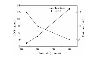
To ensure the sample is uniformly and equivalently reacted with the antibody on the NC membrane at each time, a stable flow rate is an important parameter, which can ensure precision improved. As shown in Fig. 2, the detection sensitivity of the developed reagent chips for cTnI detection at different flow rates is different. When the flow rate was 40 μL/min, the LOD value was 13 pg/mL, which is higher than a traditional immunochromatographic reagent strip (10 pg/mL). The weak result might be attributed to the very fast speed of the reaction solution passing through the NC membrane, resulting in insufficient binding of the antibody on the membrane. Moreover, at the flow rate of 20 μL/min, the detection sensitivity was able to meet the requirement, and the detection sensitivity of 5 pg/mL for cTnI determination was obtained, while the elapsed time of 10 min can also meet the demand of clinic determination. The result is consistent with the traditional immunochromatographic reagent strip. Up till now, the clinical diagnosis of cTnI abnormalities is considered to be 26 pg/mL, and the developed microfluidic assay strip can meet the needs. After the flow rate was further decreased, the detection sensitivity of the developed microfluidic system could obtain a 3 pg/mL at the flow rate of 13.3 μL/min in 15 min. Hence, except stated otherwise, the flow rate of 20 μL/min is used in the following experiments.

|
Download:
|
| Fig. 2. LOD values for cTnI detection by the developed microfluidic chip with various flow rates. | |
To evaluate the quantitative performance of the developed microfluidic chip, the cTnI antigen was diluted by 1% BSA to obtain different concentrations for determination. As shown in Fig. 3, the trend in intensity change with cTnI is illustrated, while the consistency between the calculated and detection results is shown in the inset. The quantitative results in Fig. 3 indicated good linearity between the signals of cTnI detection and the corresponding cTnI concentration (R2=0.995). Furthermore, the developed microfluidic system revealed a limit of detection (LOD) as low as 5 pg/mL for cTnI determination in the range of 5 pg/mL to 1000 pg/mL.

|
Download:
|
| Fig. 3. The performance of the developed microfluidic system cTnI determination in the range of 5–1000 pg/mL. | |
The precision of the strip was assessed via the coefficient of variation (CV). As shown in Fig. 4, the CV of cTnI determination for the developed microfluidic system is in the range of 2%−4%, while that of the traditional immunochromatographic reagent strip ranges from 8%−15%. The result showed that the microfluidic strip exhibited a satisfactory precision for cTnI detection and assessment, which could be attributed to the fact that unlike the traditional reagent strip, the microfluidic chip could fully mix samples with fluorescent microspheres, thereby avoiding the challenges such as the inconsistent reaction between the sample and microsphere at different times and batches, as well as avoiding the possible insufficient release of the microsphere on the conjugate pad. Additionally, the sample-fluorescent microspheres mixture can pass through the NC membrane in the same way through a uniform flow rate and react with the coated antibody, hence avoiding the inconsistency of pore size and surface hydrophobicity of the membrane in the traditional reagent strip.

|
Download:
|
| Fig. 4. The relationship between cancentration of sample and CVs of the development and the traditional chromatography reagent strip. | |
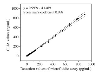
To investigate the clinical applicability and diagnostic capability of the fabricated microfluidic strip, 30 serum samples collected from hospitalized patients suffering from cTnI were analyzed by the Abbott i2000SR chemiluminescence immunoassay (CLIA) and microfluidic modified immunoassay system. To verify the as-developed microfluidic assay technique, Passing Boblok regression and Spearman's correlation coefficient were used to analyze the linear dependence between the two methods. In Fig. 5, the regression equation and Spearman's correlation coefficients of rank correlation of cTnI between CLIA and microfluidic modified strip detection values are shown. In the inset, the slopes of the regression equations (0.999) and the Spearman's correlation coefficients (0.997) are close to 1, including the 95% confidence intervals (CIs) of the corresponding parameters. The result showed that the detection values of the microfluidic modified strip displayed a good linear correlation with CLIA values for cTnI determination. Based on meeting the requirements of sensitivity, precision, testing time, and clinic detection, the microfluidic modified immunoassay system is proposed suitable for the precise cTnI diagnosis and prognosis in hospitals.

|
Download:
|
| Fig. 5. Passing Bolok regression lines between the results obtained from the Abbott i2000SR and microfluidic modified strip for cTnI determination in real serum samples. The solid lines represent the linear regression line, with the dash lines showing the range of 95% CI. | |
In this study, the developed microfluidic strip can function in the absence of the sample- and conjugate pads in the chip, while the precision of the chromatography system can be greatly improved using the same fluorescent microspheres, NC membrane and antibody compared with traditional reagent strip. More importantly, the detection time is controlled within 10 min for cTnI determination. Also, the CV is controlled within 4%, while the maximum precision of the contrast chromatographic reagent strip can reach 15%.
Moreover, for the traditional chromatographic strip, sensitivity and CVs have always been the biggest obstacles to its wider application. In the research of this chip, many materials used are the same as those applied in the traditional reagent strips, such as membrane, antibody and microspheres, the biggest change is that the process is optimized. Firstly, the microspheres and samples are mixed evenly, and then the samples pass through the NC membrane at a uniform speed. The CVs of the chip in this study is greatly improved in comparison with those of the traditional strip. The biggest improvement lies in the mixing between the samples and the microspheres, demonstrating that this is a new approach to improve the CVs of the traditional reagent strip.
Finally, the detection sensitivity can be kept at the same order of magnitudes with that of the traditional chromatographic reagent strip, while the determination correlation of the developed microfluidic chip has been maintained at a very satisfactory level with the chemiluminescence system. Hence, the microfluidic device provided in this study can greatly improve test accuracy.
Declaration of competing interestThe authors declare that they have no known competing financial interests or personal relationships that could have appeared to influence the work reported in this paper.
AcknowledgmentsThis work was financially supported by National Natural Science Foundation of China (Nos. 81902153, 61871180, 62071119 and 61971187) and Jiangsu Provincial Key Research and Development Program (No. BE 2018695).
| [1] |
K. Lewandrowski, Clin. Lab. Med. 29 (2009) 421-432. DOI:10.1016/j.cll.2009.06.015 |
| [2] |
P. Drain, E. Hyle, F. Noubary, et al., Lancet Infect. Dis. 14 (2014) 239-249. DOI:10.1016/S1473-3099(13)70250-0 |
| [3] |
L. Huang, Y. Zhang, E. Su, et al., Nanoscale Adv. 3 (2020) 1138-1143. |
| [4] |
L. Huang, D. Zhang, L. Jiao, et al., Chin. Chem. Lett. 29 (2018) 1853-1856. DOI:10.1016/j.cclet.2018.11.028 |
| [5] |
G. Yang, Z. Xiao, C. Tang, et al., Biosens. Bioelectron. 141 (2019) 111416. DOI:10.1016/j.bios.2019.111416 |
| [6] |
S. Sathish, T. Kazumi, A. Shen, View 1 (2020) e1. |
| [7] |
S. He, L. He, B. Liu, et al., Chin. Chem. Lett. 30 (2019) 1031-1034. DOI:10.1016/j.cclet.2019.03.013 |
| [8] |
Y. Wang, Z. Liu, X. Wang, et al., J. Biomed. Nanotechnol. 15 (2019) 1792-1800. DOI:10.1166/jbn.2019.2809 |
| [9] |
M. Majdinasab, M. Sheikh-Zeinoddin, S. Soleimanian-Zad, et al., J. Chromatogr. B 974 (2015) 147-154. DOI:10.1016/j.jchromb.2014.10.034 |
| [10] |
R. Huang, L. He, S. Li, et al., Nanoscale 12 (2020) 2445-2451. DOI:10.1039/C9NR08747H |
| [11] |
Z. Xiao, H. Chen, H. Chen, et al., J. Biomed. Nanotechnol. 15 (2019) 1113-1134. DOI:10.1166/jbn.2019.2782 |
| [12] |
Z. Tang, H. Karnes, Biomed. Chromatogr. 14 (2000) 442-449. DOI:10.1002/1099-0801(200010)14:6<442::AID-BMC26>3.0.CO;2-9 |
| [13] |
P. Jiang, C. Haji, X. Ye, et al., Nanosci. Nanotechnol. Lett. 11 (2019) 638-644. DOI:10.1166/nnl.2019.2933 |
| [14] |
Y. Lai, L. Wang, Y. Liu, et al., J. Biomed. Nanotechnol. 14 (2018) 44-65. DOI:10.1166/jbn.2018.2505 |
| [15] |
G. Yang, Y. Lai, Z. Xiao, et al., Chin. Chem. Lett. 29 (2018) 1857-1860. DOI:10.1016/j.cclet.2018.11.030 |
| [16] |
M. Wang, S. Meng, M. Gu, et al., J. Biomed. Nanotechnol. 15 (2019) 2100-2107. DOI:10.1166/jbn.2019.2836 |
| [17] |
X. Tian, B. Bai, Y. Chen, et al., Nanosci. Nanotechnol. Lett. 11 (2019) 279-285. DOI:10.1166/nnl.2019.2879 |
| [18] |
S. Rho, S. Kim, S. Lee, et al., Curr. Appl. Phys. 9 (2009) 534-537. DOI:10.1016/j.cap.2008.03.025 |
| [19] |
G. Yang, Y. Lai, Z. Xiao, et al., Chin. Chem. Lett. 29 (2018) 1857-1860. DOI:10.1016/j.cclet.2018.11.030 |
| [20] |
G. Yang, J. Zhang, J. Yan, et al., J. Biomed. Nanotechnol. 15 (2019) 1937-1947. DOI:10.1166/jbn.2019.2820 |
| [21] |
M. Gu, J. Liu, D. Li, et al., Nanosci. Nanotechnol. Lett. 19 (2019) 1139-1144. |
| [22] |
E. Juntunen, T. Myyrylainen, T. Salminen, et al., Anal. Biochem. 428 (2012) 31-38. DOI:10.1016/j.ab.2012.06.005 |
| [23] |
H. Tachibana, A. Kakinol, M. Kazama, et al., Parasitology 145 (2018) 1890-1895. DOI:10.1017/S0031182018000690 |
| [24] |
Q. Xie, Y. Wu, Q. Xiong, et al., Biosens. Bioelectron. 54 (2014) 262-265. DOI:10.1016/j.bios.2013.11.002 |
| [25] |
Y. Liu, T. Li, C. Ling, et al., Chin. Chem. Lett. 30 (2019) 2359-2362. DOI:10.1016/j.cclet.2019.10.033 |
| [26] |
S. Wu, F. Zhu, L. Hu, et al., Food Chem. 232 (2017) 770-776. DOI:10.1016/j.foodchem.2017.04.058 |
| [27] |
Y. Xie, L. Zhang, X. Yang, et al., Food Addit. Contam. A 34 (2017) 371-378. DOI:10.1080/19440049.2016.1277038 |
| [28] |
W. Zhang, X. He, P. Liu, et al., Anal. Lett. 49 (2016) 2165-2176. DOI:10.1080/00032719.2016.1138496 |
| [29] |
M. Mahfuz, M. Gazi, M. Hossain, et al., Toxin Rev. 39 (2020) 123-137. DOI:10.1080/15569543.2018.1514638 |
| [30] |
M. Zinggeler, P. Fosso, Y. Hao, et al., Anal. Chem. 89 (2017) 5698-5702. |
| [31] |
P. Li, Z. Zhang, Q. Zhang, et al., Electrophoresis 33 (2012) 2253-2265. DOI:10.1002/elps.201200050 |
| [32] |
B. Li, J. Ma, L. Wang, et al., Nanosci. Nanotechnol. Lett. 11 (2019) 593-599. DOI:10.1166/nnl.2019.2912 |
| [33] |
M. Liu, H. Yang, H. Jiang, et al., Nanosci. Nanotechnol. Lett. 11 (2019) 214-221. DOI:10.1166/nnl.2019.2878 |
| [34] |
C. Berli, P. Kler, Microfluid. Nanofluid. 20 (2016) 1-9. DOI:10.1007/s10404-015-1676-z |
| [35] |
J. Garcia-Cordero, D. Kurzbuch, F. Benito-Lopez, et al., Lab Chip 10 (2010) 2680-2687. DOI:10.1039/c004980h |
| [36] |
D. Zhou, J. Luo, T. Sun, et al., Nanosci. Nanotechnol. Lett. 11 (2019) 136-142. DOI:10.1166/nnl.2019.2861 |
| [37] |
T. Wang, M. Zhang, D. Dreher, et al., Lab Chip 13 (2013) 4190-4197. DOI:10.1039/c3lc50783a |
| [38] |
J. Kong, L. Jiang, X. Su, et al., Lab Chip 9 (2009) 1541-1547. DOI:10.1039/b818430e |
| [39] |
M. Abdelgawad, M.W. Watson, A. Wheeler, Lab Chip 9 (2009) 1046-1051. DOI:10.1039/b820682a |
| [40] |
C. Zheng, J. Wang, Y. Pang, et al., Lab Chip 12 (2012) 2487-2490. DOI:10.1039/c2lc40145b |
| [41] |
B. Hu, J. Li, L. Mou, et al., Lab Chip 17 (2017) 2225-2234. DOI:10.1039/C7LC00249A |
| [42] |
Y. Lai, Y. Deng, G. Yang, et al., J. Biomed. Nanotechnol. 14 (2018) 1688-1694. DOI:10.1166/jbn.2018.2617 |
| [43] |
H. Shi, W. Xiang, C. Liu, et al., Nanosci. Nanotechnol. Lett. 10 (2018) 1707-1712. DOI:10.1166/nnl.2018.2835 |