 2021, Vol. 32


b Jiangxi Key Laboratory for Microscale Interdisciplinary Study, Institute for Advanced Study, Nanchang University, Nanchang 330031, China
GSH is the most abundant non-protein thiol (1‒10 mmol/L) in the cell, and is also a necessary endogenous antioxidant [1]. It maintains intracellular redox activity, xenobiotics metabolism, intracellular signal transduction and gene regulation [2]. The levels of GSH are closely related to many diseases, including cancer, Alzheimer's disease and other diseases. It was reported that the content of GSH in tumor tissue is at least four times higher than that of normal cells [3, 4]. Therefore, GSH is often used as an important marker for disease diagnosis, especially cancer. Thus, quick and accurate detection of the level of GSH in biosystem is highly demanded [5-9].
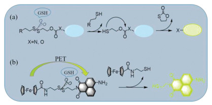
At present, fluorescent probes for detection of GSH have attracted considerable interest for the simplicity of operation, low cost, and high sensitivity of fluorescence technique [10, 11]. Thus, various fluorescent probes with different of reaction mechanisms, including Michael addition [12, 13], cyclization of aldehydes [14, 15], cleavage of disulfide bonds [16, 17], metal coordination and nucleophilic substitution [18-20], have been developed for sensing of GSH and related biothiols. Among them, the disulfide-based conjugates have attracted particular attention because these systems show some advantages such as easy synthesis, good biocompatibility and stability. The disulfide-bond has been found in proteins, oxidized glutathione, and also in natural products [21, 22]. The disulfide-based conjugates have been widely used in chemosensors, drug delivery systems, and nanomaterial carriers [23-29]. For sensing application, the conventional strategy is based on the GSH triggered cascade reaction mechanism (Scheme 1, method a). In this strategy, the disulfide bond is covalently linked to a fluorophore by a carbamate (X = N) or a carbonate (X=O) linkage. After adding GSH, the disulfide bond is first cleaved, followed by an intramolecular cyclization with the carbonyl, and releasing the linked fluorophore as a free amine or phenol. This strategy is useful for ICT-based fluorophore, such as naphthalimide, coumarin [30], BODIPY [31], and Rhodol [32], because the displacement from electronic withdrawing group carbamate/carbonate into the electronic donor NH2/OH will change the emission intensity or wavelength. Interestingly, systems with the more direct one-step disulfide cleavage mechanism are still rare for GSH recognition (Scheme 1, method b), even though this method has been widely applied in drug delivery systems and nanomaterial carriers field. Actually, the prominent Ellman's reagent is based on the disulfide bond cleavage for chromogenic sensing of thiol-containing amide acid and protein [33]. Thus, we envision if a fluorescent quencher and an organic fluorophore are linked through a disulfide bond, a useful fluorescence turn-on sensor will be constructed based on the PET mechanism. Compared with method a, this strategy may show some advantages, such as easy synthesis, high efficiency and more choices of fluorophores.

|
Download:
|
| Scheme 1. The two design mechanisms for detection of GSH. | |
In this context, herein we report a new probe Naph-SS-Fc for fluorescent sensing of GSH. As shown in Scheme 1, this probe consists of a ferrocene unit, a naphthalimide fluorophore and a disulfide bond linker. The free probe shows a weak fluorescence due to the PET (photoinduced electron transfer) quenching effect from ferrocene to naphthalimide fluorophore [34]. After reacting with GSH, the disulfide bond is cleaved, and the PET process is blocked with releasing the fluorescent active Naph-SH species. The new probe has been used for recognition of GSH and other biological thiols under physiological conditions, and also successfully applied for distinguishing cancer cells from normal cells using the confocal laser scanning microscopy cell imaging techniques.
The design route to Naph-SS-Fc is shown in Scheme S1 (Supporting information). Using 4-bromo-1, 8-naphthalene anhydride 1 as the starting material, the 4-amino-1, 8-naphthalene anhydride 3 was first synthesized, which was coupled with mono-t-Boc-cystamine 4 to give Naph-SS-Boc. Naph-SS-Boc was then subjected to deprotection under TFA solution to get Naph-SS-NH2. Finally, coupling Naph-SS-NH2 with ferrocene carboxylic acid provided Naph-SS-Fc in 40% yield.
To confirm that the ferrocene unit can quench the fluorescence of naphthalimide, the emission spectra of Naph-SS-Fc and its control compound Naph-SS-Boc were investigated in DMSO: PBS buffer solution (2:8, v/v, 10 mmol/L, pH 7.4). Both Naph-SS-Boc and Naph-SS-Fc display a similar absorption at 436 nm (Fig. S1 in Supporting information). Upon excitation at 430 nm, Naph-SS-Boc gives a strong green emission centered at 541 nm, while Naph-SS-Fc shows a weak emission, which can be easily detected by the naked eyes (Fig. 1a). The fluorescence quantum yield of Naph-SS-Fc is calculated to be 0.036, while that value for Naph-SS-Boc is 0.245. This result reveals that the efficient PET quenching process happens from the ferrocene unit to the naphthalimide fluorophore in Naph-SS-Fc [35, 36].

|
Download:
|
| Fig. 1. (a) Fluorescence spectra of Naph-SS-Fc and Naph-SS-Boc in PBS (10 mmol/L, DMSO: PBS=2:8, v/v, pH 7.4). Inset: Photographs of Naph-SS-Fc (1) and Naph-SS-Boc (2) under irradiation with 365 nm lamp. (b) The calculated HOMO/LUMO levels of FcCONH-Et and Naph-Et. | |
The PET quenching mechanism can be rationed by studying the LUMO and HOMO levels of the Naph and Fc units in Naph-SS-Fc. Here we selected Naph-Et and FcCONH-Et as the electronic acceptor and donor parts to estimate the orbital levels (Fig. 1b), which was calculated from the oxidation potential and UV absorption data (Fig. S1). As shown in Fig. 1b, the LUMO energy of FcCONH-Et is about ‒1.15 eV, which is between the HOMO and LUMO energy of Naph-Et. Therefore, facile electrons transfer from the LUMO orbital of FcCONH-Et to LUMO of Naph-Et, so called the PET process, causing fluorescence quenching [37, 38].
The detailed emission titrations for Naph-SS-Fc with GSH were then investigated. Fig. 2a shows the emission spectra changes of Naph-SS-Fc before and after addition of various amounts of GSH in PBS buffer solution. Upon the addition of various amount of GSH and incubated for 40min, the emission intensity of Naph-SS-Fc at 541 nm gradually increases, and is saturated by adding 50 equiv. of GSH. The fluorescent color of the solution also changes from dark emission to a strong yellow-green emission. In addition, the fluorescence intensity of Naph-SS-Fc at 541 nm has a good linear relationship with the concentration of GSH in a wide range of 0‒300 μmol/L. The detection limit of Naph-SS-Fc for GSH is calculated to be 134 nmol/L, based on the 3δ/k equation (Fig. S2 in Supporting information).

|
Download:
|
| Fig. 2. The GSH concentration-dependent fluorescence spectra (a) and (b)fluorescence intensity at 541 nm of Naph-SS-Fc (10 μmol/L) in PBS (10 mmol/L, DMSO: PBS=2:8, v/v, pH 7.4). Inset: Photographs of Naph-SS-Fc before and after addition of GSH under irradiation with 365 nm lamp. Time-dependent fluorescence spectra (c) and (d) fluorescence intensity at 541 nm of Naph-SS-Fc (10 μmol/L) upon addition of 50 equiv. of GSH in PBS (10 mmol/L, DMSO: PBS=2:8, v/v, pH 7.4). | |
The response time of Naph-SS-Fc toward GSH was explored by adding 50 equiv. of GSH into Naph-SS-Fc solution (Fig. 2). It was observed that the fluorescence intensity at 541 nm enhanced steadily, and reached the maximum at about 45min. Thus, Naph-SS-Fc can quickly detect GSH in a dose-dependent manner in aqueous solution, and has the potential application for detection of GSH level in bio-system.
The above fluorescence turn-on response of Naph-SS-Fc toward GSH is clearly ascribed to the disulfide bond cleavage of Naph-SS-Fc, which lead to release of the fluorescent Naph-SH species, as shown in Scheme 1. This assumption mechanism is further confirmed by the mass spectrum. Naph-SS-Fc was treated with GSH and incubated for 45minutes at 37 ℃, and then subjected to mass spectrometry analysis. A strong peak ofm/z 237.0686 corresponding to Naph-SH (Fig. S3 in Supporting information) was observed, proving that the disulfide bond of Naph-SS-Fc was successfully cleaved by GSH to release Naph-SH, resulting in an enhaned fluorescence.
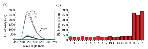
To exclude the interference of other related biological species, the fluorescent response of Naph-SS-Fc toward other amino acids was investigated (Fig. 3), including Ala, Asp, Arg, Leu, His, Lys, Glu, Gly, Met, Phe, Ser, Thr, Tyr, Trp, Val, Cys, and Hcy. Naph-SS-Fc shows barely fluorescence changes upon addition of Ala, Asp, Arg, Leu, His, Lys, Glu, Gly, Met, Phe, Ser, Thr, Tyr, Trp, Val. In contrast, a significant fluorescent enhancement was observed when addition of Hcy or Cys. The emission titration experiment revealed that the fluorescent response of Naph-SS-Fc toward Hcy or Cys behaves in a concentration-dependent manner, with the detection limit values of 113 nmol/L and 247 nmol/L, respectively (Figs. S4 and S5 in Supporting information). This similar fluorescent response of Naph-SS-Fc toward GSH, Hcy and Cys is due to the bearing of the same sulfhydryl group in their structures, which can effectively cleave the disulfide bond of Naph-SS-Fc. GSH is the most abundant species with the concentration of 1‒15 mmol/L in cells, especially for the cancer cells, the low content of Cys and Hcy is neglected.

|
Download:
|
| Fig. 3. (a) The fluorescence spectra of Naph-Fc (10 μmol/L) after treatment with 10 equiv. of various amino acids. (b) The fluorescence intensity of Naph-Fc (10 μmol/L) at 541 nm in the absence and presence of 10 equiv. of various amino acids (0-free, 1-Ala, 2-Asp, 3-Arg, 4-His, 5-Leu, 6-Lys, 7-Gly, 8-Glu, 9-Met, 10-Phe, 11-Ser, 12-Thr, 13-Tyr, 14-Trp, 15-Val, 16-GSH, 17-Cys, 18-Hcy) in PBS buffer solution. | |
Then, the GSH sensing ability of Naph-SS-Fc at different pH values was also investigated (Fig. S6 in Supporting information). The result showed that Naph-SS-Fc exhibits a desirable sensing performance within a pH range from 6 to 11, indicating that it can detect intracellular GSH in physiological pH condition. In addition, the competitive experiment of Naph-SS-Fc toward GSH in the presence of other miscellaneous amino acids was also carried out (Fig. S7 in Supporting information). Naph-SS-Fc exhibits a good anti-interference performance from non-thiol amino acids in PBS buffer (10 mmol/L, DMSO: PBS=2:8, v/v, pH 7.4) solution.
Owing to the high sensitivity of probe Naph-SS-Fc toward GSH, we envision that it may be used for detection of GSH in the cultured cells. The standard MTT assay reveals that more than 80% cells are viable after 24h of staining with the probe Naph-SS-Fc in the concentration 0–25 μmol/L (Fig. S8 in Supporting information), indicating that the probe Naph-SS-Fc gives a low cytotoxicity.
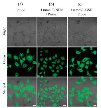
Fig. 4 shows the confocal laser scanning microscopy (CLSM) fluorescence images of living HepG2 cells incubated with Naph-SS-Fc. To control intracellular GSH concentration, three sets of cells were selected for carrying these experiments: cells treated with only probe Naph-SS-Fc (group a), cells pretreated withN-ethylmaleimide (NME) and then Naph-SS-Fc (group b), and cells pretreated with GSH and then added Naph-SS-Fc. NME can scavenge biothiols and then decrease its concentration in living cells. From Fig. 4, we can clearly observe that cells incubated with Naph-SS-Fc show a strong emission, which can be attributed to the effective disulfide bond cleavage reaction by the intracellular GSH. The emission reduces by pretreated with NME, and increases via the addition of GSH (group c). In addition, SMMC cells were used to evaluate the sensitivity of Naph-SS-Fc to endogenous GSH. As shown in Fig. S9 (Supporting information), the cells of group c treated with GSH-ester (a GSH-precursor) have stronger fluorescence than group b, which verifies the sensitivity of Naph-SS-Fc to internal GSH. The result reveals that Naph-SS-Fc can detect GSH level in living cells.

|
Download:
|
| Fig. 4. Fluorescence imaging of endogenous biothiols with Naph-SS-Fc. (a) HepG2 cells were incubated with Naph-SS-Fc only for 3h. (b) Pre-incubate HepG2 cells with NEM (1 mmol/L) for 30min, and then incubate with Naph-SS-Fc (10 μmol/L) for another 3h. (c) HepG2 cells were pretreated with 1 mmol/L GSH for 30min, and then incubated with Naph-SS-Fc. λex: 405 nm; λem: 425-575 nm. Scale bar: 20 μm. | |
Regarding the higher concentration of GSH in cancer cells than the normal cells, we thus envision the present probe Naph-SS-Fc has potential application for discriminating cancer cells from normal cells. Here two types of normal cells (HUVEC, LO2) and three types of cancer cells (SMMC, CT-26 and HepG2) were selected and incubated with Naph-SS-Fc for 30min, and then imaged the endogenous GSH levels were imagedvia the CLMS. As shown in Fig. 5 and Fig. S10 (Supporting information), the fluorescent intensity of the cancer cells is stronger than that of the normal cell lines. For example, the relative fluorescence intensity of CT-26 cancer cells is about 3.0 times of HUVEC normal cells. The above result shows that Naph-SS-Fc can differentiate cancer cells from normal cells for biological applications.

|
Download:
|
| Fig. 5. The CLSM imaging of LO2 cells (a), HUVEC cells (b), CT-26 cells (c), SMMC cells (d) and HepG2 cells (e) after incubating with probe Naph-SS-Fc (10 μmol/L) for 3h. λex: 405 nm; λem: 425‒575 nm. Scale bar: 20 μm. | |
In conclusion, we have successfully designed and synthesized a PET-based fluorescent probe Naph-SS-Fc for detection of GSH in vitro and in vivo. This probe consists of a ferrocene unit, a naphthalimide fluorophore and a disulfide bond linker. Owing to the effective quenching from ferrocene, probe Naph-SS-Fc shows very weak emission. After reacting with GSH, it exhibits a significant fluorescent enhancement for cleavage of the disulfide bond and releasing the fluorescent Naph-SH species. Probe Naph-SS-Fc shows good biocompatibility and cytomembrane penetration, and can detect intracellular GSH level. Further more, it has successfully been used for distinguishing normal cells from cancer cells.
Declaration of competing interestThe authors report no declarations of interest.
AcknowledgmentsThis work was supported by the National Nature Science Foundation of China (Nos. 21762028 and 21462027), and the Nature Science Foundation of Jiangxi Province of China (No. 20172BCB22007), which are greatly acknowledged by the authors.
Appendix A. Supplementary dataSupplementary material related to this article can be found, in the online version, at doi:https://doi.org/10.1016/j.cclet.2020.10.047.
| [1] |
C. Hwang, A.J. Sinskey, H.F. Lodish, Science 257 (1992) 1496-1502. DOI:10.1126/science.1523409 |
| [2] |
R.A. Cairns, I.S. Harris, T.W. Mak, Nat. Rev. Cancer 11 (2011) 85-95. DOI:10.1038/nrc2981 |
| [3] |
M. Gamcsik, M. Kasibhatla, S. Teeter, O.M. Colvin, Biomarkers 17 (2012) 671-691. DOI:10.3109/1354750X.2012.715672 |
| [4] |
N. Traverso, R. Ricciarelli, M. Nitti, et al., Oxid. Med. Cell. Longevity 2013 (2013) 972913. |
| [5] |
S. Lee, J. Li, X. Zhou, J. Yin, J. Yoon, Coord. Chem. Rev. 366 (2018) 29-68. DOI:10.1016/j.ccr.2018.03.021 |
| [6] |
V.S. Lin, W. Chen, M. Xian, C.J. Chang, Chem. Soc. Rev. 44 (2015) 4596-4618. DOI:10.1039/C4CS00298A |
| [7] |
L.Y. Niu, Y.Z. Chen, H.R. Zheng, et al., Chem. Soc. Rev 44 (2015) 6143-6160. DOI:10.1039/C5CS00152H |
| [8] |
H.S. Jung, X. Chen, J.S. Kim, J. Yoon, Chem. Soc. Rev. 42 (2013) 6019-6031. DOI:10.1039/c3cs60024f |
| [9] |
L. Yang, H. Xiong, Y. Su, et al., Chin. Chem. Lett. 30 (2019) 563-565. DOI:10.1016/j.cclet.2018.12.017 |
| [10] |
X. Li, X. Gao, W. Shi, H. Ma, Chem. Rev. 114 (2014) 590-659. DOI:10.1021/cr300508p |
| [11] |
J. Chan, S.C. Dodani, C.J. Chang, Nat. Chem. 4 (2012) 973-984. DOI:10.1038/nchem.1500 |
| [12] |
J. Liu, Y.Q. Sun, Y. Huo, et al., J. Am. Chem. Soc. 136 (2014) 574-577. DOI:10.1021/ja409578w |
| [13] |
J. Chen, X. Jiang, C. Zhang, et al., ACS Sens. 2 (2017) 1257-1261. DOI:10.1021/acssensors.7b00425 |
| [14] |
L.Y. Niu, Y.S. Guan, Y.Z. Chen, et al., J. Am. Chem. Soc. 134 (2012) 18928-18931. DOI:10.1021/ja309079f |
| [15] |
M. Li, P. Cui, K. Li, J. Feng, X. Yu, Chin. Chem. Lett. 29 (2018) 992-994. DOI:10.1016/j.cclet.2017.11.011 |
| [16] |
Y. Liu, S. Zhu, K. Gu, et al., ACS Appl. Mater. Interfaces 9 (2017) 29496-29504. DOI:10.1021/acsami.7b07091 |
| [17] |
M. Gangopadhyay, R. Mengji, A. Paul, et al., Chem. Commun. 53 (2017) 9109-9112. DOI:10.1039/C7CC03241B |
| [18] |
H. Zhang, L. Xu, W. Chen, et al., ACS Sens. 3 (2018) 2513-2517. DOI:10.1021/acssensors.8b01101 |
| [19] |
Y.F. Kang, L.Y. Niu, Q.Z. Yang, Chin. Chem. Lett. 30 (2019) 1791-1798. DOI:10.1016/j.cclet.2019.08.013 |
| [20] |
Q. Guo, Y. Zhang, Z.H. Lin, Q.Y. Cao, Y. Chen, Dyes and Pigments 172 (2020) 107872. DOI:10.1016/j.dyepig.2019.107872 |
| [21] |
M. Hara, K. Asano, I. Kawamoto, et al., J. Antibiotics 42 (1989) 1768-1774. DOI:10.7164/antibiotics.42.1768 |
| [22] |
I. Rahman, W. MacNee, Free Radical Biol. Med. 28 (2000) 1405-1420. DOI:10.1016/S0891-5849(00)00215-X |
| [23] |
M.H. Lee, Z. Yang, C.W. Lim, et al., Chem. Rev. 113 (2013) 5071-5109. DOI:10.1021/cr300358b |
| [24] |
P.T. Wong, S.K. Choi, Chem. Rev. 115 (2015) 3388-3432. DOI:10.1021/cr5004634 |
| [25] |
M.H. Lee, A. Sharma, M.J. Chang, et al., Chem. Soc. Rev. 47 (2018) 28-52. DOI:10.1039/C7CS00557A |
| [26] |
Q. Wang, J. Guan, J. Wana, Z. Li, RSC Adv. 10 (2020) 24397-24409. DOI:10.1039/d0ra04155f |
| [27] |
M. Ye, X. Wang, J. Tang, et al., Chem. Sci. 7 (2016) 4958-4965. DOI:10.1039/C6SC00970K |
| [28] |
M.H. Lee, J.Y. Kim, J.H. Han, et al., J. Am. Chem. Soc. 134 (2012) 12668-12674. DOI:10.1021/ja303998y |
| [29] |
S. Maiti, N. Park, J.H. Han, et al., J. Am. Chem. Soc. 135 (2013) 4567-4572. DOI:10.1021/ja401350x |
| [30] |
M.H. Lee, J.H. Han, P.S. Kwon, et al., J. Am. Chem. Soc. 134 (2012) 1316-1322. DOI:10.1021/ja210065g |
| [31] |
F. Wang, L. Zhou, C. Zhao, et al., Chem. Sci. 6 (2015) 2584-2589. DOI:10.1039/C5SC00216H |
| [32] |
S. Bhuniya, S. Maiti, E.J. Kim, et al., Angew. Chem. Int. Ed. 53 (2014) 4469-4474. DOI:10.1002/anie.201311133 |
| [33] |
G.L. Ellman, Arch. Biochem. Biophys. 82 (1959) 70-77. DOI:10.1016/0003-9861(59)90090-6 |
| [34] |
L. Zhou, L. Xie, C. Liu, Y. Xiao, Chin. Chem. Lett. 30 (2019) 1799-1808. DOI:10.1016/j.cclet.2019.07.051 |
| [35] |
L. Zhou, X.T. Fan, Y.D. Xu, Q.Y. Cao, New J. Chem. 39 (2015) 8087-8092. DOI:10.1039/C5NJ01995H |
| [36] |
M. Alfonso, A.E. Ferao, A. Tárraga, P. Molina, Inorg. Chem. 54 (2015) 7461-7473. DOI:10.1021/acs.inorgchem.5b01071 |
| [37] |
J.P. Claude, D.S. Williams, T.J. Meyer, J. Am. Chem. Soc. 118 (1996) 9782-9783. DOI:10.1021/ja960312v |
| [38] |
W. Sun, M. Li, J. Fan, X. Peng, Acc. Chem. Res. 52 (2019) 2818-2831. DOI:10.1021/acs.accounts.9b00340 |