 2020, Vol. 31


b State Key Laboratory of Applied Optics, Changchun Institute of Optics, Fine Mechanics and Physics, Chinese Academy of Sciences, Changchun 130012, China
Hydrogels are three-dimensional (3D) networks of physically or chemically cross-linked natural [1] or synthetic polymers [2], which are inflated with a water medium [3, 4]. Hydrogel is an important type of soft materials with interesting applications in tissue engineering [5], drug delivery [6], sensing [7], separation and purification technologies [8], to name just a few. Conventional hydrogels usually have poorly mechanical properties and limited functions, which hampers the practical applications of the hydrogels. The hydrogels with enhanced mechanical properties and functions were achieved by new type of hydrogels such as sliding hydrogels [9], double-network hydrogels [10] and nano-composite hydrogels [11]. Recently, nanocomposite hydrogels with enhanced functionalities were prepared to broaden the spectrum of applications of hydrogels [12].
Nanocomposite hydrogels are made from the mixture of nanoparticles (NPs) and polymers by the chemical or physical interactions [13, 14]. Nanocomposite hydrogels have collective properties of polymers and NPs, which makes nanocomposite hydrogels have interesting applications in the field of sensing [15], actuator [16], biomedicine [17], separation and purification technologies [18]. The nanocomposite hydrogels have been successfully prepared by polymers and functional NPs, such as metal/metal oxide NPs, polymeric NPs, silica NPs, semiconductor quantum dots and carbon-based nanomaterials (graphene oxide, carbon nanotubes, CDs) [19].
In particular, nanocomposite hydrogels from polymers and CDs attracted immense interest because of the excellent properties of the CDs [20]. CDs are a type of emerging fluorescent nanomaterials, mainly including graphene quantum dots (GQDs) [21], carbon quantum dots (CQDs) [22] and carbonized polymer dots (CPDs) [23]. Compared with other NPs, CDs have many advantages, such as bright luminescence, ease of preparation and surface-functionalization, good biocompatibility, low cost and low toxicity. Nanocomposite hydrogels from CDs and polymers are emerging materials with functional diversity due to collective properties inheriting from the CDs and polymers. They have attracted extensive attention in the past years due to their unique properties and important applications in the multidisciplinary research field such as sensing [24], drug delivery [25], bioimaging [26], supercapacitors [27] and environmental remediation [28].
Here, we highlight recent advances in the nanocomposite hydrogels based on CDs and polymers. We start with the preparation methods of nanocomposite hydrogels. Meanwhile, we discuss the interactions between CDs and polymers. Next, we outline the emerging applications of the nanocomposite hydrogels. We conclude with the discussion of challenges and research directions in the development of new types of nanocomposite hydrogels based on CDs and polymers. This review highlights the macroscopic hydrogels from CDs and polymers, and the micro or nanogels from polymers and carbon NPs is the subject of recent reviews [29].
2. Preparation of nanocomposite hydrogelsThere are mainly two types of methods to prepare the nanocomposite hydrogels from polymers and CDs depending on the interactions between the CDs and the network of polymers, namely CDs attached to the polymer networks by supramolecular interactions [27, 30] and CDs attached to the polymer networks by covalent interactions [20, 31]. In this section, we will summarize the current advances in the preparation of the nanocomposite hydrogels from polymers and CDs.
Nanocomposite hydrogels can be easily prepared by gelation of mixture of CDs and polymers triggered by supramolecular interactions. The interactions between CDs and polymer chains in hydrogels mainly include electrostatic interactions [32] and hydrogen bonding [33]. Hydrogen bonding plays an important role in the preparation of nanocomposite hydrogels [24, 25, 33-37]. For example, nanocomposite hydrogels have been made from CDs modified by polyethyleneimine (PEI) and microcrystalline cellulose. The strong hydrogen bonding between PEI-CDs and cellulose resulted in cellulose hydrogel with good fluorescence properties [24]. Besides, electrostatic interaction was used to prepare the nanocomposite hydrogels from the charged polymer and oppositely charged CDs [32, 38, 39]. For instance, nanocomposite hydrogels were prepared by positively charged chitosan with the negatively charged CDs (Fig. 1A). Because of the conjugated aromatic structures from CDs, the chitosan/CDs hydrogel films also possessed better UV-vis blocking properties in contrast to pure chitosan hydrogel films with reduction of transmittance up to 20% in the wavelength range of 300-600 nm [32].

|
Download:
|
| Fig. 1. (A) The procedure of preparation of chitosan-CDs nanocomposite hydrogel film by electrostatic interaction. Reproduced with permission [32]. Copyright 2014, Elsevier. (B) The preparative process and chemical structure of the nanocomposite hydrogel from GQDs and chemical hydrogels by immersing method. Reproduced with permission [44]. Copyright 2018, American Chemical Society. | |
Nanocomposite hydrogels based on CDs and polymers mentioned above are inherently physical hydrogels driven by the supramolecular interactions. These nanocomposite physical hydrogels mostly exhibited poorly mechanical properties, which hindered their practical applications. To enhance the mechanical properties, nanocomposite hydrogels have been prepared by incorporating CDs into chemically cross-linked hydrogel [40-43]. Absorption of the CDs into the chemical hydrogel network is a facile way to prepare the nanocomposite hydrogels [27, 44, 45]. The supramolecular interactions between the CDs and hydrogels, such as hydrogen-bonding and electrostatic interactions, allow CDs to retain in the chemical hydrogel networks. For instance, nano-composite hydrogels were prepared by immersing the dried cationic polymer networks prepared by radical polymerization of [2-(acryloyloxy)ethyl]trimethylammonium chloride (AETA) in the aqueous suspension of CDs (Fig. 1B). These nanocomposite hydrogels show stable fluorescence after five swelling-deswelling cycles because of the strong electrostatic interactions between the carboxylate groups of CDs and the ammonium groups of the polymers [44]. Nanocomposite hydrogels were also prepared by in situ polymerization of mixed solution of CDs and polymerizable monomers [40-43, 46-50] or polymers [30, 51]. Recently, an injectable nanocomposite hydrogel was prepared by photo-polymerization of the aqueous solution of N-methacryloyl chitosan and CDs. This hydrogel shows red fluorescence, good biocompati-bility and low photobleaching properties [30]. Besides, nano-composite hydrogel was prepared by in situ polymerization of acrylamide (AM) in the suspension of CDs. This nanocomposite hydrogel possesses the enhanced mechanical properties due to the dual role of CDs acting as physical cross-linker and lubricant [40].
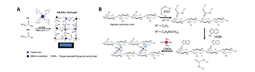
Nanocomposite hydrogels with CDs attached to the polymer networks by supramolecular interactions attracted immense interest because of the ease of their preparation and useful properties, such as fluorescence [30, 52], self-healing [35, 53] and biocompatibility [30, 53]. However, CDs tended to phase-separate from the hydrogel network, resulting in heterogeneous distribution of CDs in the final hydrogel network [48, 54]. Moreover, leaching of CDs occurred because of the relatively weak supramolecular interactions [45, 55]. Incorporating CDs in the nanocomposite hydrogels by chemically interactions may over-come the limitations above by providing homogenous distribution and non-leaching of CDs in the hydrogel network. Nanocomposite hydrogels were fabricated by polymerizable CDs modified by vinyl groups and other monomers through normal radical polymerization [20, 21, 56-58]. For example, nanocomposite hydrogel was prepared by in situ polymerization of AM, acrylic acid (AAc) and methacrylated CDs (Fig. 2A). The CDs behaved as both cross-linkers and functional nanofillers in the hydrogels which exhibited fluorescence, responsiveness to metal ions and relatively robust mechanical properties [57]. Instead of using radical polymerization, nanocomposite hydrogels were constructed by coupling amino or carboxylic groups on the surface of CDs with phosphate, carboxylic, aldehyde or amino groups on the polymer chains, respectively [31, 55, 59-62]. For example, nanocomposite hydrogels from alginate and CDs were prepared by amidation reaction between the carboxylic groups of alginate chains and the primary amine of the CDs (Fig. 2B). CDs were stable in the hydrogel without any noticeable CDs leaching out from hydrogels when the hydrogels were submerged in the aqueous solution [55].

|
Download:
|
| Fig. 2. (A) Preparative process of nanocomposite hydrogels by in situ polymerization of AM, AAc and methacrylated CDs. Reproduced with permission [57]. Copyright 2018, American Chemical Society. (B) Reaction scheme of nanocomposite hydrogels from the CDs and alginate by chemical cross-linking. Reproduced with permission [55]. Copyright 2017, Royal Society of Chemistry. | |
3. Properties of nanocomposite hydrogels
Inheriting the fluorescent properties from CDs, nanocomposite hydrogels from polymers and CDsusually have fluorescence. In contrast to the fluorescent properties of CDs solution, the nanocomposite hydrogels often have enhanced fluorescent properties in terms of fluorescent intensity and stability. For example, the nanocomposite hydrogel exhibits higher fluorescence intensity than that of the CDs solutions with the same concentration. The enhanced fluorescence intensity resulted from the CDs stabilized by the hydrogen bonding interaction between oxygen-containing groups of the CDs and the carboxyl or hydroxyl groups of nanocellulose fibrils, avoiding graphitic layer aggregation via π-π stacking interaction [33]. The fluorescence of the nano-composite hydrogel was more stable than CDs solution undergoing long term UV irradiation. This enhanced photo-stability can be attributed to the strong interactions between CDs and cellulose in the hydrogel which avoided the PEI-CDs aggregation [24]. In addition, the fluorescence of nanocomposite hydrogels shows better stability under the acidic conditions than that of the carboxylated CDs. The strong interactions between the carboxylate groups of CDs and the ammonium groups in the polymer networks will protect CDs from quenching at acidic condition [44].
Nanocomposite hydrogels also exhibit better mechanical properties than pure hydrogels because CDs behave as physical and chemical cross-linkers in the hydrogel networks. For example, nanocomposite hydrogel from polyacrylamide and CDs possesses the enhanced mechanical properties with the stretching ratio over 3700% and the fracture strength as high as 160 kPa due to the dual role of CDs acting as physical cross-linker and lubricant [40]. Besides, compared with hydrogel without CDs, the elastic modulus of the fluorescent hydrogel was increased from 19.4 kPa to 63.7 kPa due to the introduction of CDs as effective chemical cross-linkers [57].
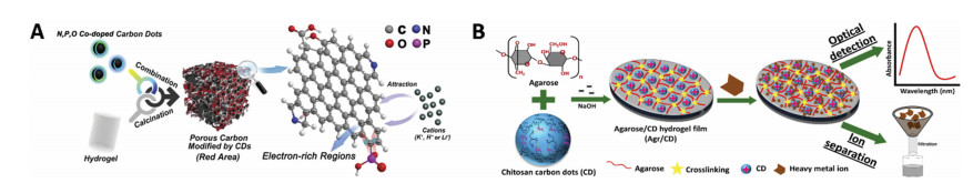
4. Emerging applications of nanocomposite hydrogelsBecause nanocomposite hydrogels made from CDs and polymers have collective properties of these two components, including abundant surface groups and large specific surface area, they have important applications in environmental remediation and energy storage. An exemplary application of nanocomposite hydrogels from CDs and polymers is separation and removal of metal ions from solution of metal ions prepared in the laboratory and natural water samples [28, 31, 38, 39]. For example, nano-composite hydrogels from CDs and agarose were used to remove efficiently and selectively the lead ions in polluted water. After adding nanocomposite hydrogels to polluted water, the Pb2+ concentration decreased from 100 nmol/L to 2.39 nmol/L, suggesting their ability of Pb2+ removal in contaminated water [28]. Recently, the porous carbon materials derived from nanocomposite hydrogels based on CDs and polyacrylamide have been used as negative electrode materials for hybrid supercapacitors with good performances (Fig. 3A). The carbon materials from nanocomposite hydrogels have a specific capacitance for 468 F/g in alkaline electrolytes [27].

|
Download:
|
| Fig. 3. (A) The porous carbon materials derived from nanocomposite hydrogels based on CDs and polyacrylamide for negative electrode materials in hybrid supercapacitors. Reproduced with permission [27]. Copyright 2018, Wiley. (B) Nanocomposite hydrogel film from agarose and CDs for optical sensor of metal ions. Reproduced with permission [38]. Copyright 2015, American Chemical Society. | |
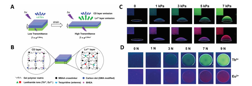
Nanocomposite hydrogels based on CDs and polymers have responsively optical properties inheriting from CDs, so they serve as sensors that detect single heavy metal ion [24, 51, 59, 63], organic pollutants [33, 64], laccase [54], nitrite [65], hypochlorite [66] and reactive oxygen species (ROS) [41], to name just a few. Several recent studies show that nanocomposite hydrogels are successfully used as sensors to detect heavy metal ions [24, 38, 51, 59, 63]. For example, nanocomposite hydrogels from CDs and agarose acted as sensing platform for detecting heavy metal ions and the detection limit was found to be 1 pmol/L for Cr6+, 0.5 μmol/L for Fe3+, Pb2+ and Mn2+, and 0.5 μmol/L for Cu2+ (Fig. 3B) [38]. Notably, nanocomposite hydrogel from CDs and polyacrylamide was prepared as the top layer of a double-layer hydrogel which shows mechanochromic properties (Figs. 4A and B). These nanocomposite double-layer hydrogels have been used as bulging pressure and contact force sensors with the sensing range from 1 kPa to 7 kPa and 0 N to 9 N, respectively (Figs. 4C and D) [67]. Except for detecting single stimulus, nanocomposite hydrogels which had ability of sensing multiple stimuli were fabricated from lanthanide (Ln) ions, CDs and polyacrylamide-co-polyacrylic acid. This hydrogel has the capacity to optically respond to chemical and physical stimuli, including pH, acetone vapors, transition-metal ions such as Zn2+, Ni2+, Mn2+, Cu2+ and Fe3+, and temperature [57].

|
Download:
|
| Fig. 4. (A) The schematics of mechanochromic hydrogels for bulging pressure and contact force sensors. (B) The chemical components and structure of the double-layer mechanochromic hydrogel. (C) The color change of the double-layer hydrogel triggered by different bulging pressures. Insets are the color of the central part of the hydrogel. (D) The pictures of the double-layer mechanochromic hydrogel under different contact forces. Reproduced with permission [67]. Copyright 2019, Wiley. | |
Nanocomposite hydrogels from CDs and polymers attracted immense interest in biomedical field, because they are biocompatible, biodegradable, low-cost, low-toxicity and sustainable. An exemplary application of nanocomposite hydrogels in biomedicine is explored as drug delivery. Some important drugs such as doxorubicin [25, 61], 5-fluorouracil [26] and vancomycin [68] were introduced into nanocomposite hydrogel networks by the supramolecular interactions, which led to the formation of anticancer and antibacterial drug delivery systems. The excellent luminescence properties of CDs might be beneficial to track the release and distribution of drugs [61]. In addition, nanocomposite hydrogels have been used as injectable hydrogels [30, 37, 69]. For example, the nanocomposite hydrogels were prepared from red-emission CDs and chitosan. The CDs are introduced as fluorescent indicator for the monitoring of degradation of hydrogels because they have advantages such as low photobleaching, red fluorescence, good homogeneity and good biocompatibility. This red fluorescent nanocomposite hydrogel was injected in mice for 288 h to achieve tracking the degradation of injectable hydrogel visually (Fig. 5). Based on the degradation profile of hydrogel in vitro, the degradation behavior of hydrogel via subcutaneous injection in vivo was depicted quantitatively by real-time and non-invasive fluorescence tracking [30].

|
Download:
|
| Fig. 5. Schematic illustration of nanocomposite hydrogels for visual monitoring of in vitro/in vivo degradation of injectable hydrogel by real-time and non-invasive fluorescence tracking. Reproduced with permission [30]. Copyright 2017, Elsevier. | |
5. Conclusions
Nanocomposite hydrogels from CDs and polymers have appealed to many researchers from different research fields. In this review, we have summarized their preparation methods into two categories based on the interaction types between CDs and polymers networks. Based on their specific properties, we have summarized the emerging applications of the nanocomposite hydrogels from CDs and polymers in the field of environmental treatment, energy storage and biomedicine. We expect this review will inspire the researchers to discover new hydrogels from CDs and polymers, and to explore new applications to solve the vital problems in chemistry, energy, environment and health.
Although the nanocomposite hydrogels from CDs and polymer showed interesting properties, such as fluorescence and good mechanical properties, these properties usually lack control over the homogeneity and robustness. Future work should focus on the prediction of properties of nanocomposite hydrogels by rational design of CDs and polymers, optimizing the condition of gelation, with the aid of computer simulation as well. The smart, or "living" nanocomposite hydrogels should be explored by designing suitable CDs and polymers and controlling the interaction between them, which will expand the spectrum of applications of the nanocomposite hydrogels.
Current researches are mostly limited in incorporating CDs to improve the mechanical properties of polymer hydrogels. The luminescent regulation of CDs with the aid of hydrogel networks lacks studies, although it is important in the field of luminescent nanomaterials and soft matters. In future studies, the fluorescent regulation of nanocomposite hydrogels may be realized by control over the aggregation of the CDs in the hydrogels network, in terms of tuning various interactions between CDs and polymers, and controlling the reactive groups, architectures, and condense structures of polymers. Furthermore, it will be possible to prepare multicolor fluorescent and even phosphorescent nanocomposite hydrogels which are advantageous for nanocomposite hydrogels to be used in bio-imaging, multi-responsive sensing and tissue engineering.
Although nanocomposite hydrogels from CDs and polymers have shown great potentials in biomedical applications, it is desired to fabricate micro-tissues or -organs in these nano-composite hydrogels by using microfabrication techniques, such as microfluidics and 3D printing. In this case, using cell-laden precursors of nanocomposite hydrogels, an organ-on-a-chip platform can be constructed by microfabrication methods above. Organ-on-a-chip platforms recapitulate the complex architectures and functions of human organs, so they have important implications in the disease models and drug screening.
AcknowledgmentsThis work was financially supported by the National Natural Science Foundation of China (NSFC, Nos. 21774041, 51433003), the National Key Research and Development Program of China (No. 2016YFB0401701), the Fundamental Research Funds for the Central Universities, Program for JLU Science and Technology Innovative Research Team (JLUSTIRT, No. 2017TD-06) and the opening funds of State Key Laboratory of Applied Optics, Changchun Institute of Optics, Fine Mechanics and Physics, Chinese Academy of Sciences.
| [1] |
Y. Li, J. Rodrigues, H. Tomás, Chem. Soc. Rev. 41 (2012) 2193-2221. DOI:10.1039/C1CS15203C |
| [2] |
X. Dai, Y. Zhang, L. Gao, et al., Adv. Mater. 27 (2015) 3566-3571. DOI:10.1002/adma.201500534 |
| [3] |
S.R. Caliari, J.A. Burdick, Nat. Methods 13 (2016) 405-414. DOI:10.1038/nmeth.3839 |
| [4] |
J. Thiele, Y. Ma, S.M.C. Bruekers, S. Ma, W.T.S. Huck, Adv. Mater. 26 (2014) 125-148. DOI:10.1002/adma.201302958 |
| [5] |
H. Wang, S.C. Heilshorn, Adv. Mater. 27 (2015) 3717-3736. DOI:10.1002/adma.201501558 |
| [6] |
J. Li, D.J. Mooney, Nat. Rev. Mater. 1 (2016) 16071. DOI:10.1038/natrevmats.2016.71 |
| [7] |
M. Qin, M. Sun, R. Bai, et al., Adv. Mater. 30 (2018) 1800468. DOI:10.1002/adma.201800468 |
| [8] |
Z. Xue, S. Wang, L. Lin, et al., Adv. Mater. 23 (2011) 4270-4273. DOI:10.1002/adma.201102616 |
| [9] |
Imran A. Bin, K. Esaki, H. Gotoh, et al., Nat. Commun. 5 (2014) 5124. DOI:10.1038/ncomms6124 |
| [10] |
J.Y. Sun, X. Zhao, W.R.K. Illeperuma, et al., Nature 489 (2012) 133-136. DOI:10.1038/nature11409 |
| [11] |
S. Merino, C. Martín, K. Kostarelos, M. Prato, E. Vázquez, ACS Nano 9 (2015) 4686-4697. DOI:10.1021/acsnano.5b01433 |
| [12] |
T. Chen, K. Hou, Q. Ren, et al., Macromol. Rapid Commun. 39 (2018) 1800337. DOI:10.1002/marc.201800337 |
| [13] |
P. Schexnailder, G. Schmidt, Colloid Polym. Sci. 287 (2009) 1-11. DOI:10.1007/s00396-008-1949-0 |
| [14] |
A.K. Gaharwar, N.A. Peppas, A. Khademhosseini, Biotechnol. Bioeng. 111 (2014) 441-453. DOI:10.1002/bit.25160 |
| [15] |
Z. Lei, Q. Wang, S. Sun, W. Zhu, P. Wu, Adv. Mater. 29 (2017) 1700321. DOI:10.1002/adma.201700321 |
| [16] |
X. Zhang, C.L. Pint, M.H. Lee, et al., Nano Lett. 11 (2011) 3239-3244. DOI:10.1021/nl201503e |
| [17] |
Y. Wu, H. Wang, F. Gao, et al., Adv. Funct. Mater. 28 (2018) 1801000. DOI:10.1002/adfm.201801000 |
| [18] |
V. Van Tran, D. Park, Y.C. Lee, Environ. Sci. Pollut. Res. 25 (2018) 24569-24599. DOI:10.1007/s11356-018-2605-y |
| [19] |
P. Thoniyot, M.J. Tan, A.A. Karim, D.J. Young, X.J. Loh, Adv. Sci. 2 (2015) 1400010. DOI:10.1002/advs.201400010 |
| [20] |
P. Zhang, W. Li, X. Zhai, et al., Chem. Commun. 48 (2012) 10431-10433. DOI:10.1039/c2cc35966a |
| [21] |
S. Zhu, S. Tang, J. Zhang, B. Yang, Chem. Commun. 48 (2012) 4527-4539. DOI:10.1039/c2cc31201h |
| [22] |
H. Li, X. He, Z. Kang, et al., Angew. Chem. Int. Ed. 49 (2010) 4430-4434. DOI:10.1002/anie.200906154 |
| [23] |
C. Xia, S. Tao, S. Zhu, et al., Chem. -Eur. J. 24 (2018) 11303-11308. DOI:10.1002/chem.201802712 |
| [24] |
C. Cheng, M. Xing, Q. Wu, J. Alloys Compd. 790 (2019) 221-227. DOI:10.1016/j.jallcom.2019.03.053 |
| [25] |
S. Javanbakht, H. Namazi, Mater. Sci. Eng. C. 87 (2018) 50-59. DOI:10.1016/j.msec.2018.02.010 |
| [26] |
A. Sachdev, I. Matai, P. Gopinath, Colloid. Surf. B:Biointerfaces 141 (2016) 242-252. DOI:10.1016/j.colsurfb.2016.01.043 |
| [27] |
J.S. Wei, C. Ding, P. Zhang, et al., Adv. Mater. 31 (2019) 1806197. |
| [28] |
J. Xu, X. Jie, F. Xie, et al., Nano Res. 11 (2018) 3648-3657. DOI:10.1007/s12274-017-1931-6 |
| [29] |
H. Wang, Q. Chen, S. Zhou, Chem. Soc. Rev. 47 (2018) 4198-4232. DOI:10.1039/C7CS00399D |
| [30] |
L. Wang, B. Li, F. Xu, et al., Biomaterials 145 (2017) 192-206. DOI:10.1016/j.biomaterials.2017.08.039 |
| [31] |
M. Alizadehgiashi, N. Khuu, A. Khabibullin, et al., ACS Nano 12 (2018) 8160-8168. DOI:10.1021/acsnano.8b03202 |
| [32] |
A. Konwar, N. Gogoi, G. Majumdar, D. Chowdhury, Carbohydr. Polym. 115 (2015) 238-245. DOI:10.1016/j.carbpol.2014.08.021 |
| [33] |
C. Ruiz-Palomero, M.L. Soriano, S. Benítez-Martínez, M. Valcárcel, Sensor. Actuator. B -Chem. 245 (2017) 946-953. DOI:10.1016/j.snb.2017.02.006 |
| [34] |
A. Khabibullin, M. Alizadehgiashi, N. Khuu, et al., Langmuir 33 (2017) 12344-12350. DOI:10.1021/acs.langmuir.7b02906 |
| [35] |
Z. Zhang, T. Li, B. Chen, S. Wang, Z. Guo, J. Mater. Sci. 52 (2017) 10614-10623. DOI:10.1007/s10853-017-1222-3 |
| [36] |
M. Hu, X. Gu, Y. Hu, Y. Deng, C. Wang, Macromol. Mater. Eng. 301 (2016) 1352-1362. DOI:10.1002/mame.201600248 |
| [37] |
H. Lu, L. Lv, J. Ma, et al., J. Mech. Behav. Biomed. Mater. 88 (2018) 261-269. DOI:10.1016/j.jmbbm.2018.08.024 |
| [38] |
N. Gogoi, M. Barooah, G. Majumdar, D. Chowdhury, ACS Appl. Mater. Interfaces 7 (2015) 3058-3067. DOI:10.1021/am506558d |
| [39] |
U. Baruah, A. Konwar, D. Chowdhury, Nanoscale 8 (2016) 8542-8546. DOI:10.1039/C6NR01129B |
| [40] |
M. Hu, X. Gu, Y. Hu, et al., Macromolecules 49 (2016) 3174-3183. DOI:10.1021/acs.macromol.5b02352 |
| [41] |
Y. Zhang, M. Wu, J. Chen, et al., ChemistrySelect 3 (2018) 5756-5765. DOI:10.1002/slct.201800497 |
| [42] |
Y. Hu, Y. Li, D. Wang, et al., Eur. Polym. J. 95 (2017) 482-490. DOI:10.1016/j.eurpolymj.2017.08.044 |
| [43] |
M. Hu, Y. Yang, X. Gu, et al., Macromol. Mater. Eng. 300 (2015) 1043-1048. DOI:10.1002/mame.201500141 |
| [44] |
A. Martín-Pacheco, A.E. Del Río Castillo, C. Martín, et al., ACS Appl. Mater. Interfaces 10 (2018) 18192-18201. DOI:10.1021/acsami.8b02162 |
| [45] |
C. Martín, A. Martín-Pacheco, A. Naranjo, et al., Nanoscale 11 (2019) 4822-4830. DOI:10.1039/C8NR09728C |
| [46] |
L. Zhou, F. Wu, J. Yu, et al., Carbon 118 (2017) 50-57. DOI:10.1016/j.carbon.2017.03.023 |
| [47] |
Y.Q. Wang, Y.N. Xue, S.R. Li, et al., J. Polym. Res. 24 (2017) 224. DOI:10.1007/s10965-017-1389-y |
| [48] |
Y.Y. Zhang, X.W. He, W.Y. Li, RSC Adv. 5 (2015) 71030-71034. DOI:10.1039/C5RA11217F |
| [49] |
S. Havanur, P.E. JagadeeshBabu, Int. J. Polym. Anal. Charact. 23 (2018) 606-620. DOI:10.1080/1023666X.2018.1484207 |
| [50] |
H. Shao, C.F. Wang, S. Chen, C. Xu, J. Polym. Sci. Part A:Polym. Chem. 52 (2014) 912-920. DOI:10.1002/pola.27086 |
| [51] |
J. Guo, M. Zhou, C. Yang, Sci. Rep. 7 (2017) 7902. DOI:10.1038/s41598-017-08353-8 |
| [52] |
P. Li, L. Huang, Y. Lin, et al., Nanotechnology 25 (2014) 055603. DOI:10.1088/0957-4484/25/5/055603 |
| [53] |
J. Chen, S. Li, Y. Zhang, et al., Adv. Healthc. Mater. 6 (2017) 1700746. DOI:10.1002/adhm.201700746 |
| [54] |
C. Ruiz-Palomero, S. Benítez-Martínez, M.L. Soriano, M. Valcárcel, Anal. Chim. Acta 974 (2017) 93-99. DOI:10.1016/j.aca.2017.04.018 |
| [55] |
R. Wijayapala, S.M. Hashemnejad, S. Kundu, RSC Adv. 7 (2017) 50389-50395. DOI:10.1039/C7RA09805G |
| [56] |
M.-J. Cho, S.-Y. Park, Sensor. Actuator. B -Chem. 282 (2019) 719-729. DOI:10.1016/j.snb.2018.11.055 |
| [57] |
Q. Zhu, L. Zhang, K. Van Vliet, A. Miserez, N. Holten-Andersen, ACS Appl. Mater. Interfaces 10 (2018) 10409-10418. DOI:10.1021/acsami.7b17016 |
| [58] |
J. Wang, X. Ma, L. Wei, et al., Colloid Polym. Sci. 296 (2018) 745-752. DOI:10.1007/s00396-018-4287-x |
| [59] |
S. Yu, K. Chen, F. Wang, Y. Zhu, X. Zhang, Luminescence 32 (2017) 970-977. DOI:10.1002/bio.3279 |
| [60] |
C. Shen, Y. Zhao, H. Liu, et al., Polym. Chem. 9 (2018) 2478-2483. |
| [61] |
S. Singh, A. Mishra, R. Kumari, et al., Carbon 114 (2017) 169-176. DOI:10.1016/j.carbon.2016.12.020 |
| [62] |
K. Junka, J. Guo, I. Filpponen, J. Laine, O.J. Rojas, Biomacromolecules 15 (2014) 876-881. DOI:10.1021/bm4017176 |
| [63] |
S. Hu, Q. Zhao, Y. Dong, et al., Langmuir 29 (2013) 12615-12621. DOI:10.1021/la402647t |
| [64] |
Z. Jiao, J. Li, L. Mo, J. Liang, H. Fan, Microchim. Acta 185 (2018) 473. DOI:10.1007/s00604-018-2996-y |
| [65] |
Y. Zhan, Y. Zeng, L. Li, et al., ACS Sens. 4 (2019) 1252-1260. DOI:10.1021/acssensors.9b00125 |
| [66] |
Y. Zhan, F. Luo, L. Guo, et al., ACS Sens. 2 (2017) 1684-1691. DOI:10.1021/acssensors.7b00601 |
| [67] |
Q. Zhu, K. Van Vliet, N. Holten-Andersen, A. Miserez, Adv. Funct. Mater. 29 (2019) 1808191. DOI:10.1002/adfm.201808191 |
| [68] |
N. Sarkar, G. Sahoo, R. Das, G. Prusty, S.K. Swain, Eur. J. Pharm. Sci. 109 (2017) 359-371. DOI:10.1016/j.ejps.2017.08.015 |
| [69] |
Y. Xiang, C. Mao, X. Liu, et al., Small 15 (2019) 1900322. DOI:10.1002/smll.201900322 |