 2020, Vol. 31


b State Key Laboratory of the Discovery and Development of Novel Pesticide, Shenyang Sinochem Agrochemicals Research and Development Co., Ltd., Shenyang 110021, China;
c School of Bioengineering, Dalian University of Technology, Dalian 116024, China
Heterocyclic compounds are an important role building block in medicinal chemistry. Thiazolidinones have been extensively investigated over recent decades for a varied range of biological activities, including antifungal [1], antiparasitic [2], antimicrobial [3], antioxidant [4], anticonvulsant [5], anti-HIV [6], anti-inflamma-tory [7], anti-tuberculosis [8], and anti-tumour activities [9]. Thiazolylhydrazone derivatives, with a R1R2C = N-N = substituent in the 2-position of thiazolidin-4-one, have been studied for their biological properties, such as antiparasitic [2, 10], antifungal [11], antiurease [12] and antiviral activities [13].
Chitin, a linear homopolymer of N-acetyl-β-D-glucosamines and a major structural component of insect cuticles, plays an important role in the molting [14-16]. In the degradation system of insect chitin, two glycoside hydrolase family members, the glycosyl hydrolase family 18 (GH18) chitinase (EC 3.2.1.14) [17] and the glycosyl hydrolase family 20 (GH20) β-N-acetylhexosaminidase (Hex; EC 3.2.1.52) [18], cooperate with each other to facilitate the degradation of chitin. The chitinase can act on random positions of the chitin polymer chain to produce chitooligosaccharides [19, 20], while Hex is responsible for hydrolyzing chitooligosaccharides to N-acetyl-D-glucosamine during chitin degradation [18, 21]. Be-cause of the absence of chitin from vertebrates and higher plants, small molecules inhibitors against chitinase and Hex are promising potential targets for pesticide development.
Reported GH20 inhibitors have varied chemical structures. Some are carbohydrate-based inhibitors, such as DNJNAc and its derivatives [22-25], GDL and its derivative PUGNAC [26-29], NTA-glucal [30], 6-Ac-CAS [31, 28], NAG-thiazoline and its derivative NMAGT [32, 33], LNB-thiazoline [34] and TMG-chitotriomycin [35]. Non-carbohydrate-based GH20 inhibitors include naphthalimides [36], pyrimethamine and its derivatives [37]. Small molecules against GH18 chitinases have been reported. The pseudotrisac-charide allosamidin isolated from the Streptomyces sp. [38], cyclopentapeptides argifin isolated from Gliocladium sp. FTD-0668 [39], argadin isolated from Clonostachys sp. FO-7314 [40], cyclo(L-Arg-D-Pro) from Pseudomonas sp. IZ208 [41], chitobiose and chitotriose thiazolines [42], xanthine derivatives [43], and CI-4 [44, 45], all exhibit effective inhibitory activities. However, it is more challenging to drive these leads into large-scale application because of poor physicochemical properties or complex synthetic chemistry.
OfHex1 [46] and OfChi-h [47], from the agricultural pest Ostrinia furnacalis, have been demonstrated to function exclusively in chitin degradation. Previously, our group reported N3 substituted thiazolylhydrazone compounds as OfHex1 inhibitors [48], and we were surprised to observe the by-product N2 substituted thiazolylhydrazone compounds during the synthesis of N3 substituted thiazolylhydrazone. Only relatively few researches on the exocyclic N substituted thiazolylhydrazone compounds have been reported. In a continued effort in search for biologically active molecules, here we report the synthesis of thiazolylhy-drazone derivatives and their inhibitory activities against OfHex1 and OfChi-h. We also investigated the inhibitory mechanisms of these compounds using structure-based molecular docking.
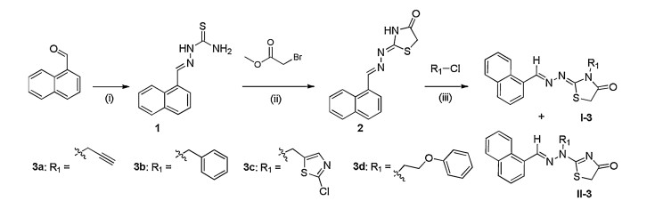
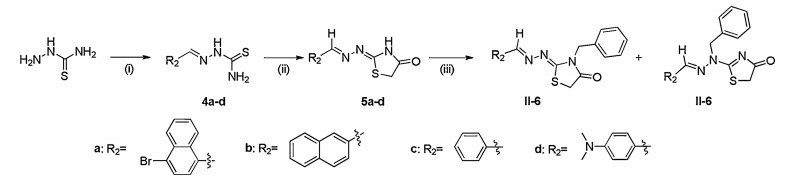
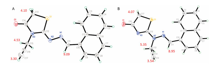
General experimental, synthesis, characterizations of new compounds can be found in Supporting information. The N2 substituted thiazolylhydrazone II were isolated as by-product during the synthesis of thiazolyhydrazone I according to previously reported method [48] (Schemes 1 and 2). Compounds I-3a, I-3c and I-3d had been reported in our previous work [48]. The Rf values of thiazolyllhydrazone I and II is about 0.8 and 0.2 in solvent of ethyl acetate-petroleum ether (1:1), respectively, and the ratio of thiazolylhydrazone I to II afforded was approximately 9:1. To confirm the regiochemistry, the N3 substituted thiazo-lylhydrazones were synthesised by the route illustrated in Scheme 3. The geometric configuration of double bond and the regiochemistry were further established by X-ray analysis of compounds I-3a (CCDC No. 1851329) and II-3a (CCDC No. 1912786). As shown in Fig. 1, for thiazolylhydrazone I, the substituted group R1 was at N3 position of thiazoline ring, and the configurations of the double bonds N(2)=C(12) and N(1)=C(11) were Z and E, respectively. For thiazolylhydrazone II, the substituted group R1 was at the exocyclic N2 position, and the configuration of the N(1)=C(11) double bonds was E. When comparing the 1H NMR data of compounds II with I, the chemical shift of CH2 protons of substituted R1 of II (such as II-3a, 5.35 ppm) appeared at a higher frequency due to the conjugative effect of the N(1)=C(11) double bonds with naphthalene ring than one of compound I (such as I-3a, 4.53 ppm).

|
Download:
|
| Scheme 1. Overview of synthetic methods for 3a-d. Reagents and conditions: (i) thiosemicarbazide, EtOH, acetic acid (cat.), r.t., 15 h; (ii) MeOH, CH3COONa, reflux, 5 h; (iii) CH3CN, K2CO3, reflux, 5 h. | |

|
Download:
|
| Scheme 2. Overview of synthetic methods for 6a-d. Reagents and conditions: (i) R2CHO, EtOH, acetic acid (cat.), r.t., 15 h; (ii) BrCH2COOEt, MeOH, CH3COONa, reflux, 5 h; (iii) chloromethylbenzene, CH3CN, K2CO3, reflux, 5 h. | |

|
Download:
|
| Scheme 3. Overview of synthetic methods for I-3b. Reagents and conditions: (i) H2NNH2 H2O, EtOH, reflux, 6 h; (ii) isothiocyanatomethylbenzene, EtOH, reflux, 3 h; (iii) BrCH2COOEt, MeOH, CH3COONa, reflux, 5 h. | |

|
Download:
|
| Fig. 1. The crystal structures and chemical shifts of 1H NMR of compounds I-3a (A) and II-3a (B). | |
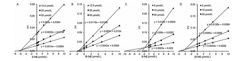
The inhibitory activities of thiazolylhydrazone derivatives I and II against OfHex1 and OfChi-h are outlined in Table 1. Some of the compounds exhibited good inhibitory activities at 1 μmol/L and 10 μmol/L. Our previous work investigating the structure-activity relationships of the thiazolylhydrazone derivatives I against OfHex1 reported the stretched conformation. Interestingly, the branched conformation of thiazolylhydrazone derivatives II, with aromatic groups at the N2 atom, such as benzyl (3b) and phenoxyethyl (3d), exhibited potent inhibitory activity. Further studies then focused on the naphthalene ring R2. The inhibitory activities of the resultant derivatives were weakly influenced by either the substitutes or their position on the naphthalene ring (compare 3b with 6a and 6b). However, replacement of the naphthalene ring with benzene, as in compounds 6c and 6d, resulted in a significant inhibitory activity decrease. Gratifyingly, it was found that the thiazolylhydrazone derivatives I and II exhibited promising inhibitory activity against OfChi-h. In particular, compounds I-3d and II-3d were found to display considerable activity against OfHex1 and OfChi-h (Fig. 2).
|
|
Table 1 Inhibitory activities of compounds I and II against OfHex1 and OfChi-h. |


|
Download:
|
| Fig. 2. Inhibitory kinetics of compounds I-3d (A), II-3d (B) against OfHex1 and I-3d (C), II-3d (D) against OfChi-h. | |
To further explore possible binding modes of these compounds, molecular docking studies were carried out using the OfHex1 (PDB code: 3NSN) and OfChi-h (PDB code: 5GQB) as templates. The predicted docking results reveal that the thiazolylhydrazone derivatives bind in the active sites of OfHex1 and OfChi-h with different modes (Fig. 3). As shown in Fig. 3A, compound I-3d binds the entire active pocket of OfHex1 via hydrogen bonds, π-π stacking and van der Waals interactions. The naphthalene and thiazolinone rings binds the -1 and +1 subsites and stack with Trp524 and Trp490, respectively. The oxygen atom of the thiazolinone ring forms a hydrogen bond with the Glu328 and Glu526 residue. The phenoxyethyl group binds a subpocket formed by Trp483 and Asn489 on Loop478-496, and Gln527 via van der Waals interactions (Fig. 3A). In the docked structure of OfHex1 in complex with II-3d, the N1 and N2 atoms of the linker form hydrogen bonds with the Glu328, Glu526 and Trp490 residues, respectively. The N3 atom forms a hydrogen bond with Glu526, and the oxygen atom of thiazolinone ring forms a hydrogen bond with the Gln527 and Asn489 residues (Fig. 3B). Compound II-3d forms more hydrogen bonds with OfHex1 than compound I-3d. The superimposition of compound I-3d and II-3d reveals that two compounds bind in the 1 and +1 subsites of OfHex1 (Fig. 3C). The predicted different binding modes of I-3d (Ki = 5.9 μmol/L, Fig. 2A) and II-3d (Ki = 1.5 μmol/L, Fig. 2B) could explain the difference in inhibitory activity.

|
Download:
|
| Fig. 3. Proposed binding modes of compounds I-3d and II-3d to the enzymes OfHex1 and OfChi-h respectively: (A) Binding mode I-3d in the active pocket of OfHex1. The compound I-3d was shown in green. The hydrogen bonds were shown in black dashes. (B) Binding mode of II-3d in the active pocket of OfHex1. The compound II-3d was shown in magenta. (C) Superimposition of compound I-3d and II-3d in the active pocket of OfHex1. (D) Binding mode I-3d in the active pocket of OfChi-h. (E) Binding mode of II-3d in the active pocket of OfChi-h. (F) Superimposition of compounds I-3d and II-3d in the active pocket of OfChi-h. | |
Interestingly, experiments showed that compound I-3d (Ki = 1.9 μmol/L, Fig. 2C) has better inhibitory activity against OfChi-h than compound II-3d (Ki = 412.1 μmol/L, Fig. 2D). As shown in Fig. 3D, the compound I-3d binds the entire active pocket of OfChi-h in a stretched conformation. The naphthalene ring is sandwiched by Trp268 and Trp389, and the phenoxyethyl group occupies the hydrophobic patch comprised by Phe184, Thr269 and Leu270. The oxygen atom of the phenoxyethyl group forms a hydrogen bond with the Trp268 residue. The N1 and N2 atoms form hydrogen bonds with Glu308, stabilizing the binding conformations. While the mechanism of interaction of II-3d with OfChi-h is different from that of I-3d, the naphthalene ring occupied the hydrophobic patch comprised by Phe184, Thr269 and Leu270, the oxygen atom of the thiazolinone ring only forms a hydrogen bond with the Leu352 residue (Fig. 3E). The superimposition of compound I-3d and II-3d reveals that the differences in the compounds' binding conformations with OfChi-h explained the differences in their inhibitory activity (Fig. 3F).
In summary, we have designed, prepared and evaluated a series of thiazolylhydrazone derivatives I and II as potential inhibitors of OfHex1 or OfChi-h. To further explore possible binding modes of these compounds, molecular docking studies were carried out using the OfHex1 and OfChi-h as templates. Docking results reveal the mechanism of improved inhibitory activity against OfHex1 and OfChi-h of compounds I-3d and II-3d. This work provides a novel scaffold for developing specific Hex and Chi-h inhibitors.
Declaration of competing interestThe authors declare that they have no known competing financial interests or personal relationships that could have appeared to influence the work reported in this paper.
AcknowledgmentThe authors acknowledge the National Key Research and Development Program of China (Nos. 2017YFD0200500, 2017YFD0201400, 2018YFD0200100) for the financial support.
Appendix A. Supplementary dataSupplementary material related to this article can be found, in the online version, at doi:https://doi.org/10.1016/j.cclet.2019.11.035.
| [1] |
C.D. Monte, S. Carradori, B. Bizzarri, et al., Eur. J. Med. Chem. 107 (2016) 82-96. DOI:10.1016/j.ejmech.2015.10.048 |
| [2] |
D.M. Ascenzio, B. Bizzarri, D.C. Monte, et al., Eur.J.Med. Chem. 86 (2014) 17-30. DOI:10.1016/j.ejmech.2014.08.046 |
| [3] |
D. Patel, P. Kumari, N. Patel, Eur. J. Med. Chem. 48 (2012) 354-362. DOI:10.1016/j.ejmech.2011.11.041 |
| [4] |
D. Secci, S. Carradori, B. Bizzarri, et al., Eur. J. Med. Chem. 117 (2016) 144-156. DOI:10.1016/j.ejmech.2016.04.012 |
| [5] |
A. Verma, S.K. Saraf, Eur. J. Med. Chem. 3 (2008) 897-905. |
| [6] |
A. Mastrolorenzo, S. Rusconi, A. Scozzafava, et al., Curr. Med. Chem. 14 (2007) 2734-2748. DOI:10.2174/092986707782360141 |
| [7] |
S. Bari, D. Firake, Anti-Inflammatory Anti-Allergy Agents Med. Chem. 15 (2016) 44-53. DOI:10.2174/1871523015666160524141630 |
| [8] |
K. Babaoglu, M.A. Page, V.C. Jones, et al., Bioorg. Med. Chem. Lett. 13 (2003) 3227-3230. DOI:10.1016/S0960-894X(03)00673-5 |
| [9] |
N. Krall, F. Pretto, W. Decurtins, et al., Angew. Chem. Int. Ed. 53 (2014) 4231-4235. DOI:10.1002/anie.201310709 |
| [10] |
M. Asadollahi-Babolia, A. Mani-Varnosfaderani, Eur. J. Pharm. Sci. 70 (2015) 117-124. DOI:10.1016/j.ejps.2015.01.014 |
| [11] |
F. Goktas, N. Cesur, D. Satana, M. Uzun, Turk. J. Chem. 38 (2014) 581-591. DOI:10.3906/kim-1307-14 |
| [12] |
R. Fazal, Z. Khalid, U. Hayat, et al., Bioorg. Chem. 63 (2015) 123-113. DOI:10.1016/j.bioorg.2015.10.005 |
| [13] |
C.Ü. Gökçe, G. Elif, N. Lieve, U.G. Nuray, Ç. Gültaze, Bioorg. Med. Chem. 24 (2016) 240-246. DOI:10.1016/j.bmc.2015.12.008 |
| [14] |
J.F. Vincent, U.G. Wegst, Arthropod Struct. Dev. 33 (2004) 187-199. DOI:10.1016/j.asd.2004.05.006 |
| [15] |
H. Merzendorfer, L. Zimoch, J. Exp. Biol. 206 (2003) 4393-4412. DOI:10.1242/jeb.00709 |
| [16] |
K.Y. Zhu, H. Merzendorfer, W. Zhang, J. Zhang, Annu. Rev. Entomol. 61 (2016) 177-196. DOI:10.1146/annurev-ento-010715-023933 |
| [17] |
S. Adrangi, M.A. Faramarzi, Biotechnol. Adv. 31 (2013) 1786-1795. DOI:10.1016/j.biotechadv.2013.09.012 |
| [18] |
K. Slamova, P. Bojarova, L. Petraskova, V. Kren, Biotechnol. Adv. 28 (2010) 682-693. DOI:10.1016/j.biotechadv.2010.04.004 |
| [19] |
M. Karlsson, J. Stenlid, J. Mol. Microbiol. Biotechnol. 16 (2009) 208-223. DOI:10.1159/000151220 |
| [20] |
Q.S. Huang, X.L. Xie, G. Liang, et al., Glycobiology 22 (2012) 23-34. DOI:10.1093/glycob/cwr092 |
| [21] |
T. Liu, Y. Duan, Q. Yang, Biotechnol. Adv. 36 (2018) 1127-1138. DOI:10.1016/j.biotechadv.2018.03.013 |
| [22] |
B.J. Ayers, A.F. Glawar, R.F. Martinez, et al., J. Organomet. Chem. 79 (2014) 3398-3409. |
| [23] |
E.V. Crabtree, R.F. Martinez, S. Nakagawa, et al., Org. Biomol. Chem. 12 (2014) 3932-3943. DOI:10.1039/c4ob00097h |
| [24] |
A. Fuente, T. Mena-Barragan, R.A. Farrar-Tobar, et al., Org. Biomol. Chem. 13 (2015) 6500-6510. DOI:10.1039/C5OB00507H |
| [25] |
A.F. Glawar, D. Best, B.J. Ayers, S. Miyauchi, et al., Chem. -Eur. J. 18 (2012) 9341-9359. DOI:10.1002/chem.201200110 |
| [26] |
M. Horsch, L. Hoesch, A. Vasella, D.M. Rast, FEBS J. 197 (1991) 815-818. |
| [27] |
M. Hattie, N. Cekic, A.W. Debowski, D.J. Vocadlo, K.A. Stubbs, Org. Biomol. Chem. 14 (2012) 3193-3197. |
| [28] |
T. Sumida, K.A. Stubbs, M. Ito, S. Yokoyama, Org. Biomol. Chem. 10 (2012) 2607-2612. |
| [29] |
T. Liu, H. Zhang, F. Liu, et al., Biochem. J. 438 (2011) 467-474. DOI:10.1042/BJ20110390 |
| [30] |
A.G. Santana, G. Vadlamani, B.L. Mark, S.G. Withers, Chem. Commun. 52 (2016) 7943-7946. DOI:10.1039/C6CC02520J |
| [31] |
E. Kappes, G. Legler, J. Carbohydr. Chem. 8 (1989) 71-388. |
| [32] |
S. Knapp, D. Vocadlo, Z. Gao, et al., J. Am. Chem. Soc. 118 (1996) 419-424. |
| [33] |
T. Liu, M. Xia, H. Zhang, et al., FEBS Lett. 589 (2015) 110-116. DOI:10.1016/j.febslet.2014.11.032 |
| [34] |
M. Hattie, T. Ito, A.W. Debowski, et al., Chem. Commun. 51 (2015) 15008-15011. DOI:10.1039/C5CC05494J |
| [35] |
H. Usuki, T. Nitoda, M. Ichikawa, et al., J. Am. Chem. Soc. 130 (2008) 4146-4152. DOI:10.1021/ja077641f |
| [36] |
M.B. Tropak, J. Blanchard, S.G. Withers, E. Brown, D. Mahuran, Chem. Biol. 14 (2007) 153-164. DOI:10.1016/j.chembiol.2006.12.006 |
| [37] |
G.H. Maegawa, M. Tropak, J. Buttner, et al., J. Biol. Chem. 92 (2007) 9150-9161. |
| [38] |
S. Sakuda, A. Isogai, S. Matsumoto, A. Suzuki, K. Koseki, Tetrahedron Lett. 27 (1986) 2475-2478. DOI:10.1016/S0040-4039(00)84560-8 |
| [39] |
S. Omura, N. Arai, Y. Yamaguchi, et al., J. Antibiot. 53 (2000) 603-608. DOI:10.7164/antibiotics.53.603 |
| [40] |
N. Arai, K. Shiomi, Y. Yamaguchi, et al., Chem. Pharm. Bull. 48 (2000) 1442-1446. DOI:10.1248/cpb.48.1442 |
| [41] |
H. Izumida, N. Imamura, H. Sano, J. Antibiot. 49 (1996) 76-80. DOI:10.7164/antibiotics.49.76 |
| [42] |
J.M. Macdonald, C.A. Tarling, E.J. Taylor, et al., Angew. Chem. Int. Ed. 49 (2010) 2599-2602. DOI:10.1002/anie.200906644 |
| [43] |
A.W. Schuttelkopf, O.A. Andersen, F.V. Rao, et al., J. Biol. Chem. 281 (2006) 27278-27285. DOI:10.1074/jbc.M604048200 |
| [44] |
D.R. Houston, I. Eggleston, B. Synstad, V.G. Eijsink, D.M. Aalten, Biochem. J. 368 (2002) 23-27. DOI:10.1042/bj20021034 |
| [45] |
D.R. Houston, B. Synstad, V.G. Eijsink, et al., J. Med. Chem. 47 (2004) 5713-5720. DOI:10.1021/jm049940a |
| [46] |
Q. Yang, T. Liu, F. Liu, M. Qu, X. Qian, FEBS J. 275 (2008) 5690-5702. DOI:10.1111/j.1742-4658.2008.06695.x |
| [47] |
T. Liu, L. Chen, Y. Zhou, et al., J. Biol. Chem. 292 (2017) 2080-2088. DOI:10.1074/jbc.M116.755330 |
| [48] |
H. Yang, T. Liu, H. Qi, et al., Bioorg. Med. Chem. 26 (2018) 5420-5426. DOI:10.1016/j.bmc.2018.09.014 |