 2020, Vol. 31


b School of Life Science&Bio-pharmaceutics, Shenyang Pharmaceutical University, Shenyang 110016, China
In recent years, mesoporous silica nanoparticles (MSNs) have aroused a great deal of attention in drug delivery, owing to the advantages of large specific surface area, controllable size and shape, easily modified surface, large cargo loading capacity, etc. A great number of drugs have been used to load into MSNs, such as iodomethane (IMC), ibuprofen (IBU), doxorubicin hydrochloride (Dox), paclitaxel, gemcitabine, leading to a high oral bioavailability [1-4]. It is not very difficult to synthesize MSNs from different templates and study on drug delivery efficiency. Generally, tetraethylorthosilicate (TEOS) based sol-gel process was mostly applied to fabricate MSNs, including hard template and soft template methods. Anodized aluminum oxide (AAO) template is a commonly used hard template, which provides varieties of pore structure and morphology [5]. AAO template was applied to prepare orderly assembled organic molecules for improving Raman scattering. Of the soft templates, surfactant has a long history of preparing MSNs. Cationic surfactant cetyltrimethylam-monium bromide (CTAB) not only can stabilize colloidal system, but also be easily removed by washing. Pluronic F127, F123, pluronic poly(ethylene oxide)-poly(propylene oxide)-poly(ethyl-ene oxide) (PEO-PPO-PEO) oligomer, etc. were also favorable for fabrication [6, 7]. Template-assisted fabrication, especially com-bined with self-assembled process, provides an alternative choice for producing porous nanoparticles in more economical and convenient ways [8]. In comparison of traditional approach, biomimetic synthesis of MSNs is essential and widely exists in nature, which is actively catalyzed the condensation of TEOS by amines [9]. It is reported that using chiral surfactant N-palmityl-L-alanine (N-PLA) as template can successfully prepared chiral MSNs and a series of factors on preparation were studied [10]. Nanoflakes with perpendicular pore channels and nanoworms with horizontal pore channels were prepared using surfactants with amines L-18Ala6PyBr and L-18Ala11PyBr, respectively [11]. C16-L-tryptophan, C16-L-proline and C16-L-histidine were also applied to synthesize MSNs with different morphology in a biomimetic approach [9]. Amines are crucial for the bioformation of mesoporous structure, while aminopropyltriethoxysilane (APTES) is always modified on the surface of MSNs with a non-negatively affected drug loading capacity. Additionally, introducing amino groups may be useful to adjust surface charge or pH, which can be protonated after entering into tumor environment and is benefit to endocytosis [12].
Hence, we synthesized a new-structured template molecule using L-leucine methyl ester hydrochloride (H-Leu-OMe HCl) for the first time. Then TEOS was condensed on the new template and finally formed a core-shell structure, named LMSNs. Morphological characterization and drug delivery evaluation demonstrated great potential in future study. Furthermore, we found that the core-shell structure mesoporous silicon increased the drug loading to 30.5% compared with the general synthesis method, but the intrinsic relationship between the high drug loading capacity and core-shell structure still need further study.
Initially, 43.0 mmol of H-Leu-OMe HCl, and 90-mL aliquot of CHCl3 and 11.0 mL of triethylamine were mixed at 25℃. Secondly, 47.3 mmol of myristic acid chloride in CHCl3 were slowly added under an ice water bath and then reacted at 25℃. The organic phase obtained is extracted with H2O, saturated NaHCO3 aq. and saturated NaCl aq., finally the lower layer was dried to obtain a crude product. 5 mmol of the above crude product was dissolved by methanol completely and 1 mol/L NaOH was added dropwise into the solution. The developer and the developing agent are the same as above, then adjust the pH 2–3 with HCl and add water. The resultant white precipitate was filtered and dried to obtain myristoyl-L-leucine (MLL). MLL was confirmed by nuclear mag-netic resonance spectroscopy analysis (1H NMR).
0.5 mmol of the MLL was dissolved in distilled water, followed by a mixture of TEOS and APTES adding dropwise and stirring at 80℃ (Table S1 in Supporting information). The product was immediately centrifuged (5000 g, 20 min) and 1 mL HCl was added and stirred overnight to remove the template. Finally, LMSNs were obtained. Tecnai G2 20 TEM instrument (FEI, Hillsboro, OR, USA) was used to characterize structures of LMSNs at 200 kV. Small-angle XRD (SAXRD) pattern was obtained by an X-ray diffractom-eter with diffraction angle ranged from 1°to 6°. The specific surface area (SBET) and the pore size distributions (PSDs) were evaluated by nitrogen adsorption/desorption measurement. Doxorubicin hy-drochloride (Dox) was loaded into LMSNs by a previously described method and in vitro release of samples was performed in 50 mL phosphate buffer at pH 5.0 and pH 7.4, respectively [13]. Also, MTT assay was applied to investigate the anti-tumor effect of Dox-loaded LMSNs [14].
We have firstly synthesized a surfactant template from H-Leu-OMe HCl and 1H NMR (600 MHz, DMSO) spectra showed different bands at δ 12.44 (s, 1 H), 8.01 (d, 1H, J = 8.0 Hz), 4.21 (ddd, 1H, J = 10.3, 8.1, 4.9 Hz), 2.14–2.04 (m, 2 H), 1.65–1.59 (m, 1 H), 1.48 (s, 4 H), 1.24 (s, 20 H), 0.92–0.79 (m, 9 H). The data of 1H NMR spectra confirmed the amination and the hydrolysis reactions were carried out and the template MLL was successfully fabricated (Fig. S1 in Supporting information).
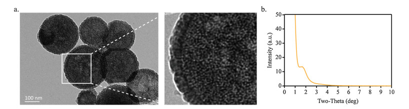
LMSNs were prepared by fabricating a new surfactant template, using APTES as co-structure directing agent and TEOS as silica source. The positively charged ammonium ion of APTES interacts with the negatively charged head group of hydrophilic part through neutralization, which induces favorable electrostatic interactions. Meanwhile, the alkoxysilane sites of APTES are polymerized with TEOS to form silica framework. TEM Image of LMSNs turned out to be a spherical core-shell shape, as shown in Fig. 1a. Surprisingly, definite lattice fringes on the surface of LMSNs showed helical architecture. We also assumed that it may have certain prospects for the loading of chiral drugs for further study. The small-angle XRD image of LMSNs was shown in Fig. 1b. A maximal peak at 1.3°–1.7° (2θ) were identified in the SAXRD pattern, demonstrating that a well-ordered mesostructured formed successfully with the surfactant template.

|
Download:
|
| Fig. 1. TEM (a) and SAXRD (b) images of LMSNs. | |
Nitrogen adsorption/desorption isotherms and pore size distribution curves of LMSNs are presented in Figs. 2a and b, respectively. A clear hysteresis loop was shown in the nitrogen adsorption/desorption isotherm of LMSNs, indicating a slit-like mesopore structure, which corresponded to typical type Ⅳ isotherm in the IUPAC classification. Nitrogen BET surface area and pore volume were calculated at 473.5 cm2/g and 0.765 cm3/g, which demonstrated great potential to load and release model drugs. To study the drug delivery efficiency, we chose Dox as model drug with poorly permeability and loaded into LMSNs with loading capacity of 30.5%. After loading Dox into pores, nitrogen BET surface area and pore volume decreased to 201.5 cm2/g and 0.143 cm2/g, respectively. These results suggested that large pore volume of LMSNs we synthesized has great potential on drug loading and drug delivery process.

|
Download:
|
| Fig. 2. Nitrogen adsorption/desorption isotherm (a) and pore size distribution pattern (b) of LMSNs and Dox-loaded LMSNs. | |
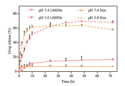
The drug dissolution profiles in PBS 7.4 and PBS 5.0 at 37℃ were shown in Fig. 3. The total drug release from LMSNs at pH 7.4 and 5.0 reached 16.5% and 67.9%. It was obviously that in comparison of at physiological neutral condition, larger amount of Dox was released from LMSNs. Compared to Dox, there was almost no burst release for Dox-loaded LMSNs in both two-pH medium in the first 10 h. Also, Dox released more from LMSNs than naked drug after 24 h in PBS 5.0. It is well known that Dox is of positive charge, and LMSNs is in negative charge, leading to rapidly absorb together and fill full of the pore. When pH value decreases to 5.5, the electrostatic attraction between the nanoparticles and doxorubicin decreases, the electrostatic repulsion increases, and then drug releases [13, 15, 16]. Owing to the acidic condition at tumor and lysosome sites, LMSNs were able to maintain a long-term controlled release and enhance the delivery efficiency of Dox. Furthermore, it is speculated that Dox could be delivered to nuclei efficiently and disturb DNA double helix [17-19].

|
Download:
|
| Fig. 3. Drug release profiles of LMSNs and Dox in PBS 7.4 and PBS 5.0 (mean ± SD, n = 3). | |
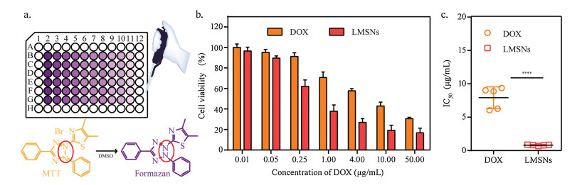
For further investigation on the inhibitory effect of Dox-loaded LMSNs and Dox to cancer cells, here we chose human colon carcinoma cell line (Caco-2). The anti-tumor effect of samples was evaluated by measuring the cell viability using an MTT (3-(4, 5-dimethylthiazol-2-yl)-2, 5-diphenyltetrazolium bromide) assay. As depicted in Fig. 4, both Dox and Dox-loaded LMSNs were observed dose-dependent cytotoxicity on Caco-2 at the concentration of 0.01, 0.05, 0.10, 0.25, 1.00, 4.00, 10.00, 50.00 mg/mL. Half maximal inhibitory concentration (IC50) was also calculated using GraphPad prism 6.07 software [14]. IC50 values of free Dox and Dox-loaded LMSNs were 6.74±1.62 mg/mL and 0.81±0.08 mg/mL, respective-ly. It is suggested that Dox-loaded LMSNs demonstrated great promise to anti-tumor.

|
Download:
|
| Fig. 4. (a) Scheme of MTT assay. Cell viability (b) and IC50 values (c) in Caco-2 cell line, respectively. ****P < 0.0001. | |
In this study, LMSNs were prepared by a firstly-synthesized template and TEM, nitrogen adsorption/desorption, as well as SAXRD were applied to confirm the structure of mesopores. Notably, LMSNs conferred an excellent drug loading capacity, dissolution-enhancing effect and anti-tumor effect. It is therefore that LMSNs can be considered a good candidate for drug delivery
AcknowledgmentThis work was supported by the Career Development Support Plan for Young and Middle-aged Teachers in Shenyang Pharma-ceutical University (No. ZQN2018005).
Appendix A. Supplementary dataSupplementary material related to this article can be found, in the online version, at doi:https://doi.org/10.1016/j.cclet.2019.05.059.
| [1] |
H. Meng, M. Wang, H. Liu, et al., ACS Nano 9 (2015) 3540-3557. DOI:10.1021/acsnano.5b00510 |
| [2] |
M. Yu, L. Xu, F. Tian, et al., Nat. Commun. 9 (2018) 2607-2618. DOI:10.1038/s41467-018-05061-3 |
| [3] |
F. Rehman, K. Ahmed, A. Rahim, et al., J. Mol. Liq. 258 (2018) 319-326. DOI:10.1016/j.molliq.2018.03.057 |
| [4] |
W. Zhang, N. Zheng, L. Chen, et al., Pharmaceutics 11 (2018) 4. |
| [5] |
C. Mijangos, R. Hernández, J. Martín, Prog. Polym. Sci. 54- 55 (2016) 148-182. |
| [6] |
B. Du, Z. Cao, Z. Li, et al., Langmuir 25 (2009) 12367-12373. DOI:10.1021/la902531p |
| [7] |
S.T. Mahmud, L.D. Wilson, Cogent Chem. 11 (2016) 1132984. |
| [8] |
X. Hou, Q. Wang, G. Mao, et al., Appl. Surf. Sci. 437 (2018) 92-97. DOI:10.1016/j.apsusc.2017.12.157 |
| [9] |
H. Li, J. Ke, H. Li, et al., Mater. Sci. Eng. C 93 (2018) 407-418. DOI:10.1016/j.msec.2018.07.081 |
| [10] |
J. Li, L. Xu, B. Yang, et al., Mater. Sci. Eng. C 55 (2015) 367-372. DOI:10.1016/j.msec.2015.05.039 |
| [11] |
L. Wu, Y. Shan, Y. Chen, et al., J. Nanosci. Nanotechnol. 12 (2012) 2037-2044. DOI:10.1166/jnn.2012.5178 |
| [12] |
Y. Wang, Y. Sun, J. Wang, et al., ACS Appl. Mater. Interf. 8 (2016) 17166-17175. DOI:10.1021/acsami.6b05370 |
| [13] |
J. Li, X. Du, N. Zheng, et al., Colloid Surface B 141 (2016) 374-381. DOI:10.1016/j.colsurfb.2016.02.009 |
| [14] |
L. Fan, F. Li, H.T. Zhang, et al., Biomaterials 31 (2010) 5634-5642. DOI:10.1016/j.biomaterials.2010.03.066 |
| [15] |
J. Gu, S. Su, M. Zhu, et al., Microporous Mesoporous Mater. 161 (2012) 160-167. DOI:10.1016/j.micromeso.2012.05.035 |
| [16] |
M. Xie, H. Shi, Z. Li, et al., Colloid. Surface B 110 (2013) 138-147. DOI:10.1016/j.colsurfb.2013.04.009 |
| [17] |
N. Zheng, J. Li, C. Xu, et al., Artifi. Cell. Nanomed. B 46 (2018) 1132-1140. DOI:10.1080/21691401.2017.1362414 |
| [18] |
J.S. Suk, S.K. Lai, N.J. Boylan, et al., Nanomedicine 6 (2011) 365-375. DOI:10.2217/nnm.10.123 |
| [19] |
M. Schenk, C. Mueller, Best Prac. Res. 22 (2008) 391-409. |