 2020, Vol. 31


b Department of Chemistry and Nano Science, Ewha Womans University, Seoul 120-750, Republic of Korea;
c Department of Pharmaceutical Chemistry, V. L. College of Pharmacy, Raichur 584103, India
Fluorescence detection is a trustworthy approach to monitor metal ions, especially in chemical reactions, natural environments and life activities [1-3]. Trace metals are some of the metallic elements which are essential for human beings while excessive amount is found to be particularly prone to cause poisoning, cancer and even death, especially heavy metals such as copper and mercury. Heavy metal pollution has become increasingly severe [4-7]. Copper ion (Cu2+) is an indispensable trace element in living organisms, takes part in a lot of life activities, promoting the synthesis of hemoglobin and the development of red blood cells. However, the lack of copper in human body can cause the diseases such as anemia and vitiligo [8-10]. But no extreme will hold long. Excessive copper ion tend to bring a great burden on human organs, especially the gallbladder and liver, leading to disease and even death [11]. Similarly, the less-common metal, mercury, has long time been considered to be highly dangerous to human beings. Oral, inhalation or exposure to mercury can cause severe brain and liver damage [12]. Mercury ion is also a serious environmental pollutant. When it accumulates in plants, soil and water, it is a serious hazard to Earth's organisms such as fish and some other aquatic organism [13-16].
Therefore, it is of great necessity to monitor the content of copper and mercury ions. A variety of detection methods have emerged, such as high-performance liquid chromatography, atomic absorption, electrochemical analysis [17, 18]. Among these reported analytical methods, UV absorption and fluorescence spectroscopy are the most common due to their inherent advantages of easy operation, high sensitivity and selectivity [19-22]. A new type of probe capable of simultaneously detecting a variety of different analytes appeared, which uses a single detection probe to analyze multiple analytes via different spectra [23-28]. For example, a chemosensor employed more than two spectra like UV–vis and fluorescence has become increasingly popular, since this method does not need tedious receptor synthesis and can detect multiple analytes with high selectivity [29, 30].
Many chemosensors based on rhodamine have been employed as fluorescence or colorimetric detection of metal ions [31-33]. Due to a classic open-loop and closed-loop structure of rhodamine, the differences in fluorescence, absorption and even macroscopic color are obvious [34]. In addition, due to the longer emission wavelength of rhodamine structure endow rhodamine-based sensors with a wider application in detecting metal ions [35-38].
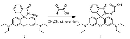
A new rhodamine hydrazone derivative named sensor 1 bearing carboxylic acid group as a chemosensor has been reported in this article (Scheme 1), for recognizing Cu2+ and Hg2+ respectively through different devices. After adding aqueous solution of Cu2+ into the buffer solution containing sensor 1, colorless of the solution turned pink, indicating that 1 and Cu2+ were effectively combined. Differently, when Hg2+ was added to 1 solution, no similar phenomenon was observed, but the fluorescence of the solution was greatly enhanced.

|
Download:
|
| Scheme 1. Synthesis of sensor 1. | |
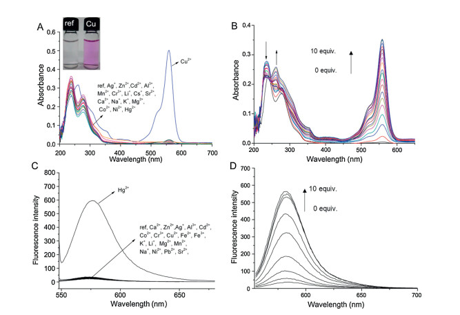
Stock solutions of metal ions (10 mmol/L) such as Cu2+ and Hg2+ were used to detect the ability of sensor 1 to bind with different metal ions. Other test metal ions were presented in Supporting information. Separate sensor 1 remained colorless in CH3CN-HEPES buffer solution (20 mmol/L, pH 7.4) (1:9, v/v) and exhibited no absorption above 450 nm, indicating that the 1 is mainly in the form of spirolactam. The absorption spectra of sensor 1 (10 μmol/L) with different metal ions (100 μmol/L) were collected (Fig. 1A). In the absorption spectra, there was no apparent change in sensor 1 with test metal ions except Cu2+, and when bound with Cu2+, the absorption intensity at 560 nm of the solution increased by a large margin. The solution also has a distinct color change from colorless to pink by the naked eye. All metal ions with solution of 1 were presented in Fig. S4 (Supporting information). The above results showed sensor 1 can effectively distinguish Cu2+ and other metal ions by the naked eye.

|
Download:
|
| Fig. 1. (A) Absorbance spectra of sensor 1 (10 μmol/L) with various metal ions (100 μmol/L). Insert: Color change of sensor 1 (10 μmol/L) with Cu2+ (100 μmol/L) in CH3CN-HEPES buffer solution (20 mmol/L, pH 7.4) (1:9, v/v). (B) Absorbance spectra of 1 (10 μmol/L) in CH3CN-HEPES buffer solution (20 mmol/L, pH 7.4) (1:9, v/v) in presence of increasing amounts of Cu2+. (C) Fluorescent changes of 1 (10 μmol/L) with various metal ions (50 μmol/L) in CH3CN-HEPES buffer solution (20 mmol/L, pH 7.4) (1:9, v/v). λex = 545 nm, Slit: 5 nm/5 nm. (D) Fluorescent titrations of 1 (10 μmol/L) with Hg2+ in CH3CN-HEPES buffer solution (20 mmol/L, pH 7.4) (1:9, v/v). λex = 545 nm, Slit: 5 nm/ 5 nm. | |
With titration of 10 equiv. of Cu2+ to sensor 1 (10 μmol/L), a gradual enhancement was observed at 560 nm in the absorption band (Fig. 1B). In the Job's Plot curve, the absorption intensity reaches the maximum when the ratio of Cu2+/(1+Cu2+) is 0.5 (Fig. S5 in Supporting information), from where we got the information that sensor 1 had a 1:1 ratio relationship with Cu2+. In addition, the association constant of 1 with Cu2+ was calculated as 1.2 × 10⁵ L/mol (error < 15%) [39]. The absorbance titrations curve of 1 with Hg2+ in CH3CN-HEPES buffer solution (20 mmol/L, pH 7.4) (1:9, v/v) was shown in in Fig. S6 (Supporting information).
Then, we collected the fluorescence spectra of sensor 1 (10 μmol/L) with above metal ions (100 μmol/L) (Fig. 1C) under ambient condition. Like many literature reported, 545 nm was chosen as the excitation wavelength. From the spectra, there was a great increase in the fluorescence intensity of sensor 1 with Hg2+, up to about 70 times. But no similar increase was observed of 1 with other metal ions even with Cu2+. As for the reason for fluorescence enhancement, it is clearly found that 1 formed coordination with Hg2+, resulting in rhodamine spirolactam ringopened form. Due to the well-known paramagnetic effect of Cu2+, when 1 coupled with Cu2+, although ring-opened form occurred, fluorescence intensity was greatly quenched.
Fluorescence titration experiments were then conducted by increasing the amounts of Hg2+ into the solution of sensor 1 (Fig. 1D). After adding 10 equiv. of Hg2+, 70-fold enhancement in the fluorescence intensity was observed. The association constant of 7.2 × 10⁵ L/mol (error < 15%) was obtained in sensor 1 solution within Hg2+. In order to determine the binding mode of 1 and Hg2+, ESI mass spectroscopy of the solution of 1 with Hg2+ added was obtained (Fig. S7 in Supporting information). The peak at m/z 513.2 refers to [sensor 1+H]+, the peak at m/z 1225.2 corresponded to Hg(1)2 and m/z 443.4 accompanied by hydrolysis reaction was observed, finally, we acquired the ratio of Hg2+ with 1 to be 1:2.
Although S2- has a strong binding affinity for Hg2+, and after adding sodium sulfide to a mixed solution of sensor 1 (10 μmol/L) with Hg2+ (100 μmol/L), the fluorescence intensity was quenched a little. Even more 20 equiv. of Na2S was added, the fluorescence intensity at 585 nm quenched only 40% (Fig. S8 in Supporting information). It is thus obtained that the combination of sensor 1 with Hg2+ can be classified as an irreversible chemosensor for irreversible bond between 1 and Hg2+. On the contrary, it was believed that 1 bound with Cu2+ was reversible, which has been proved by reversible experiments (Figs. S9 and S10 in Supporting information). When 20 equiv. of EDTA was added in the solution mixture of 1 (10 μmol/L) and Cu2+ (100 μmol/L), the pink solution again returned the initial colorless. With reference to absorbance intensity at 552 nm, about 95% was quenched because of a spirolactam ring closure. Based on the stoichiometry constant of 1 with Cu2+ and Hg2+, Job's Plot and ESI-mass data, the proposed binding modes of 1 with Cu2+ and Hg2+ have been shown in Scheme 2.

|
Download:
|
| Scheme 2. Proposed binding modes of sensor 1 with Cu2+ and Hg2+. | |
Finally, considering the dual capacity of sensor 1 to detect Cu2+ and Hg2+ selectively via absorbance spectra (naked eye) and fluorescence spectra respectively, so it is also necessary to test the interference between the two metal ions. We tested the fluorescence and absorption spectra of the four solutions containing the sensor 1, Cu2+, Hg2+, and Cu2++Hg2+ (Fig. 2 and Fig. S11 in Supporting information). Similar to previous experimental results, upon adding Hg2+ into solution containing sensor 1, fluorescence intensity at 580 nm showed a great enhancement. But the enhancement remained stable after adding both Cu2++Hg2+ into 1 solution (Fig. 2A). As for sensor detecting Cu2+ progress, absorbance spectra was carried out in a similar way. At the wavelength of 560 nm, the absorbance had a great enhancement, but only a small increase with Hg2+ after adding Cu2+ into the solution of 1, the absorbance intensity did not change a lot after adding both Cu2+ and Hg2+ (Fig. 2B). During the measurement of fluorescence and absorbance spectra, both Cu2+ and Hg2+ were 10 equiv. of 1. From the obtained cross-interference data, it was concluded that when detecting Cu2+ or Hg2+, the detection was not affected by another metal ion.

|
Download:
|
| Fig. 2. The fluorescence intensity of solutions containing sensor 1, Cu2+, Hg2+, and Cu2+ + Hg2+ (A), the absorption intensity of solutions containing sensor 1, Cu2+, Hg2+, and Cu2++Hg2+ (B). | |
In conclusion, a bifunctional chemosensor named 1 to selectively detect Cu2+ and Hg2+ ions via different spectra has been reported in this article. Sensor 1 binds with Cu2+ and Hg2+ in the ratio of 1:1 and 1:2 respectively. In the solution containing 1 after adding Cu2+ and Hg2+, absorbance and fluorescence spectra were employed to monitor the complex formation. And the color change of sensor 1 with Cu2+ could be detected by the naked eye. There was an enhancement in fluorescence at 580 nm in the solution of 1 and Hg2+. The ability of 1 sensing Cu2+ and Hg2+ does not interfere with each other according to experiment data.
Declaration of competing interestThe authors declare that they have no known competing financial interests or personal relationships that could have appeared to influence the work reported in this paper.
AcknowledgmentsThis work was supported by the National Key Research and Development Program of China (No. 2018YFA0902200), the National Natural Science Foundation of China (Nos. 21978131, 21722605 and 21878156), the Six Talent Peaks Project in Jiangsu Province(No.XCL-034) and the Project of Priority Academic Program Development of Jiangsu Higher Education Institutions (PAPD).
Appendix A. Supplementary dataSupplementary material related to this article canbefound, in the online version, at doi:https://doi.org/10.1016/j.cclet.2019.11.013.
| [1] |
F. Wang, L. Wang, X. Chen, J. Yoon, Chem. Soc. Rev. 43 (2014) 4312-4324. DOI:10.1039/c4cs00008k |
| [2] |
K.A. Morales, Y. Yang, Z. Long, et al., J. Am. Chem. Soc. 135 (2013) 12980-12983. DOI:10.1021/ja406958k |
| [3] |
L. Chen, S.J. Park, D. Wu, H.M. Kim, J. Yoon, Chem. Commun. (Camb.) 55 (2019) 1766-1769. DOI:10.1039/C8CC08608G |
| [4] |
K. Villa, J. Parmar, D. Vilela, S. Sánchez, ACS Appl. Mater. Interfaces 10 (2018) 20478-20486. DOI:10.1021/acsami.8b04353 |
| [5] |
S. Tang, J. Sun, Y. Li, et al., Talanta 187 (2018) 287-294. DOI:10.1016/j.talanta.2018.04.102 |
| [6] |
L. Wang, B. Chen, P. Peng, et al., Chin. Chem. Lett. 28 (2017) 1965-1968. DOI:10.1016/j.cclet.2017.07.016 |
| [7] |
M. Nagy, S.L. Kovács, T. Nagy, et al., Talanta 201 (2019) 165-173. DOI:10.1016/j.talanta.2019.04.007 |
| [8] |
J.C. Claessens, P.V. Cappellen, Environ. Sci. Technol. 41 (2007) 909-914. DOI:10.1021/es0620944 |
| [9] |
W. Lva, M. Lina, R. Lia, et al., Chin. Chem. Lett. 30 (2019) 1410-1414. DOI:10.1016/j.cclet.2019.04.011 |
| [10] |
J.L. Ma, B.C. Yin, X. Wu, B.C. Ye, Anal. Chem. 88 (2016) 9219-9225. DOI:10.1021/acs.analchem.6b02465 |
| [11] |
Y. Wang, C. Zhang, X. Chen, et al., Nanoscale 8 (2016) 5977-5984. DOI:10.1039/C6NR00430J |
| [12] |
S. Ding, M. Dong, Y. Wang, et al., J. Am. Chem. Soc. 138 (2016) 3031-3037. DOI:10.1021/jacs.5b10754 |
| [13] |
X.H. Yang, S. Sun, P. Liu, et al., Chin. Chem. Lett. 25 (2014) 9-14. DOI:10.1016/j.cclet.2013.10.032 |
| [14] |
S. Yoon, A.E. Albers, A.P. Wong, C.J. Chang, J. Am. Chem. Soc. 127 (2005) 16030-16031. DOI:10.1021/ja0557987 |
| [15] |
L. Lan, Q. Niu, T. Li, Anal. Chim. Acta 1023 (2018) 105-114. DOI:10.1016/j.aca.2018.03.023 |
| [16] |
B. Zhu, G. Ren, M. Tang, et al., Dyes Pigments 149 (2018) 686-695. DOI:10.1016/j.dyepig.2017.11.041 |
| [17] |
J. Becker, M. Zoriy, J. Becker, et al., Anal. Chem. 77 (2005) 5851-5860. DOI:10.1021/ac0506579 |
| [18] |
X. Xia, T. Wang, X. Yuan, Chin. Chem. Lett. 25 (2014) 1403-1406. DOI:10.1016/j.cclet.2014.05.033 |
| [19] |
J. Chen, W. Liu, B. Zhou, et al., J. Org. Chem. 78 (2013) 6121-6130. DOI:10.1021/jo400783x |
| [20] |
L. Zeng, Y. Yuan, C. Jiang, et al., Dyes Pigments 165 (2019) 408-414. DOI:10.1016/j.dyepig.2019.02.049 |
| [21] |
F. Ali, S. Aute, S. Sreedharan, et al., Chem. Commun. (Camb.) 54 (2018) 1849-1852. DOI:10.1039/C7CC09433G |
| [22] |
W. Chen, Y. Pan, J. Chen, et al., Chin. Chem. Lett. 29 (2018) 1429-1435. DOI:10.1016/j.cclet.2018.08.011 |
| [23] |
T. Pinkert, D. Furkert, T. Korte, et al., Angew. Chem. Int. Ed. 56 (2017) 2790-2794. DOI:10.1002/anie.201611706 |
| [24] |
M. Kaur, M.J. Cho, D.H. Choi, Dyes Pigments 125 (2016) 1-7. DOI:10.1016/j.dyepig.2015.09.030 |
| [25] |
M. Schmittel, H.W. Lin, Angew. Chem. Int. Ed. 46 (2007) 893-896. DOI:10.1002/anie.200603362 |
| [26] |
L. Tang, F. Li, M. Liu, et al., Spectrochimica Acta Part A 78 (2011) 1168-1172. DOI:10.1016/j.saa.2010.12.072 |
| [27] |
Z. Guo, Y. Ma, Y. Liu, et al., Sci. China Chem. 61 (2018) 1293-1300. DOI:10.1007/s11426-018-9240-6 |
| [28] |
Z. Xu, X. Huang, X. Han, et al., Chem 4 (2018) 1609-1628. DOI:10.1016/j.chempr.2018.04.003 |
| [29] |
N. Singh, R.C. Mulrooney, N. Kaur, J.F. Callan, Chem. Commun. (Camb.) 40 (2008) 4900-4902. |
| [30] |
L.L. Chang, Q. Gao, S. Liu, et al., Dyes Pigments 153 (2018) 117-124. DOI:10.1016/j.dyepig.2018.02.013 |
| [31] |
N. Lin, J. Li, Z. Lu, et al., Nanoscale 7 (2015) 4971-4977. DOI:10.1039/C5NR00515A |
| [32] |
H.N. Kim, M.H. Lee, H.J. Kim, et al., Chem. Soc. Rev. 37 (2008) 1453-1744. DOI:10.1039/b811182k |
| [33] |
S. Fernández-Alonso, T. Corrales, J.L. Pablos, et al., Sens. Actuators B-Chem. 270 (2018) 256-262. DOI:10.1016/j.snb.2018.05.030 |
| [34] |
Y. Jiao, L. Zhou, H. He, et al., Talanta 184 (2018) 143-148. DOI:10.1016/j.talanta.2018.01.073 |
| [35] |
C. Liu, T. Xiao, Y. Wang, et al., Tetrahedron 73 (2017) 5189-5193. DOI:10.1016/j.tet.2017.07.012 |
| [36] |
E. Yoshioka, M. Inoue, Y. Nagoshi, et al., J. Org. Chem. 83 (2018) 8962-8970. DOI:10.1021/acs.joc.8b01099 |
| [37] |
H. Chen, J. Wang, D. Shan, et al., Anal. Chem. 90 (2018) 7056-7063. DOI:10.1021/acs.analchem.8b01455 |
| [38] |
Z. Zhang, S. Lu, C. Sha, et al., Sens. Actuators B-Chem. 208 (2015) 258-266. DOI:10.1016/j.snb.2014.10.136 |
| [39] |
Association constants were obtained using the computer program ENZFITTER, available from Elsevier-BIOSOFT, 68 Hills Road, Cambridge CB21LA, United Kingdom.
|