 2019, Vol. 30


b Department of Chemistry, Taiyuan Normal University, Taiyuan 030031, China;
c Institute for NanoScience Design, Osaka University, Osaka 560-8531, Japan
Surface plasmon coupled emission (SPCE), which was widely known as an interaction between the fluorophores in close vicinity of the metallic films and surface plasmons (SPs), has received extensive attention from researchers [1-5]. The fluorophores placed on the metal substrate within a few hundred nanometers could couple with SPs, and lead to a directional fluorescence emission with high p-polarization [6-10]. Because of the distance-sensitive coupling property of SPCE which could suppress the background signal and purify fluorescence signal, SPCE has been successfully implemented in DNA detection [11, 12] and immunoassays [13, 14]. With the increasing complexity and diversification of analytes, the improvement of the detection sensitivity becomes the first priority. Therefore, various approaches, such as development of instruments [15], employment of nanomaterials [16, 17] and assistance of an external field [18] have been introduced in SPCE-based systems to enhance the signal and increase detection sensitivity. Magnetic field, one of the most widely used external fields, has been employed in field-enhanced related systems such as surface plasmon resonance (SPR) and surface enhanced Raman scattering (SERS) to improve detection sensitivity [19-24]. Our group has investigated the pulsed magnetic field modulated SPCE system: The SPCE intensity is dramatically modulated by applying a pulsed magnetic field to the noble metal/ferromagnetic metal substrate. This modulation is due to the change of the SPs properties by the external magnetic field and the high magneto-optical (MO) activity of the metal substrate [18].
Along with the rapid development of nanotechnology, the application of magnetic nanoparticles (MNPs) in biological field has aroused great interest of researchers. MNPs are a kind of intelligent nano-magnetic materials, which have the characteristics of nano-materials such as small size, large surface area and high loading, and in the meantime, have magnetic response which could be orientated and migrated by external magnetic field easily [25]. Iron oxide nanoparticles are the most commonly used MNPs, such as Fe3O4 and γ-Fe2O3. Considering the easy aggregation of MNPs caused by the large surface area and high surface activity, the chemical modification and coating are normally introduced in the synthesis process [26]. After these procedures, abundant functional groups are existed on the surface of MNPs, which could improve the biocompatibility and simplify the further chemical modification. Moreover, induced by the strong magnetic response, MNPs can be easily separated and purified through external magnetic field, which could greatly simplify the separation and cleaning procedure during nano-particles synthesis. Benefiting from these features, MNPs are widely used in the biological field including biomarkers and separation, biosensing and detection, drug carriers and disease diagnosis [27-30].
The modulation of magnetic field on SPCE depends on the effect of magnetic field on the property of SPCE and the MO activity of the substrate [18]. The introduction of MNPs is expected to improve the MO activity of the substrate, which might have the potential to further enhance the effect of the magnetic field modulation. Herein, we report a significant enhancement effect of MNPs on magnetic field modulated-SPCE by modifying MNPs on the substrate. Furthermore, we have designed a biosensor based on this strategy. To our knowledge, this is the first application of the magnetic field modulated SPCE into the biosensor and detection field.
The experimental substrate was chosen based on the magnetic field modulated SPCE system produced in multilayers of noble and ferromagnetic metals, glass/2 nm Cr/7.5 nm Co/3 nm Cr/23 nm Au [18, 31]. The Cr layer existed in the experimental substrate to improve the adherence between two different materials. To our knowledge, ferromagnetic layers feature a vital drawback in their strong absorption, which leads to the broad plasmon resonance and high propagation loss. A suitable way to overcome this drawback is to introduce the noble metal into the ferromagnetic metal substrate to maintain both magnetic and plasmonic properties [18]. SPCE signal was detected with a home-made spectrofluorimeter. The experimental substrate was attached with a semi-cylindrical prism fastened on a rotary stage. In the reverse Kretschmann (RK) configuration, the incident light (532 nm, 5 mW) was normal to the substrate interface and SPCE signal was collected at a directional emission angle from the prism side. The external magnetic field was generated by an air-core coil with a 4 mm inner diameter connected to a condenser bank of 10, 000 F, which was operated under the charging voltage of 0-400 V, and its direction was perpendicular to the substrate. When the capacitor was charged in a given voltage, the magnetic field was applied by turning on the current flow. The peak magnetic flux density of the pulsed magnetic field was varied in the range of 0-2.25 T, and the peak magnetic flux density of the pulsed magnetic field was 1.8 T for all the magnetic field modulation. The pulse duration time was approximately 4 ms which was sufficiently long to magnetize the metallic multilayer substrate.
The information for all the regents and materials is shown in Supporting information. The substrate was incubated in 1 mmol/L cysteamine for 24 h to form amino-modified multilayer film on Au surface and then gently rinsed with ethanol and dried in pure N2. Then, the substrate was immersed into an aqueous dispersion of MNPs for 30 min and rinsed with ultrapure water and dried in N2 to obtain MNPs modified substrate. The synthesis and surface modification procedures of MNPs are described in Supporting information. After that, 1 mmol/L rhodamine B (RhB) was dissolved in 2% polymethyl methacrylate (PMMA), and the spin-coating process was carried out on the substrate at 3000 rpm for 40 s and dried in air to obtain dye-doped MNPs modified multilayer. For comparison, 1 mmol/L RhB in 2% PMMA solution was spin-coated on the multilayer to obtain the dye-doped blank substrate without MNPs modification.
The TEM images of MNPs and MNPs with amino group functionalization are shown in Fig. S1 (Supporting information). As can be seen, the TEM image (Fig. S1a) demonstrated that the MNPs employed sodium citrate as the dispersant were aggregated a lot which could be due to the magnetic isolation process, but showed good uniformity in size for the core MNPs (~12 nm). Fig. S1b shows the TEM images of MNPs with amino group. A silica layer successfully coated by decomposition of tetraethyl orthosi-licate (TEOS) and several MNPs were wrapped into "ball" to make the nanoparticles with size about 50 nm. To our knowledge, when the fluorophore is sufficiently close to the surface (usually less than 10 nm), the energy of the fluorophore transfers to the metal surface in a non-radiative method, and quenching effect is dominant. When the interaction distance between fluorophore and surface plasmon is less than 50 nm, the coupling efficiency increases rapidly with the increasing distance. After the distance exceeds 200 nm, coupling efficiency decreases. It is generally believed that the effective coupling distance between fluorophore and surface plasmon is about 50 nm [12]. Therefore, the size of MNPs selected in our manuscript was about 50 nm, which could position the fluorophores on the MNPs in the region with effective coupled enhancement and facilitate the application of magnetic field modulated SPCE in biosensing and detection field.
As mentioned earlier, the modulation of SPCE by magnetic field depends on the effect of magnetic field and the MO activity of the substrate. The introduction of MNPs with strong magnetic response on the multilayer substrate is plausible to enhance the magnetic field modulation effect of SPCE (Fig. 1a). The response of the SPCE signal with MNPs modification to the pulsed magnetic field is shown in Fig. 1b. As shown in Fig. 1b, the response was obvious and the signal enhanced rate was over 100%. The signal enhanced rate is defined as 100(IMF-I0)/I0 (IMF is the maximum intensity of SPCE when a magnetic field is applied, and I0 is the normal SPCE intensity before the application of a magnetic field). For comparison, the response of the SPCE signal without MNPs to the pulsed magnetic field gave a similar shape but the intensity was low and the enhanced rate was about 26% (Fig. 1c and Fig. S2 in Supporting information). This indicates that the introduction of MNPs greatly improved the effect of magnetic field on SPCE signal, with the rate of signal enhancement increased by a factor of four. Therefore, the employment of MNPs can effectively enhance the magnetic field modulation. Besides, as mentioned before, the duration time for the pulse magnetic field was about 4 ms. The magnetic flux density of the pulsed magnetic field was increased rapidly to the peak value and then decreased back to zero with a "pulse" shape. The SPCE response to the magnetic field also exhibited the similar shape but clearly with a longer duration time (more than 30 s). The observed recovery delay may correspond to slow annihilation of magnetization of the multilayer substrate with ferromagnetic metal. Moreover, the peak magnetic flux density of the pulsed magnetic field (1.8 T) was much higher than the saturated magnetization of the multilayer (about 5 m T) [31] because the role of the magnetic field utilized to modulate the SPCE is not only to saturate the magnetization of the substrate for the MO activity, but also to affect the properties of SPs.

|
Download:
|
| Fig. 1. (a) The schematic diagram for MNPs enhanced magnetic field modulation of SPCE; (b) The response of SPCE with MNPs on the substrate to the pulsed magnetic field (1.8 T) and (c) the percentage of signal changes with and without MNPs on the substrate after the magnetic field application. | |
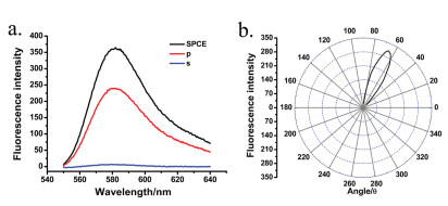
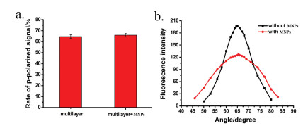
Due to the dependence of SPCE property on the composition of experimental substrate and the weak surface plasmon resonance effect of MNPs, the introduction of MNPs is possible to influence the properties of SPCE. The basic properties of SPCE with MNPs modification were investigated. The SPCE signal possessed highly p-polarized emission with a good angular distribution around 66° (Fig. 2). The comparison of SPCE properties with and without MNPs modification is shown in Fig. 3. It can be seen that the polarization was remained about the same, but the emission distribution angel showed a light displacement with an obvious peak broadening after the modification (the half peak width changed from 14.2° to 23.9°), which was attributed to the change in refractive index and the weak surface plasmon resonance effect of MNPs.

|
Download:
|
| Fig. 2. (a) The polarization and (b) angular distribution for the SPCE signal of multilayer with MNPs (1 mmol/L RhB-2% PMMA spin-coated on 2 nm Cr/7.5 nm Co/3 nm Cr/23 nm Au/Fe3O4 substrate). | |

|
Download:
|
| Fig. 3. The comparisons of (a) the rate of p-polarized signal to total SPCE signal and (b) the angular distribution for multilayer with and without MNPs modification. | |
Considering the enhancement effect of magnetic field modulation and the high loadings, easy modification and purification of MNPs, ssDNA detection system based on the magnetic field modulated SPCE with MNPs modification was established to demonstrate the feasibility of this system used in biodetecting and biosensing technology. The schematic diagram of the modification procedure for ssDNA detection is shown in Fig. 4a and the modification processes are described in Supporting information. After the different amount of target DNA and probe DNA were prepared on the substrates, the SPCE properties were detected. The SPCE properties for concentration of 250 nmol/L were shown as the model. As shown in Fig. S3 (Supporting information), the signal of SPCE held high p-polarization and the emission angle was distributed around 53°. Besides, the angular distributions for other concentrations remained almost constant. The emission angles for other concentrations are shown in Fig. S4 (Supporting information).

|
Download:
|
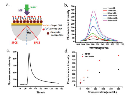
| Fig. 4. (a) The schematic diagram of the modification procedure for ssDNA detection; (b) The SPCE spectra for the different concentration of target DNA modified on magnetic multilayer with the magnetic nanoparticles; (c) The response of the SPCE signal to a pulsed magnetic field for target DNA of 500 nmol/L; (d) The relationship between the SPCE intensity with and without a pulsed magnetic field and the concentration of target DNA. | |
The SPCE signal was detected in real-time followed by the application of a pulsed magnetic field on the substrate to enhance the SPCE signals. Then, the peak value of SPCE signal was compared to the normal SPCE signal without magnetic field application. The SPCE spectra of the target DNA- probe DNA complex prepared on the substrates modified with MNPs are shown in Fig. 4b. The fluorescence signals of SPCE were increased with the increasing concentrations of target DNA after the modification of comple-mentary DNA with fluorophore labeled (Fig. 4b). Moreover, the SPCE signal with MNPs and target DNA modification exhibited a strong response to pulsed magnetic field and the peak intensity was as twice as the normal SPCE intensity (Fig. 4c), which indicates that the magnetic field could enhance SPCE signal. The responses of SPCE signal to pulsed magnetic field for different concentrations of target DNA were shown in Fig. S5 (Supporting information). The dependences of the peak intensity of SPCE after the magnetic field application and the normal SPCE signal without magnetic field application on the concentration of target DNA are shown in Fig. 4d. The apparent enhancement effect of magnetic field on SPCE signal was observed, which proves the sensitivity improvement by a MNPs-assisted magnetic field modulation for the SPCE based biodetector.
In summary, MNPs were introduced in the pulse magnetic field modulated SPCE to enhance the magnetic field modulation with the enhancement factor of 4 when comparing with the magnetic field modulation SPCE without MNPs modification, which should be due to the improvement the MO activity of the metal substrate. The MNPs-assisted magnetic field modulation has the potential to develop a universal method to enhance the signal of SPCE based systems. Utilizing the signal enhancement, a biodetection technique for ssDNA was established, which is a new attempt for using magnetic field modulated SPCE to design a biosensor. After the electrostatic adsorption of MNPs on the multilayer substrate, the magnetic field modulation effect was obvious and the detection signal was enhanced over 100%. There is a possibility to further increase the modulated effect by improving the MO activity and optimizing the properties of SPs, such as introducing new material to the substrate to enhance the SPCE signal and modulation effect in the meantime to improve the performance of magnetic field modulation method. This work proves the enhancement of MNPs on the magnetic field modulation and the feasibility of magnetic field modula-tion SPCE to design a new type of stimulus-response system, which lays the foundation for the application of the magnetic field-modulated SPCE in the field of biosensing and detection.
AcknowledgmentsFinancial support from the National Natural Science Foundation of China (Nos. 21874110, 21375111, 21505109, 21521004 and 21804098), the Fund of the Ministry of Education of China (No. IRT17R66), and Scientific and Technological Innovation Programs of Higher Education Institutions in Shanxi (No. 201802104) is gratefully acknowledged.
Appendix A. Supplementary dataSupplementary material related to this article can be found, in the online version, at doi:https://doi.org/10.1016/j.cclet.2019.06.045.
| [1] |
I. Gryczynski, J. Malicka, Z. Gryczynski, et al., Anal. Biochem. 324 (2004) 170-182. DOI:10.1016/j.ab.2003.09.036 |
| [2] |
H. Mishra, A. Dragan, C.D. Geddes, J. Phys. Chem. C 115 (2011) 17227-17236. DOI:10.1021/jp106671t |
| [3] |
D.G. Zhang, K.J. Moh, X.C. Yuan, Opt. Express 18 (2010) 12185-12190. DOI:10.1364/OE.18.012185 |
| [4] |
D.G. Zhang, X.C. Yuan, J.H. Teng, Appl. Phys. Lett. 97 (2010) 231117. DOI:10.1063/1.3525576 |
| [5] |
P.K. Badiya, S.G. Patnaik, V. Srinivasan, et al., Chem.Phys.Lett. 685 (2017) 139-145. DOI:10.1016/j.cplett.2017.07.056 |
| [6] |
S.H. Cao, Z.C. Wang, Y.H. Weng, et al., Methods Appl. Fluoresc. 5 (2017) 024006. DOI:10.1088/2050-6120/aa6ab4 |
| [7] |
Y.H. Weng, L.T. Xu, Q. Liu, et al., Phys. Status Solidi A 215 (2018) 1800373. |
| [8] |
P.K. Badiya, V. Srinivasan, S.P. Naik, et al., Plasmonics 13 (2018) 519-524. DOI:10.1007/s11468-017-0538-9 |
| [9] |
J.S. Yuk, E.F. Guignon, M.A. Lynes, Chem. Phys. Lett. 591 (2014) 5-9. DOI:10.1016/j.cplett.2013.10.081 |
| [10] |
J.S. Yuk, E.F. Guignon, M.A. Lynes, Analyst 138 (2013) 2576-2582. DOI:10.1039/c3an00135k |
| [11] |
T. Nht, T. Ktl, J.H. Lee, et al., Small 14 (2018) 1801385. DOI:10.1002/smll.201801385 |
| [12] |
S.H. Cao, Z.X. Zou, Y.H. Weng, et al., Biosens. Bioelectron. 58 (2014) 258-265. DOI:10.1016/j.bios.2014.02.067 |
| [13] |
K.X. Xie, S.H. Cao, Z.C. Wang, et al., Sens. Actuators B:Chem. 253 (2017) 804-808. DOI:10.1016/j.snb.2017.06.099 |
| [14] |
K. Aslan, C.D. Geddes, Anal. Chem. 81 (2009) 6913-6922. DOI:10.1021/ac900973r |
| [15] |
W.P. Cai, Y.Y. Zhai, S.H. Cao, et al., Rev. Sci. Instrum. 87 (2016) 013705. DOI:10.1063/1.4940193 |
| [16] |
K.X. Xie, L.T. Xu, Y.Y. Zhai, et al., Talanta 195 (2019) 752-756. DOI:10.1016/j.talanta.2018.11.112 |
| [17] |
P.K. Badiya, V. Srinivasan, S.P. Naik, et al., Plasmonics 13 (2017) 519-524. DOI:10.1007/s11468-017-0538-9 |
| [18] |
K.X. Xie, S.H. Cao, Q. Liu, et al., Chem. Commun. 51 (2015) 12320-12323. DOI:10.1039/C5CC03400K |
| [19] |
M.G. Manera, E. Ferreiro-Vila, J.M. Garcia-Martin, et al., Biosens. Bioelectron. 58 (2014) 114-120. DOI:10.1016/j.bios.2014.02.003 |
| [20] |
M.G. Manera, E. Ferreiro-Vila, J.M. García-Martín, et al., Sens. Actuators B:Chem. 182 (2013) 232-238. DOI:10.1016/j.snb.2013.02.057 |
| [21] |
Q. Wu, Y. Sun, D. Zhang, et al., Biosens. Bioelectron. 96 (2017) 288-293. DOI:10.1016/j.bios.2017.05.023 |
| [22] |
H. Zhang, C.P. Fu, S.T. Wu, et al., Anal. Methods 11 (2019) 783-793. DOI:10.1039/C8AY02423E |
| [23] |
H. Zhang, C.P. Fu, Y. Yu, Anal. Methods 10 (2018) 624-633. DOI:10.1039/C7AY02727C |
| [24] |
H. Watarai, M. Suwaa, Anal. Chim. Acta 690 (2011) 137-147. DOI:10.1016/j.aca.2011.02.019 |
| [25] |
Z. Chen, C. Wu, Z.F. Zhang, et al., Chin. Chem. Lett. 11 (2018) 1601-1608. |
| [26] |
R. Kawabata, T. Mizoguchi, A. Kandori, Rev. Sci. Instrum. 87 (2016) 035112. DOI:10.1063/1.4943256 |
| [27] |
X. Yu, Y. Sun, C.Z. Jiang, et al., J. Sep. Sci. 25 (2012) 3403-3411. |
| [28] |
W.H. Ji, Z.B. Xiao, G.Y. Liu, et al., Chin. Chem. Lett. 28 (2017) 1829-1834. DOI:10.1016/j.cclet.2017.06.024 |
| [29] |
M. Dowaidar, H.N. Abdelhamid, M. Hällbrink, et al., Sci. Rep. 7 (2017) 9159. DOI:10.1038/s41598-017-09803-z |
| [30] |
Y. Yang, Y. Zhang, J.C. Shen, et al., Chin. Chem. Lett. 27 (2016) 891-895. DOI:10.1016/j.cclet.2016.01.060 |
| [31] |
B. Sepúlveda, A. Calle, L.M. Lechuga, et al., Opt. Express 31 (2006) 1085-1087. DOI:10.1364/OL.31.001085 |