 2019, Vol. 30


b Key Laboratory of Organo-Pharmaceutical Chemistry, Gannan Normal University, Ganzhou 341000, China;
c Beijing Advanced Innovation Center for Soft Matter Science and Engineering, Beijing University of Chemical Technology, Beijing 100029, China
Photodynamic therapy (PDT), which combines a photosensitizer (PSs) and light to generate reactive oxygen species (ROS) to bring about phototoxicity, has been widely studied and applied in anticancer and antibacterial field during the past decades [1-4]. As ROS (mainly singlet oxygen, 1O2) are highly reactive, and can degrade a majority of cellular components like proteins, DNA and RNA, resistance is not likely to be developed in photodynamic cancer therapy and photodynamic antibacterial therapy, which makes PDT an ideal alternative for traditional clinical cancer treatment and pathogen infection therapy that always suffered from drug-resistance [5]. Moreover, by utilizing the fluorescence of PSs, fluorescence image-guided PDT has received more and more attention because of the intrinsic advantages of fluorescence imaging, including low cost, superb sensitivity, excellent temporal resolution, noninvasive and real-time imaging ability [6-8].
However, the ROS generated by PSs in PDT is mainly 1O2, which is a short-living species (< 0.04 μs) with a low apparent diffusion coefficient (< 0.02 mm) [9], and thus the active sphere of 1O2 is quite limited in biological systems, implying the great significance of the binding ability of PSs toward 1O2 biotargets. Intracellular proteins are reported to be the major 1O2 biotarget, in view of their abundance within cells [10, 11]. Thus studies on the interactions between PSs and intracellular proteins and the subsequent degradation of intracellular proteins by 1O2 are of great interest for the development of PSs with high efficiency and have become a research hotspot in PDT [12, 13].
Unfortunately, traditional PSs such as porphyrins and phthalocyanines usually suffered from aggregation caused quenching (ACQ) effect due to hydrophobic π-π stacking, resulting in aggregate formation in aqueous media or after they accumulated in cells and tissues, which can dramatically reduce their fluorescence emission and ROS production. Worse more, the formed aggregates will also impede the interaction between PSs and intracellular proteins and further reduce the photo-sensitizing efficiency of PSs, leading to low PDT efficiency. To disrupt the intermolecular π-π stacking of PSs, researchers try to decorate porphyrins with various bulky groups [14-16]. Mean-while, some incident problems also start to emerge prominently, such as poor water solubility, poor chemical/photo stability or complicated synthetic processes. Based on the aforementioned situation, it is highly challenging and rewarding to develop a simple and highly efficient PS that can overcome the above disadvantages.
Aggregation-induced emission (AIE) is a unique photophysical phenomenon, providing high possibility to solve the ACQ problem. Generally, luminogens with AIE feature (AIEgens) are non-emissive or weakly emissive in solutions but become strong emitters with excellent photoluminescence (PL) quantum yields in aggregated state or the solid state [17-21]. Most AIEgens possess a twisted molecular structure with weakened intermolecular π-π inter-actions in the aggregated state, which is favorable to alleviate emission quenching. Motivated by the structural trait of AIEgens, we envision confidently that the introduction of AIEgen to porphyrin derivatives will be conducive to eliminate porphyrin's ACQ problem and increase its ROS efficiency.
In this contribution, we try to use commercially available AIEgen, 1, 1, 2-triphenyl-2-(4-bromomethylphenyl)ethylene (TPE CH2Br), to decorate commercially available porphyrin, 5, 10, 15, 20-tetra(4-pyridyl)porphyrin (TPyP), with the aim to eliminate the intermolecular π-π interaction of porphyrin and increase the ROS generation efficiency. An AIEgen decorated prophyrin (TPETPyP) can be easily obtained via a simple one-step reaction to form organic salts with moderate water solubility (Fig. 1A and Scheme S1 in Supporting information). The ROS generation efficiency of TPETPyP can be enhanced to 0.85 due to the inhibition of π-π stacking and the introduction of pyridinium salts which renders TPETPyP water solubility. TPETPyP exhibited great binding affinity to model proteins (BSA and lysozyme), which is favorable in PDT considering the limited active sphere of 1O2. Furthermore, TPETPyP also displayed efficient protein photo-cleaving ability with the aid of visible light, indicating high application potential in PDT. Future work to investigate the photodynamic anticancer and photodynamic antimicrobial ability of TPETPyP was on the way in our lab. This work will not only provide an easy way to decorate traditional prophyrins to obtain good PSs with high photosensitizing efficiency, but also open up a new application point of AIEgens in PDT.

|
Download:
|
| Fig. 1. (A) Structure of TPETPyP. (B) UV–vis and PL spectra of TPETPyP in PBS solution. | |
The synthetic route to TPETPyP was shown in Scheme S1. Generally, TPE-CH2Br and TPyP in DMF was heated at 80 ℃ under a nitrogen atmosphere. TPETPyP was easily obtained by recrystalli-zation. We conducted 1H NMR, 13C NMR and HRMS to confirm the structure of TPETPyP. Details can be found in Supporting information (Figs. S1-S3 in Supporting information).
With TPETPyP in hand, we first investigated its UV-vis and PL spectra in PBS (5 mmol/L, pH 7.4). As shown in Fig. 1B, TPETPyP showed a typical intense Soret band at 439 nm (ε = 1.27 × 10⁵ L mol -1 cm -1) and four Q bands at 527, 563, 594 and 651 nm, respectively. TPETPyP also showed typical TPE absorption band at 317 nm. Upon light excitation, TPETPyP exhibited two emission peaks at 668 and 723 nm, which is consistent with reported porphyrin derivatives [22, 23].
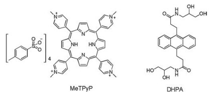
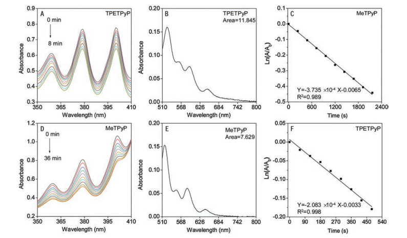
To test whether the bulky TPE pendants can help to reduce the intermolecular π-π stacking of porphyrin and as a result enhance the ROS generation, we examined the 1O2 production ability of TPETPyP in PBS by measuring the degradation rate of N, N'-di-(2, 3-dihydroxypropyl)-9, 10-anthracenedipropanamide (DHPA) using 5, 10, 15, 20-tetrakis(1-methyl-4-pyridinio)porphyrin tetra-(p-toluenesulfonate) (MeTPyP) as a reference (Scheme 1). As displayed in Fig. 2, upon visible light irradiation, both MeTPyP and TPETPyP can generate 1O2, which can react with DHPA, leading to the decrease of its absorbance. And TPETPyP gave a more rapid degradation rate of DHPA. According to the 1O2 quantum yield of MeTPyP (0.74) [24] and Eq. (1) (where Φ is the 1O2 quantum yield, A represents the light absorbed by the PSs and K is the decomposition rate constant of DHPA by PSs), the 1O2 quantum yield of TPETPyP was calculated to be 0.85 in PBS, which is much higher than the reference compound MeTPyP. The high 1O2 quantum yield of TPETPyP can be attributed to the following two aspects: firstly, the bulky TPE pendants may isolate the porphyrin core and greatly impede the intermolecular π-π stacking of porphyrin, which can be proved by comparing the 1O2 quantum yield of TPETPyP (0.85) to that of MeTPyP (0.74); and secondly, the four pyridinium salts rendered the whole TPETPyP molecule water solubility, which further enhanced its ROS generation ability, in comparison to the low 1O2 quantum yield of TPyP (0.03, Fig. S4 in Supporting information). Additionally, TPETPyP maintains high ROS generation ability with pH values varied from 4 to 7.4 (Fig. S5 in Supporting information), indicating its potential application in different biological samples. Moreover, the photostability of TPETPyP was further examined according to reference method [25]. Results in Fig. S6 (Supporting information) demonstrated that, the emission intensity of TPETPyP exhibited slight decrease (10%) after 80 min light irradiation, indicating the good photo-stability of TPETPyP.

|
(1) |

|
Download:
|
| Scheme 1. Structures of MeTPyP and DHPA. | |

|
Download:
|
| Fig. 2. Chemical trapping measurements of the 1O2 quantum yield. Photodegradation of DHPA with TPETPyP (A) and MeTPyP (D). The absorption peak area of TPETPyP (B) and MeTPyP (E). The decomposition rate constants of DHPA by TPETPyP (C) and MeTPyP (F). | |
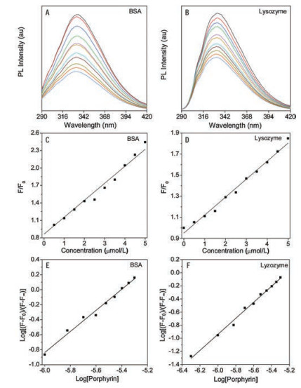
Encouraged by the high 1O2 quantum yield of TPETPyP in PBS, we inferred that TPETPyP could act as an efficient PS in PDT. Accordingly, we try to test the interactions between TPETPyP and the intracellular proteins, the major 1O2 biotarget, and further quantitatively analyze the interactions. Here, we chose bovine serum albumin (BSA) as the model protein to do the related experiments, because BSA is a serum albumin that not only is the most common model protein but also has been utilized in targeting delivery of phototherapeutic sensitizers. The fluorescence titration experiments showed that the binding of TPETPyP to proteins can greatly quench the intrinsic emissions of both BSA and lysozyme, from which the binding mode and binding strength can be deduced (Fig. 3). The fluorescence quenching rate constants can be calculated based on the Stern-Volmer equation (Eq. (2)) [26, 27], where κq, τ0 and [Q] are the fluorescence quenching rate constant, the fluorescence lifetime of protein in the absence of the quencher, and the concentration of the quencher, i.e., the concentration of TPETPyP here, respectively. F0 and F are the fluorescence intensities in the absence and presence of the quencher, respectively.

|
(2) |

|
Download:
|
| Fig. 3. Fluorescence quenching of BSA (A) or lysozyme (B) by TPETPyP. Stern-Volmer plot of the BSA (C) or lysozyme (D) fluorescence quenching by TPETPyP. Double logarithmic plots of BSA (E) and lysozyme (F) fluorescence quenching by TPETPyP. | |
It is well known that the fluorescence quenching mechanism can be proceeded via different mechanisms, such as dynamic and static quenching. According to the fluorescence titration data and the lifetime of 10 -8 s for BSA [28], the quenching rate constant of TPETPyP is calculated to be 3.09×10¹³ L mol-1 s-1. Similarly, 3.09 the quenching rate constant of TPETPyP toward lysozyme is deter-mined to be 1.706×10¹³ L mol-1 s-1. Since these quenching rate constants are 3 orders of magnitude higher than the collision quenching constant 2.0×10¹⁰ L mol -1 s -1) [29] for various quenchers with biomolecules, a static quenching mechanism, that is, the binding of TPETPyP towards BSA or lysozyme can be concluded.
For static quenching mechanism, the binding constant and the number of binding sites can be calculated from Eq. (3) [26, 30]:

|
(3) |
where K and n are the binding constant and the number of binding sites, F0 and F are the fluorescence intensities in the absence and presence of the quencher, respectively, [TPETPyP] is the concen-tration of the quencher, F∞ is the fluorescence at the infinite concentration of TPETPyP (calculated as the intercept of 1/(F0-F) vs. 1/[TPETPyP]. Therefore, K and n can be calculated by plotting log [(F0-F)/(F-F∞)] versus log[TPETPyP]. Results in Figs. 3E and F exhibited that the binding constants were 1.13×10⁵ L/mol and 1.70×10⁵ L/mol for BSA and lysozyme, respectively. The binding numbers of TPETPyP with BSA and lysozyme are 1.0 and 1.2, respectively. These results indicated that TPETPyP can interact with BSA and lysozyme with high binding affinity, which is favorable for the photosensitizing effect considering the limited active sphere of 1O2 in biological systems.
Encouraged by the high 1O2 quantum yield of TPETPyP and high binding affinity of TPETPyP toward proteins, we further evaluated the photosensitizing ability of TPETPyP by testing its protein photo-cleaving ability, since intracellular proteins are the main biotarget of 1O2. Protein electrophoresis experiments were carried out using BSA as the model protein. As shown in Fig. 4A, TPETPyP displayed a concentration dependent photocleaving ability toward BSA. TPET-PyP lead to almost complete degradation of BSA at a concentration of 10 μmol/L. While in stark contrast (Fig. 4B), the photocleavage was either not observed or greatly inhibited in the dark or by the addition NaN3, indicating the involvement of 1O2. All these results indicated that the AIEgen decorated porphyrin TPETPyP can act as an efficient protein photocleaving agent, and is a promising PS in PDT.

|
Download:
|
| Fig. 4. Photocleavage of BSA in 20% DMSO/PBS solution upon 2 h of irradiation (λ≥470 nm) and analyzed by 8% SDS-PAGE: (A) lane 1: BSA alone; lane 2: BSA + TPETPyP (10 μmol/L) in the dark; lane 3: BSA + TPETPyP (1 μmol/L); lane 4: BSA + TPETPyP (2 μmol/L); lane 5: BSA + TPETPyP (5 μmol/L); lane 6: BSA + TPETPyP (7 μmol/L); lane 7: BSA + TPETPyP (10 μmol/L); lane 8: BSA alone. (B) lane 1: BSA alone; lane 2: BSA + TPETPyP (5 μmol/L) in the dark; lane 3: BSA + TPETPyP (5 μmol/L); lane 4: BSA + TPETPyP (5 μmol/L) + NaN3 (1 μmol/L); lane 5: BSA alone. | |
In conclusion, we have developed an AIEgen decorated porphyrin photosensitizer with water solubility and high 1O2 generation ability. TPETPyP can be easily obtained through a one-step reaction. The bulky AIEgen (TPE pendants) greatly impeded the intermolecular π-π stacking of the porphyrin core, thus eliminated the aggregation and enhanced the 1O2 quantum yield. Meanwhile, the four pyridinium salts formed in TPETPyP rendered the whole molecule water solubility, which further reduced aggregation in aqueous solution and contributed to the high 1O2 generation ability in PBS. TPETPyP exhibited high binding affinity to proteins, which is favorable for the photosensitizing process. Protein electrophoresis experiments demonstrated that upon visible light irradiation, TPETPyP can efficiently photocleave BSA, indicating that TPETPyP can act as an efficient PS in PDT. Further work to evaluate its photodynamic anticancer and photodynamic antimicrobial activity is ongoing in our lab. The easy decorating procedure plus the unique performance of TPETPyP will not only provide a facile method to utilize the bulky AIEgens to eliminate π-π stacking of the porphyrin core but also broaden the application field of AIEgens in PDT.
AcknowledgmentsWe thank the National Natural Science Foundation of China (Nos. 21663005, 21871060, 21804022 and 21702016), the Natural Science Foundation of Jiangxi Province (Nos. 2018ACB21009, 20181BAB213007), the Science and Technology Project of the Education Department of Jiangxi Province of China (No. GJJ170846), the Special Graduate Student Innovation Fund of Jiangxi Province (No. YCX18B007) and Beijing National Laboratory for Molecular Sciences (No. BNLMS201813) for the financial support.
Appendix A. Supplementary dataSupplementary material related to this article can be found, in the online version, at doi:https://doi.org/10.1016/j.cclet.2019.08.037.
| [1] |
M. Lan, S. Zhao, W. Liu, et al., Adv. Healthcare Mater. (2019) 1900132. |
| [2] |
X. Li, S. Lee, J. Yoon, Chem. Soc. Rev. 47 (2018) 1174-1188. DOI:10.1039/C7CS00594F |
| [3] |
M. Wainwright, J. Antimicrob. Chemother. 42 (1998) 13-28. DOI:10.1093/jac/42.1.13 |
| [4] |
N. Tian, Y. Feng, W. Sun, et al., Dalton Trans. 48 (2019) 6492-6500. DOI:10.1039/C9DT00441F |
| [5] |
S. Noimark, C.W. Dunnill, M. Wilson, I.P. Parkin, Chem. Soc. Rev. 38 (2009) 3435-3448. DOI:10.1039/b908260c |
| [6] |
A.S. Klymchenko, Acc. Chem. Res. 50 (2017) 366-375. DOI:10.1021/acs.accounts.6b00517 |
| [7] |
J.F. Lovell, T.W.B. Liu, J. Chen, G. Zheng, Chem. Rev. 110 (2010) 2839-2857. DOI:10.1021/cr900236h |
| [8] |
H.Q. Peng, L.Y. Niu, Y.Z. Chen, et al., Chem. Rev. 115 (2015) 7502-7542. DOI:10.1021/cr5007057 |
| [9] |
P.R. Ogilby, Chem. Soc. Rev. 39 (2010) 3181-3209. DOI:10.1039/b926014p |
| [10] |
F. Kratz, J. Control. Release 132 (2008) 171-183. DOI:10.1016/j.jconrel.2008.05.010 |
| [11] |
M.J. Hawkins, P. Soon-Shiong, N. Desai, Adv. Drug Deliv. Rev. 60 (2008) 876-885. DOI:10.1016/j.addr.2007.08.044 |
| [12] |
G.Y. Jiang, W.H. Lei, Q.X. Zhou, Y.J. Hou, X.S. Wang, Photochem. Photobiol. Sci. 11 (2012) 715-723. DOI:10.1039/c2pp05352g |
| [13] |
S. Tanimoto, S. Matsumura, K. Toshima, Chem. Commun. (2008) 3678-3680. |
| [14] |
K. Liu, Y. Liu, Y. Yao, et al., Angew. Chem. Int. Ed. 52 (2013) 8285-8289. DOI:10.1002/anie.201303387 |
| [15] |
C.C. Chang, M.C. Hsieh, J.C. Lin, T.C. Chang, Biomaterials 33 (2012) 897-906. DOI:10.1016/j.biomaterials.2011.10.018 |
| [16] |
B. Guo, X. Cai, S. Xu, et al., J. Mater. Chem. B 4 (2016) 4690-4695. DOI:10.1039/C6TB01159D |
| [17] |
X. You, H. Ma, Y. Wang, et al., Chem. -Asia J. 12 (2017) 1013-1019. DOI:10.1002/asia.201700243 |
| [18] |
F. Hu, S. Xu, B. Liu, Adv. Mater. 30 (2018) 1801350. DOI:10.1002/adma.201801350 |
| [19] |
D. Wang, M.M.S. Lee, W. Xu, et al., Angew. Chem. Int. Ed. 58 (2019) 5628-5632. DOI:10.1002/anie.201900366 |
| [20] |
C. Chen, H. Ou, R. Liu, D. Ding, Adv. Mater. (2019), doi:http://dx.doi.org/10.1002/adma.201806331.
|
| [21] |
X. Ni, X. Zhang, X. Duan, et al., Nano Lett. 19 (2019) 318-330. DOI:10.1021/acs.nanolett.8b03936 |
| [22] |
K.G. Yu, D.H. Li, C.H. Zhou, J.L. Diao, Chin. Chem. Lett. 20 (2009) 411-414. DOI:10.1016/j.cclet.2008.11.030 |
| [23] |
Q. Zhang, X. Dong, K.P. Wang, et al., Chin. Chem. Lett. 28 (2017) 777-781. DOI:10.1016/j.cclet.2017.03.001 |
| [24] |
F. Wilkinson, W.P. Helman, A.B. Ross, J. Phys. Chem. Ref. Data 22 (1993) 113-262. DOI:10.1063/1.555934 |
| [25] |
X. Zhu, J.X. Wang, L.Y. Niu, Q.Z. Yang, Chem. Mater. 31 (2019) 3573-3581. DOI:10.1021/acs.chemmater.9b01338 |
| [26] |
P. Athina, J.G. Rebecca, A.F. Richard, J. Agric. Food Chem. 53 (2005) 158-163. DOI:10.1021/jf048693g |
| [27] |
Y.Q. Wang, H.M. Zhang, G.H. Tao, S.H. Tang, J. Lumin. 126 (2007) 211-218. DOI:10.1016/j.jlumin.2006.06.013 |
| [28] |
B. Valeur, Molecular fluorescence, Principles and Applications, Wiley Press, New York, 2001 p. 84.
|
| [29] |
J.R. Lakowicz, G. Weber, Biochemistry 12 (1973) 4161-4170. DOI:10.1021/bi00745a020 |
| [30] |
J. Xiao, X. Chen, L. Zhang, et al., J. Agric. Food Chem. 56 (2008) 910-915. DOI:10.1021/jf073036k |