 2019, Vol. 30


Polydiacetylenes (PDAs) are a family of conjugated polymers that have received great interest in the past several decades [1]. It is known that assembled diacetylene monomers (DAs) polymerize to form PDAs under 254 nm UV irradiation or γ-irradiation [2]. Upon formation, PDAs are nonfluorescent and they have a deep-blue color associated with an absorption band centered at ca. 640 nm [3]. Fluorescent probes grow rapidly based on high sensitivity, realtime monitoring and simple operation [4-6]. PDAs are functionalized with various head group most commonly as chemosensors and biosensors. Upon the interaction with metal ions [7-9], organic vapor [10-12], surfactants [13, 14], biomolecules [15-17], etc., PDAs undergo nonfluorescent-to-fluorescent and blue-to-red color transitions [18, 19]. Moreover, temperature [20-22], pH [23-25] changes as well as mechanical stress [26] can also cause emission and color changes of selected PDAs. Also, conductive materials have been combined with PDAs to fabricate materials that are useful for electrochemical sensing [27] or as energy devices [28]. Furthermore, by incorporating selected polymers into PDAs, it is possible to fabricate new materials by using surface coating [29], ink-jet printing [30] and electrostatic spinning [31] techniques. Kim et al. have carried out seminal studies focusing on applications of these types of PDA systems, which have led to the development of novel materials that are humidity-responsive [32] or that can be utilized for fingerprint-recognition [33] and solvent-sensing [34]. In 1993, Charych et al. first reported the development of a PDA-based biosensor for the detection of influenza virus [35]. Since that time, numerous PDA-based biosensors have been developed, and examples have been provided showing their use in monitoring biological process and systems, as well as diagnosing and treating diseases. Emphasis is given in this review to recent advances made in the development of PDA-based biosensors for detection and imaging of disease sites or substances and for delivering therapeutic drugs.
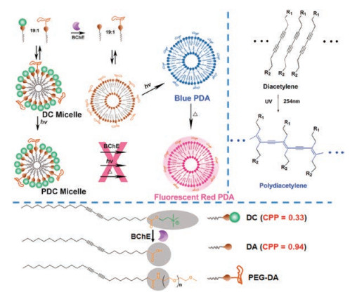
2. PDAs for sensing enzyme activityEnzymes are biocatalysts that promote a wide variety of biological process and, consequently, their functions are closely related to numerous diseases. For example, butyrylcholinesterase (BChE), which is mainly distributed in peripheral tissues, catalyzes hydrolysis of the neurotransmitter acetylcholine (ACh). Overexpression of BChE is known to be associated with Alzheimer's disease (AD) [36-38]. Guo et al. designed a BChE detection system, which is derived from a choline (DC) modified DA and a poly- (ethylene glycol) linked DA (PEG-DA) (Fig. 1) [39]. PEG doping in an optimized DC/PEG-DA molar percent ratio of 95% was found to greatly improve the solubility of the DA vesicles, the polymerization process and the colorimetric response Two methods were assessed to fabricate the PDA based BChE sensing system. The first involves incubation of the enzyme with a mixture of the DA vesicles followed by 254 nm UV irradiation to induce polymerization. This process results in formation of a deep blue colored PDA, which undergoes a blue-to-red color and nonfluorescent-to-fluorescent transition at 37 ℃. However, an alternative procedure in which polymerization of the DA vesicles occurs prior to incubation with BChE, produces a PDA that does not undergo an emission and color change at 37 ℃. By using this protocol, BChE overexpressed in the cytoplasm of HepG2 cells was detected in the form of red fluorescence images. In addition, movement of overexpressed BChE from the cytoplasm to cell nucleus during cell apoptosis was monitored successfully utilizing the probe.

|
Download:
|
| Fig. 1. Programmed process of photopolymerization and generating red fluorescent PDA by controlling the order of enzyme (BChE), UV light and heat. | |
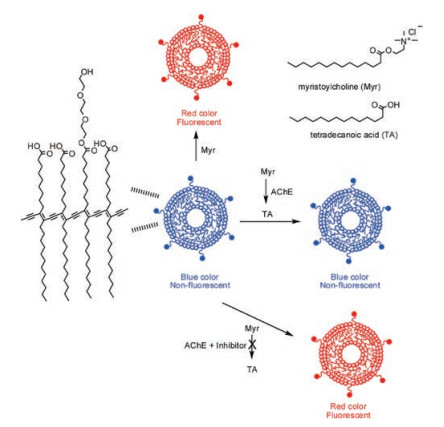
Our group developed a novel PDA-based probe for detection of acetylcholinesterase (AChE) (Fig. 2), created by using irradiation induced polymerization of an assembled 3:7 mixture of 2-(2-(2- hydroxyethoxy)-ethoxy)ethyl pentacosa-10, 12-diynoate (PCDA-HEP) and pentacosa-10, 12-diynoic acid (PCDA) [40]. Addition of myristoylcholine (Myr) and AChE to the PDA liposomes does not promote a blue-to-red color change in response to AChE catalyzed hydrolysis of Myr. The reason for this is that the hydrolysis product tetradecanoic acid (TA) possesses a negative charge that is present in the carboxylate headgroups in the PDA. However, addition Myr and AChE along with an AChE inhibitor causes the PDA to undergo a blue-to-red color change that can be easily observed by using the naked eye. Thus, it appears that interaction between the positively charged head group in Myr and the negatively charges carboxylates in the PDA stimulate the blue-to-red color transition.

|
Download:
|
| Fig. 2. Schematic illustration of AChE recognition by PDAs in the presence of inhibitor and in the absence of inhibitor. Copied with permission [40]. Copyright 2013, American Chemical Society. | |
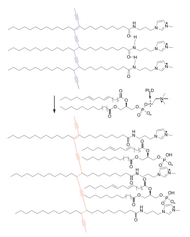
Phospholipase D (PLD) is a cellular enzyme that regulates important biological processes [41, 42]. In another study in our group, we developed a PDA-based sensor that detects the activity of PLD (Fig. 3) [43]. The probe was formed by polymerization of imidazolium group terminated DAs, which generates a PDA that contains positively charged headgroups. Treatment of a phospholipid with PLD in the presence of the PDA leads to production of a negatively charged phosphate monoester that promotes the color transition and fluorescence enhancement of the PDA. However, the original blue color of the PDA is preserved when a PLD inhibitor is present in the mixture. Interestingly, a test strip loaded with the imidazolium group functionalized PDA serves as convenient PLD detection system.

|
Download:
|
| Fig. 3. Proposed mechanism of PLD activity detection based on synthesized PDAs. Copied with permission [43]. Copyright 2019, Elsevier. | |
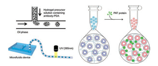
The phosphinothricin acetyltransferase (PAT) gene is a marker in transgenic plants [44]. Jeon et al. designed a PAT probe that is comprised of PDAs encapsulated within immunohydrogel beads to prevent their typical instability and low sensitivity (Fig. 4) [45], In this effort, the probe was generated by first polymerizing a 8:2 mixture of 10, 12-tricosadiynoic acid (TCDA) and 1, 2-dimyristoyl-phosphatidylcholine (DMPC) and then combining the resulting PDA with an antibody of PAT. It was found that addition of different concentrations of PAT to a solution of the probe promotes a blue-to-red color transition only when the PAT concentration is greater than 2 μmol/L. Thus, the PDA based probe itself does not meet the sensitivity requirements of GM biosensors. However, embedding the PDA-antibody conjugate in immunohydrogel beads generates a PAT probe that has improved stability and a greatly enhanced limit of PAT detection of 20 nmol/L.

|
Download:
|
| Fig. 4. The preparation process of immunohydrogel beads encapsulating PDAs and colorimetric response in the presence of PAT protein. Copied with permission [45]. Copyright 2015, American Chemical Society. | |
3. PDAs for tumor-targeting
Cancer is a world-wide problem [46, 47] that results in a larger number of deaths than those caused by all other maladies. Among the major hurdles in developing cures for cancer is the lack of clarity about mechanisms for tumor growth and the serious side effects associated with most cancer chemotherapies [48, 49]. The ability to identify and therapeutically target tumors at early stages is particularly critical for survival. As a result, a great effort has been given to devising theranostic agents that simultaneously detect and treat tumors [50, 51]. Tumor-targeting, PDA-based fluorescence probes have been designed for this purpose. For example, Wu and co-workers fabricated a system comprised of PDA and encapsulated camptothecin (CPT) for treatment of ovarian cancer (Fig. 5) [52]. The probe PDA-CPT was synthesized by 254 nm UV irradiation induced copolymerization of a 1:1 mixture of the peptide functionalized diacetylene monomer PA1 and diacetylene monomer PA2, which possesses a morpholine-substituted 1, 8- naphthalimide as a fluorophores in the presence of CPT. Absorption and fluorescence spectroscopy was used to show that effective polymerization had occurred and that CPT was successfully loaded in the PDA micelles. In addition, a study of the micelles demonstrated that CPT release takes place more rapidly under acid rather than neutral conditions. Significantly, the green fluorescence of PDA-CPT following incubation with SKOV-3 cells well overlaps with the red fluorescence of Lyso-Tracker Red showing that the probe becomes located in the acidic environment of lysosome lumen. Moreover, the results of MTT assays indicate that the viability of cells treated with PDA-CPT is far lower than those treated with CPT alone, and that. treatment of mice containing a SKOV-3 tumor with PDA-CPT causes a larger decrease in tumor volume than those treated only with CPT.

|
Download:
|
| Fig. 5. Polymerization of PDAs with packaged antitumor drug CPT and delivery mechanism of PDAs-CPT in ovarian cancer cells. Reproduced with permission [52]. Copyright 2017, Royal Society of Chemistry. | |
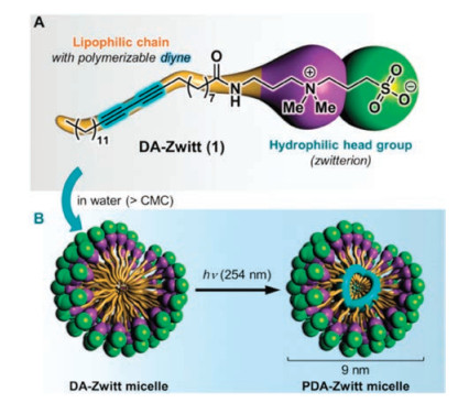
Doris and Ducongé et al. showed that a zwitterionic DA monomer, containing dimethylamine and sulfonyl hydroxide moieties (Fig. 6) [53] form self-assembled PDA micelles (PDA-Zwitt), which have a tumor-targeting ability in a murine model for breast cancer. Near Infrared (NIR) planar imaging of mice, intravenously injected with PDA-Zwitt micelles loaded with the carbocyanine dye DiR, shows that DiR is released from the probe and diffuses throughout mice tissue surrounding the tumor area. As a result, the area surrounding the tumor becomes intensely fluorescent 24 h post-injection and a sharp contrast develops between non-fluorescing normal tissues. After 24 h, the fluorescence decreases gradually until it completely disappears owing to excretion of PDA-Zwitt micelles. In addition, inspection of computed tomography (CT) and free-space fluorescence diffuse optical tomography (fDOT) images indicate that PDA-Zwitt is distributed at the border of tumor cells.

|
Download:
|
| Fig. 6. Structure of DA-Zwitt and generated PDA-Zwitt micelle under 254 nm UV. Copied with permission [53]. Copyright 2015, CCC Republication. | |
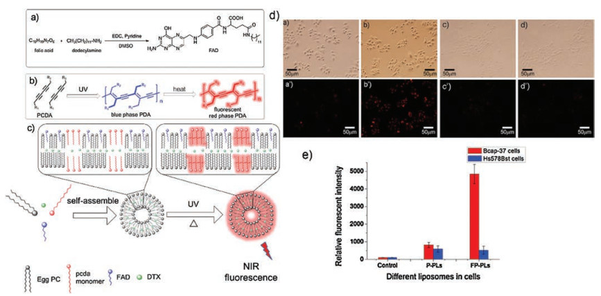
An et al. prepared the PDA liposomes P-PLs and the folate containing analog FP-PLs, both containing encapsulated Docetaxel (DTX) as a model drug (Fig. 7) [54]. P-PLs and FP-PLs containing different concentrations of DTX were incubated with both Bcap-37 breast cancer cells and Hs578Bst normal cells. MTT assays indicate that P-PLs does not differentiate between the two kinds of cells whereas FP-PLs, as a consequence of the tumor-targeting ability arising from the presence of folate receptors overexpressed in cancer cells, recognizes the Bcap-37.

|
Download:
|
| Fig. 7. (a) Structure of FAD. (b) Photopolymerization process of PDAs. (c) Composition of FP-PL. Fluorescence images of P-PLs (a, a'), FP-PLs (b, b'), P-PLs (c, c'), FP-PLs (d, d') upon incubation with Bcap-37 (a, b, a', b') and Hs578Bst (c, d, c', d') cells. (d) Fluorescence intensity of P-PLs and FP-PLs in Bcap-37 and Hs578Bst cells. Reproduced with permission [54]. Copyright 2015, Elsevier. | |
Chen et al. synthesized lactoferrin-conjugated magnetic PDA-assembled nanocarriers (Lf-Cur-PDNC) for tumor imaging and therapy (Fig. 8) [55]. Curcumin (Cur) in the probe serves as an antitumor drug and Lf plays dual roles of delivering Cur by assisting its crossing of the blood-brain barrier (BBB) and targeting tumors. Lf-Cur-PDNC displays a higher toxicity to cancer cells than that of its analog PNDC, which does not contain the Lf moiety. Moreover, the red fluorescence of Lf-PNDC enables tumor-targeting and monitoring of endocytosis. Analysis of magnetic resonance (MR) images shows that the intensity of the signal associated with Lf-Cur-PNDC in tumors decreases 72 h post-incubation, and that Cur from Lf-Cur-PNDCs is more greatly localized in the brain tumor. Finally, the volume of a tumor treated with Lf-Cur-PDNC decreases significantly.

|
Download:
|
| Fig. 8. Synthesis of drug nanocarriers (a) and preparation of Lf-Cur-PDNCs by EDC and NHS (b). Copied with permission [55]. Copyright 2016, John Wiley and Sons. | |
The long polypetide magainin Ⅱ was linked to PDAs in a system designed by Wu and Tian et al. for tumor treatment (Fig. 9) [56]. Cell images indicate that the PDA micelles mainly localize in the cytoplasm and on cell membranes. The IC50 value of the unique PDA is 5-fold higher in A549 cells compared with other literatures and the PDAs are highly biocompatible. Studies with A549 xenograft mice showed that treatment with the Magainin-linked PDA in phosphate buffered saline (PBS) leads to inhibition of tumor growth while causing nearly no effects on normal tissues.

|
Download:
|
| Fig. 9. Structure of MGN-Ⅱ-PDA micelles (a) and scheme illustration of tumor-targeting (b). Copied with permission [56]. Copyright 2014, Royal Society of Chemistry. | |
4. PDAs for cell-imaging and monitoring cellular activity
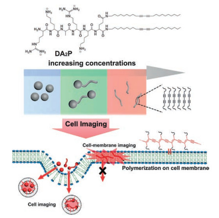
Cell imaging is a necessary component of analyzing biological systems and it is particularly beneficial for obtaining accurate clinical diagnoses [57]. Schmuck et al. synthesized an interesting DA monomer functionalized with the peptide DA2P (Fig. 10) [58]. At low concentrations this DA forms tadpole- and bola-shaped nanostructures and at high concentrations it assembles into nanofibers. The DA monomers polymerize on HeLa cells to form a red fluorescing PDA. At a concentration of 50 μmol/L, the PDA well co-localizes with lysosome and at of 250 μmol/L it becomes located in the cell membrane. As a result, concentration-dependent cell imaging is made possible by using this probe.

|
Download:
|
| Fig. 10. Structure of DA2P and different nanostructures based on increasing concentrations and cell-imaging of different nanostructures in HeLa cells. Copied with permission [58]. Copyright 2017, John Wiley and Sons. | |
Chan et al. designed a flexible bioimaging system that contains PDA encapsulated semiconducting polymer dots (Fig. 11) [59]. In this construct, carboxylate headgroups on the PDA attach to the P-dots, which is employed to create a FRET system comprised of PFBT-DBT polymer and the photostable NIR dye NIR695. Inspection of cell images shows that the PDA, containing enclosed NIR685- embedded P-dots, aggregate in the acidic environments of endosomes or lysosomes. Owing to the presence of the carboxylate groups on the surfaces of these assemblies, it is possible to covalently link streptavidin for recognition of a biotinylated antibody on the cell surface. As a result of the red fluorescence arising from the PDA, this probe can be used for selective bioimaging.

|
Download:
|
| Fig. 11. Preparation of PDA-enclosed NIR dye-doped P-dots and SA modified PDA-coated P-dots and application for cell surface labeling. Copied with permission [59]. Copyright 2014, American Chemical Society. | |
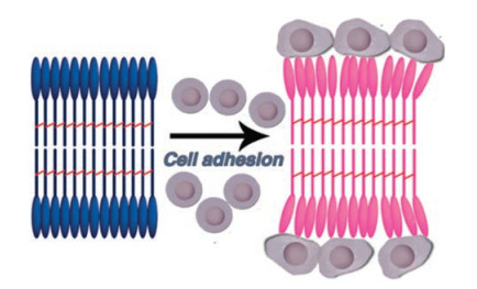
Löwik et al. developed a peptide modified PDA that can be utilized to sense cell adhesion (Fig. 12) [60]. The amphiphile PDA fibers, containing Glu-terminal amino acid moieties and the tripeptide Arg-Gly-Asp (RGDS) amphiphile in a ratio of 6:1, were found to undergo a blue-to-red color change when the temperature is increased or when they adhere to cells. The cytotoxicity of these amphiphile fibers is lower than those of other terminal amino acid modified amphiphiles or when other ratios of the Glu- and RGDS-amphiphile fibers are used. This sensor has the potential of being employed for monitoring cellular activity.

|
Download:
|
| Fig. 12. Schematic illustration of colorimetric response toward cell adhesion. Copied with permission [60]. Copyright 2015, Royal Society of Chemistry. | |
5. PDAs for microorganism detection
Microorganisms, including bacteria, virus and fungi, are the causes of many human diseases. Because of their involvement in human health, these microscopic species are important targets in the development of new biosensor systems [61]. Especially important in this regard are strategies for the design of theranostic agents that detect bacteria or viruses and simultaneously deliver antimicrobial drugs. In a study aimed at this goal, Thet et al. developed PDA liposomes that contain carboxyfluorescein for fluorescence detection of bacterial toxins (Fig. 13) [62]. Liposomes consisted of lipids, cholesterol and TCDA, three kinds of lipids with different chain length had an effect on polymerization. 1, 2- Distearoyl-sn-glycero-3-phosphocholine (DSPC) introduced in liposomes made the polymerization easier compared with other two lipids due to the similar chain length with TCDA, the liposomes show good stability accompanied with very little fluorescence leakage 7 days later. Three kinds of liposomes all responded to various bacterial supernatants and toxins. The degree of polymerization influenced the response to S. aureus LAC supernatant and δ-toxin, however show negligible affection to P. aeruginosa PAO1 supernatant and rhamnolipid.

|
Download:
|
| Fig. 13. Schematic illustration of liposomes consisted of lipids, cholesterol, carboxyfluorescein and TCDA in response to pathogenic bacteria. Copied with permission [62]. Copyright 2014, American Chemical Society. | |
A new PDA based probe for sensing and killing bacteria was designed by Yoon et al. (Fig. 14) [63]. The probe PDA-I-I was generated by photopolymerization of a 1:1 mixture of PCDA-Imethanol (monomer 1) and PCDA-Im (monomer 2). An investigation of antibacterial activities of the PDAs showed that PDA-I-I at a concentration of 10 μmol/L inhibits growth of Gram-positive pathogens and that its inhibition of Gram-negative pathogens does not occur until its concentration reaches at least 100 μmol/L. Analysis of TEM images shows that the antimicrobial activity of the probe is associated with electrostatic interaction induced destruction of the cell membrane. In addition, color changed from blue to red with the enhancement of fluorescence. This probe had promising application prospect as antimicrobial agent.

|
Download:
|
| Fig. 14. Structure of polymerized PDA-I-I and TEM images of (a) S. aureus, (b) MRSA, (c) ESBL-EC cells in the presence/absence of PDA-I-I. Reproduced with permission [63]. Copyright 2016, Elsevier. | |
Lee and Mooney et al. conducted an investigation exploring the bacterial detection properties of amine-functionalized PDA liposomes (Fig. 15) [64]. After the incubation with three bacterial strains, NCIB3610 show obvious color transition, however little color change could be observed in PA14 even that the growth of bacteria in PA14 was more than NCIB3610, therefore indirect interaction between PDAs and bacteria existed. The difference between NCIB3610 and SSB466 was that NCIB3610 released surfactin but SSB466 did not, PDAs show color changes toward surfactin alone. While NCIB3610 solution was dropped to PDA solution, fluorescence appearedin the region without bacteria, which indicated the indirect interaction between PDAs and surfactin. The detection method was also a good choice for detection of bacteria.

|
Download:
|
| Fig. 15. Proposed mechanism of PDAs for bacteria-sensing. Copied with permission [64]. Copyright 2016, Royal Society of Chemistry. | |
Jenkins and Fan et al. developed a dual functioning wound dressing based method for bacterial detection (Fig. 16) [65]. The system is comprised of a multilayer network in which one layer consists of a hydrogel encapsulating PDA vesicles containing an antibacterial agent. The other layer is also comprised of a hydrogel encapsulating vesicles, but in this case it contains the dye carboxyfluorescein for fluorescence visualization. The system undergoes a naked eye observable color change when it inhibits a bacteria inflection. Silver nitrate was chosen as the antimicrobial agent in this PDA-based system, which effectively kills and inhibits Gram-negative and Gram-positive bacteria, respectively. The results of studies indicate that use of this wound dressing approach accelerates cell network formation and that cell viability exceeds 85% over one week. An assessment of the use of Gentamicin sulphate as antimicrobial agent demonstrates that the wound dressing approach accelerates wound healing in mice tissue. The PDA probe has the beneficial feature that it can be utilized to both detect and inhibit bacterial infections.

|
Download:
|
| Fig. 16. Construction diagram of nanocomposite wound dressing and schematic illustration of wound dressing respond to pathogenic bacteria. Copied with permission [65]. Copyright 2018, Elsevier. | |
Ahn et al. synthesized PDA/anti-HBs complexes for detection of the hepatitis B surface antigen (HBsAg) (Fig. 17) [66]. PDA-ABA vesicles polymerize under UV irradiation to produce PDAs that were then coated with the hepatitis B surface antigen (HBsAb) and BSA was utilized to load PDA/HBsAb on the NC membrane. PDA/ HBsAb undergo a color and fluorescence change after being combined with HBsAg. Red fluorescence of PDA complexes could be observed clearly upon addition of only 0.1 ng/mL of HBsAg. In contrast, fluorescence of a commercial PS bead occurs when 1 ng/mL of HBsAg is present and no visible color transition takes place at this concentration. The observations suggest that the PDA complexes are highly sensitive and, thus, they can be used in a commercial kit application.

|
Download:
|
| Fig. 17. (a) Preparation of PDA/HBsAb complexes. (b) Schematic diagram of PDA/HBsAb complexes for HBsAg detection on NC membrane. Copied with permission [66]. Copyright 2017, John Wiley and Sons. | |
6. PDAs for biomolecule detection
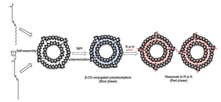
The basic amino acid L-arginine plays a key role in several biological functions including cell division, wound healing, immune function and release of hormones, the monitor of L-arginine is conducive to disease diagnosis [67-70], thus the detection of L-arginine is significant. L-Lysine is an essential amino acid needed for proper growth of humans and the production of carnitine. The absence of balanced amounts of lysine in the diet can cause several severe diseases [71-73]. Therefore, the recognition of lysine is necessary. Jung et al. fabricated a unique L-lysine and L-arginine probe by polymerizing vesicles comprised of a 1:9 mixture of amine group containing β-cyclodextrin (β-CD) derivatives and a PCDA monomer (Fig. 18) [74]. The PDA vesicles undergo a transition from blue-to-red upon addition of arginine or lysine. The cause of this change is insertion of the amino acids into the CD cavity of CD through interactions between their carboxylate groups with the amine moieties in the CD containing PDA network.

|
Download:
|
| Fig. 18. Schematic illustration of β-CD conjugated PDA for arginine (R) and lysine (K) recognition. Reproduced with permission [74]. Copyright 2016, Nature. | |
Our group developed a PDA that is formed from a 9:1 mixture of PCDA and a pyrene containing PCDA-PY (Fig. 19) [75]. Photopolymerization of this mixture generates a PDA in which fluorescence from the pyrene fluorophore is quenched. The binary system PDAs/Mg2+, created by addition of Mg2+ to the PDA undergoes a color change and fluorescence response to both arginine and lysine while the Mg2+ complex of the PDA formed by polymerization of PCDA alone experiences only negligible photophysical changes. This finding indicates that pyrene moiety is required for the conformation change of the conjugated backbone of the PDA.

|
Download:
|
| Fig. 19. Proposed mechanism of PDAs/Mg2+ for the detection of L-Arg/ L-Lys. Copied with permission [75]. Copyright 2018, Elsevier. | |
Biosensors for detecting DNA have attracted much attention because of their utility in guiding biological diagnoses of and therapies for diseases [76-78]. Park et al. prepared a PDA probe for sensing clinical DNA samples (Fig. 20) [79] by polymerization of a 1.5:6.5:2.0 mixture of PCDA-9AA, PCDA and DMPC. In the presence of PCR-amplified dsDNA, the PDA sensor undergoes a blue-to-red color change with a detection limit of ca. 20 nmol/L. In contrast, only a negligible color change is promoted by ssDNA. These phenomena are likely consequences of the greater force associated with interaction between dsDNA and the conjugated chain.

|
Download:
|
| Fig. 20. (a) Structures of PDAs containing PCDA-9AA, PCDA and DMPC. (b) Proposed mechanism of PDAs for sensing DNA samples. Copied with permission [79]. Copyright 2015, Elsevier. | |
Vascular endothelial growth factor selectively stimulates angiogenesis of endothelial cells [80]. Kim and co-workers devised PDA based solid-state sensory systems for efficient detection of the vesicular growth factor (VEGF) (Fig. 21) [81]. Two strategies were used to fabricate the mussel-inspired PDA liposomes. The first begins by generating liposomes through assembly of a 4:1 mixture of 10, 12-pentacosadiynoic acid (PCDA) and 1, 2-dimyristoyl-snglycero-3-phosphate (DMPA). The liposomes are then conjugated to dopamine before being coated on a polytetrafluoroethylene (PTFE) membrane. In another protocol, a 3:7 mixture of PCDA-catechol, generated by reaction of PCDA with dopamine, and the PCDA-aminoethyl amide are assembled and then immobilized on the substrate. Photopolymerization of the DA monomers in conjugates, formed between these materials and appropriate ssDNA receptors, generates blue colored assemblies that undergo a blue-to-red and nonfluorescent-to-fluorescent transitions upon interaction with VEGF. The first probe senses VEGF with a detection limit of 50 nmol/L while the second systems recognizes VEGF with a detection limit of 10 nmol/L.

|
Download:
|
| Fig. 21. (A) Two strategies for fabricating mussel-inspired PDA liposome. (B) Material-independent immobilization, receptor modified PDAs and targeted detection. Copied with permission [81]. Copyright 2017, American Chemical Society. | |
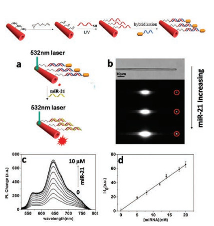
MicroRNAs (miRNAs) are a class of endogenous noncoding noncoding RNA molecules that regulate functions. miRNA-21 is overexpressed in human tumors and, consequently, it is a biomarker for targeted cancer therapy [82-84]. Thus, various sensors have been developed for detecting miRNA-21. In one example, Zou et al. fabricated the 1D Au@PDA microtube for detection (Fig. 22) [85]. Addition of miRNA-21 to a buffered solution of Au@PDA results in a concentration dependent fluorescence enhancement caused by the displacement of DNA functionalized nanorods from the PDA surface. The detection limit of this sensor for miRNA-21 is 2 nmol/L and it has a high selectivity in that it undergoes negligible changes in the presence of other miRNAs. Also, Au@PDA microtube immobilized on a hydrophobic substrate detects miRNA-21 with a limit of 0.01 nmol/L, which is two orders of magnitude lower than that of the non-immobilized material. Moreover, Au@PDA microtube immobilized on a hydrophobic substrate was successfully used for miRNA-21 detection in a biological environment.

|
Download:
|
| Fig. 22. Design of single Au@PDA microtube waveguide system: (a) Au@PDA microtube for miRNA-21 detection; (b) White-field image of PDA microtube with addition of miRNA-21; (c) PL changes of PDA microtube with increasing concentration of miRNA-21; (d) Fluorescence enhancement with increasing concentration of miRNA-21. Reproduced with permission [85]. Copyright 2016, Elsevier. | |
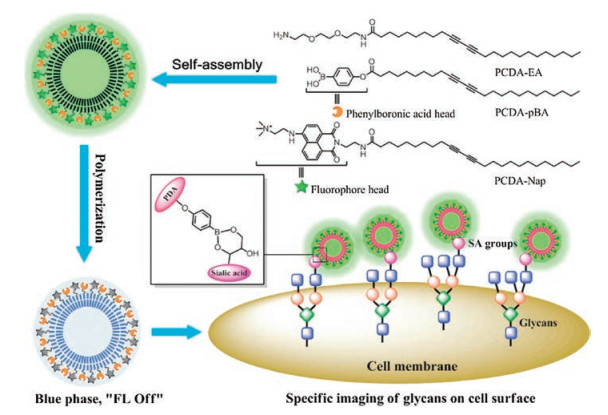
The concentrations of the carbohydrate sialic acids (SA), which normally exist in blood serum and as a glycan on cell surfaces, serve as indicators of several diseases [86-89]. Therefore, methods for quantitative detection of SA have potential applications in clinical diagnostics. Wang et al. reported the results of a study leading to the development of a PDA-based sensor for detecting SA glycans (Fig. 23) [90]. The probe is formed from liposomes comprised of a 4.5:4.5:1 mole ratio of PCDA-pBA, which contains a carbohydrate sensitive phenylboronic acid headgroup as a SA receptor, PCDA-EA, which is used for enhancement of binding, and PCDA-Nap, which contains a naphthalimide fluorophore as a headgroup. Copolymerization of this mixture induced by 254 nm UV irradiation forms a blue colored PDA and is accompanied by fluorescence quenching. Free SA in PBS can be directly detected using this probe in the form of a blue-to-red color transition and turn-on fluorescence. Furthermore, the results of MTT assay indicate that the PBDA liposomes have low cytotoxicity toward MCF-7 cells. Moreover, SA on cell surfaces was also imaged using this probe.

|
Download:
|
| Fig. 23. Polymerization of PDAs consisted of PCDA-EA, PCDA-pBA and PCDA-Nap and scheme illustration of PDA liposomes for cell surface imaging. Copied with permission [90]. Copyright 2018, Royal Society of Chemistry. | |
7. Conclusion
In this review, we summarized recent advances that have occurred in the development of PDA-based biosensors. The results of studies discussed in the review demonstrate that the new sensors are useful in enzyme activity sensing, tumor targeting, cell imaging, cellular activity monitoring and microorganism and biomolecule. However, the efforts have uncovered some shortcomings of these probes and have shown that further studies are required to transform the simple detection systems into ones that can be utilized in real-time biological applications. In addition, the exact nature of factors that cause blue-to-red color transitions and enhancement of red fluorescence of the PDAs in the sensors are unknown. Thus, although it is clear that the transitions are a consequence of perturbations taking place in conformations of conjugated backbone in the PDA polymers, more investigations are needed to identify the detailed mechanism by which the changes take place. Also, although the use of PDA-based biosensors for detecting diseases is at a very early stage of development, it is clear that these systems have the potential of serving as theranostic agents for simultaneously detecting tumors and stemming their growth. Although several challenges exist, we believe that PDA-based biosensors will have future applications to the diagnosis and treatment of diseases.
AcknowledgmentsThis study was financially supported by the National Key Research and Development Program of China (No. 2018YFA0902200), the National Natural Science Foundation of China (Nos. 21722605, 21978131 and 21878156), the Six Talent Peaks Project in Jiangsu Province (No. XCL-034) and the Project of Priority Academic Program Development of Jiangsu Higher Education Institutions (PAPD).
| [1] |
D.E. Wang, L. Zhao, M.S. Yuan, et al., ACS Appl. Mater. Interfaces 8 (2016) 28231-28240. DOI:10.1021/acsami.6b10794 |
| [2] |
C.F. Chen, J. Chen, T.Y. Wang, M.H. Liu, ACS Appl. Mater. Interfaces 8 (2016) 30608-30615. DOI:10.1021/acsami.6b10392 |
| [3] |
X.Q. Chen, J. Lee, M.J. Jou, J.M. Kim, J. Yoon, Chem. Commun. (2009) 3434-3436. |
| [4] |
L.J. Tang, M.Y. Tian, H.B. Chen, et al., Dyes Pigments 158 (2018) 482-489. DOI:10.1016/j.dyepig.2017.12.028 |
| [5] |
L.J. Tang, D. Xu, M.Y. Tian, X.M. Yan, J. Lumin. 208 (2019) 502-508. DOI:10.1016/j.jlumin.2019.01.022 |
| [6] |
Y.H. Chen, T.W. Wei, Z.Z. Zhang, et al., Chin. Chem. Lett. 28 (2017) 1957-1960. DOI:10.1016/j.cclet.2017.05.010 |
| [7] |
Y. Li, L.H. Wang, X. Yin, et al., J. Mater. Chem. A 2 (2014) 18304-18312. DOI:10.1039/C4TA04547E |
| [8] |
P. Narkwiboonwong, G. Tumcharern, A. Potisatityuenyong, S. Wacharasindhu, M. Sukwattanasinitt, Talanta 83 (2011) 872-878. DOI:10.1016/j.talanta.2010.10.054 |
| [9] |
J. Lee, H. Jun, J. Kim, Adv. Mater. 21 (2009) 3674-3677. DOI:10.1002/adma.200900639 |
| [10] |
V.K. Rao, N.L. Teradal, R. Jelinek, ACS Appl. Mater. Interfaces 11 (2019) 4470-4479. DOI:10.1021/acsami.8b20930 |
| [11] |
X.N. Wang, X.L. Sun, P.A. Hu, et al., Adv. Funct. Mater. 23 (2013) 6044-6050. DOI:10.1002/adfm.201301044 |
| [12] |
B.W. Davis, A.J. Burris, N. Niamnont, et al., Langmuir 30 (2014) 9616-9622. DOI:10.1021/la5017388 |
| [13] |
X.Q. Chen, S. Kang, M.J. Kim, et al., Angew. Chem. Int. Ed. 49 (2010) 1422-1425. DOI:10.1002/anie.200905041 |
| [14] |
Y.Y. Zhang, J. Northcutt, T. Hanks, et al., Food Chem. 221 (2017) 515-520. DOI:10.1016/j.foodchem.2016.09.168 |
| [15] |
P. Boullanger, D. Lafont, M.N. Bouchu, et al., C. R. Chim. 11 (2008) 43-60. DOI:10.1016/j.crci.2007.03.007 |
| [16] |
C.X. Guo, P. Boullanger, T. Liu, L. Jiang, J. Phys. Chem. B 109 (2005) 18765-18771. DOI:10.1021/jp052580y |
| [17] |
Q.L. Nie, Y. Zhang, J. Zhang, M.Q. Zhang, J. Mater. Chem. 16 (2006) 546-549. DOI:10.1039/B511474H |
| [18] |
J. Lee, H.J. Kim, J. Kim, J. Am. Chem. Soc. 130 (2008) 5010-5011. DOI:10.1021/ja709996c |
| [19] |
X.M. Sun, T. Chen, S.Q. Huang, L. Li, H.S. Peng, Chem. Soc. Rev. 39 (2010) 4244-4257. DOI:10.1039/c001151g |
| [20] |
S. Wacharasindhu, S. Montha, J. Boonyiseng, et al., Macromolecules 43 (2010) 716-724. DOI:10.1021/ma902282c |
| [21] |
X.Q. Chen, J. Yoon, Dyes Pigments 89 (2011) 194-198. DOI:10.1016/j.dyepig.2009.12.015 |
| [22] |
I.S. Park, H.J. Park, J.M. Kim, ACS Appl. Mater. Interfaces 5 (2013) 8805-8812. DOI:10.1021/am402701n |
| [23] |
S.J. Kew, E.A.H. Hall, Anal. Chem. 78 (2006) 2231-2238. DOI:10.1021/ac0517794 |
| [24] |
D.J. Ahn, E.H. Chae, G.S. Lee, et al., J. Am. Chem. Soc. 125 (2003) 8976-8977. DOI:10.1021/ja0299001 |
| [25] |
S. Seo, D. Kim, G. Jang, et al., React. Funct. Polym. 73 (2013) 451-456. DOI:10.1016/j.reactfunctpolym.2012.11.016 |
| [26] |
S. Chae, J.P. Lee, J.M. Kim, Adv. Funct. Mater. 26 (2016) 1769-1776. DOI:10.1002/adfm.201504845 |
| [27] |
W. Zhang, H.B. Xu, Y. Chen, S. Cheng, L.J. Fan, ACS Appl. Mater. Interfaces 5 (2013) 4603-4606. DOI:10.1021/am401099s |
| [28] |
M. Ulaganathan, R.V. Hansen, N. Drayton, et al., ACS Appl. Mater. Interfaces 8 (2016) 32643-32648. DOI:10.1021/acsami.6b12171 |
| [29] |
J. Lee, O. Yarimaga, C.H. Lee, Y.K. Choi, J.M. Kim, Adv. Funct. Mater. 21 (2011) 1032-1039. DOI:10.1002/adfm.201002042 |
| [30] |
B. Yoon, D.Y. Ham, O. Yarimaga, et al., Adv. Mater. 23 (2011) 5492-5497. DOI:10.1002/adma.201103471 |
| [31] |
J. Yoon, Y.S. Jung, J.M. Kim, Adv. Funct. Mater. 19 (2009) 209-214. DOI:10.1002/adfm.200800963 |
| [32] |
D.H. Park, W. Jeong, M. Seo, B.J. Park, J.M. Kim, Adv. Funct. Mater. 26 (2016) 498-506. DOI:10.1002/adfm.201504088 |
| [33] |
J. Lee, M. Pyo, S.H. Lee, et al., Nat. Commun. 5 (2014) 3736. DOI:10.1038/ncomms4736 |
| [34] |
J. Lee, H.T. Chang, H. An, et al., Nat. Commun. 4 (2013) 2461. DOI:10.1038/ncomms3461 |
| [35] |
D.H. Charych, J.O. Nagy, W. Spevak, M.D. Bednarski, Science 261 (1993) 585-588. DOI:10.1126/science.8342021 |
| [36] |
K.D. Green, M.Y. Fosso, S. Garneau-Tsodikova, Molecules 23 (2018) 3552. |
| [37] |
D.S. Guo, K. Wang, Y.X. Wang, Y. Liu, J. Am. Chem. Soc. 134 (2012) 10244-10250. DOI:10.1021/ja303280r |
| [38] |
P. Anand, B. Singh, Arch. Pharm. Res. 36 (2013) 375-399. DOI:10.1007/s12272-013-0036-3 |
| [39] |
S. Peng, Y.C. Pan, Y.L. Wang, et al., Adv. Sci. 4 (2017) 1700310. DOI:10.1002/advs.201700310 |
| [40] |
G.D. Zhou, F. Wang, H.L. Wang, et al., ACS Appl. Mater. Interfaces 5 (2013) 3275-3280. DOI:10.1021/am400260y |
| [41] |
F. Speranza, M. Mahankali, K.M. Henkels, J. Gomez-Cambronero, J. Biol. Chem. 289 (2014) 28885-28897. DOI:10.1074/jbc.M114.597146 |
| [42] |
C. Spencer, H.A. Brown, Biochemistry 54 (2015) 1208-1218. DOI:10.1021/bi501291t |
| [43] |
Z.Z. Zhang, J. Li, F. Wang, et al., Sens. Actuators B:Chem. 282 (2019) 636-643. DOI:10.1016/j.snb.2018.11.117 |
| [44] |
M. Du, T. Yang, Y.C. Zhang, K. Jiao, Electroanalysis 21 (2009) 2521-2526. DOI:10.1002/elan.200900187 |
| [45] |
S.H. Jung, H. Jang, M.C. Lim, et al., Anal. Chem. 87 (2015) 2072-2078. DOI:10.1021/ac501795x |
| [46] |
S.K. Golombek, J.N. May, B. Theek, et al., Adv. Drug Deliv. Rev. 130 (2018) 17-38. DOI:10.1016/j.addr.2018.07.007 |
| [47] |
P. Kesharwani, A.K. Iyer, Drug Discov. Today 20 (2015) 536-547. DOI:10.1016/j.drudis.2014.12.012 |
| [48] |
M. Gao, F.B. Yu, C.J. Lv, J. Choo, L.X. Chen, Chem. Soc. Rev. 46 (2017) 2237-2271. DOI:10.1039/C6CS00908E |
| [49] |
W.T. Yang, W.S. Guo, W.J. Le, et al., ACS Nano 10 (2016) 10245-10257. DOI:10.1021/acsnano.6b05760 |
| [50] |
Y.A. Zhong, F.H. Meng, C. Deng, Z.Y. Zhong, Biomacromolecules 15 (2014) 1955-1969. DOI:10.1021/bm5003009 |
| [51] |
X.G. Wang, Z.Y. Dong, H. Cheng, et al., Nanoscale 7 (2015) 16061-16070. DOI:10.1039/C5NR04045K |
| [52] |
D.H. Yao, S. Li, X.M. Zhu, J.C. Wu, H. Tian, Chem. Commun. 53 (2017) 1233-1236. DOI:10.1039/C6CC08581D |
| [53] |
I. Theodorou, P. Anilkumar, B. Lelandais, et al., Chem. Commun. 51 (2015) 14937-14940. DOI:10.1039/C5CC05333A |
| [54] |
L.L. Li, X.Q. An, X.J. Yan, Colloids Surf. B 134 (2015) 235-239. DOI:10.1016/j.colsurfb.2015.07.008 |
| [55] |
J.H. Fang, T.L. Chiu, W.C. Huang, et al., Adv. Healthc. Mater. 5 (2016) 688-695. DOI:10.1002/adhm.201500750 |
| [56] |
D.L. Yang, R.F. Zou, Y. Zhu, et al., Nanoscale 6 (2014) 14772-14783. DOI:10.1039/C4NR04405C |
| [57] |
D. Wang, H.F. Su, R.T.K. Kwok, et al., Chem. Sci. 9 (2018) 3685-3693. DOI:10.1039/C7SC04963C |
| [58] |
H. Jiang, X.Y. Hu, S. Schlesiger, et al., Angew. Chem. Int. Ed. 56 (2017) 14526-14530. DOI:10.1002/anie.201708168 |
| [59] |
P.J. Wu, S.Y. Kuo, Y.C. Huang, C.P. Chen, Y.H. Chan, Anal. Chem. 86 (2014) 4831-4839. DOI:10.1021/ac404237q |
| [60] |
B.E.I. Ramakers, S.A. Bode, A.R. Killaars, J.C.M. van Hest, D.W.P.M. Löwik, J. Mater. Chem. B 3 (2015) 2954-2961. |
| [61] |
P.F. Xu, R.B. Zhang, N. Yang, et al., Biomicrofluidics 13 (2019) 024110-024118. DOI:10.1063/1.5086087 |
| [62] |
N.T. Thet, W.D. Jamieson, M. Laabei, J.D. Mercer-Chalmers, A.T.A. Jenkins, J. Phys. Chem. B 118 (2014) 5418-5427. DOI:10.1021/jp502586b |
| [63] |
S. Lee, H. Cheng, M. Chi, et al., Biosens. Bioelectron. 77 (2016) 1016-1019. DOI:10.1016/j.bios.2015.10.090 |
| [64] |
J. Park, S.K. Ku, D. Seo, et al., Chem. Commun. 52 (2016) 10346-10349. DOI:10.1039/C6CC03116A |
| [65] |
J. Zhou, D.Y. Yao, Z.Y. Qian, et al., Biomaterials 161 (2018) 11-23. DOI:10.1016/j.biomaterials.2018.01.024 |
| [66] |
J. Roh, S.Y. Lee, S. Park, D.J. Ahn, Chem.-Asian J. 12 (2017) 2033-2037. DOI:10.1002/asia.201700769 |
| [67] |
T. Berninger, C. Bliem, E. Piccinini, O. Azzaroni, W. Knoll, Biosens. Bioelectron. 115 (2018) 104-110. DOI:10.1016/j.bios.2018.05.027 |
| [68] |
W.J. Lu, Y.F. Gao, Y. Jiao, et al., Nanoscale 9 (2017) 11545-11552. DOI:10.1039/C7NR02336G |
| [69] |
M.M. Yu, W.W. Du, H.X. Li, H.Y. Zhang, Z.X. Li, Biosens. Bioelectron. 92 (2017) 385-389. DOI:10.1016/j.bios.2016.10.090 |
| [70] |
T. Liu, N. Li, J.X. Dong, et al., Biosens. Bioelectron. 87 (2017) 772-778. DOI:10.1016/j.bios.2016.08.098 |
| [71] |
T. Yu, C.N. Xu, J. Qiao, R.Y. Zhang, L. Qi, Chin. Chem. Lett. 30 (2019) 660-663. DOI:10.1016/j.cclet.2018.10.001 |
| [72] |
Y.L. Zhou, Z.C. Yang, M.T. Xu, Anal. Methods 4 (2012) 2711. DOI:10.1039/c2ay25475a |
| [73] |
A. Arendowski, T. Ruman, Anal. Methods 10 (2018) 5398-5405. DOI:10.1039/C8AY01677A |
| [74] |
E. Cho, H. Kim, Y. Choi, S.R. Paik, S. Jung, Sci. Rep. 6 (2016) 31115. DOI:10.1038/srep31115 |
| [75] |
Z.Z. Zhang, T.W. Wei, Y.H. Chen, et al., Sens. Actuators B:Chem. 255 (2018) 2211-2217. DOI:10.1016/j.snb.2017.09.027 |
| [76] |
X.X. Xiang, Y. Li, L. Ling, et al., Sens. Actuators B:Chem. 290 (2019) 68-72. DOI:10.1016/j.snb.2019.03.111 |
| [77] |
C.P. Chen, W.F. Liu, S.P. Tian, T.T. Hong, Sensors 19 (2019) 1712. DOI:10.3390/s19071712 |
| [78] |
U.D. Kamaci, M. Kamaci, A. Peksel, Spectrochim. Acta A 212 (2019) 232-239. DOI:10.1016/j.saa.2019.01.011 |
| [79] |
Y.K. Jung, H.G. Park, Biosens. Bioelectron. 72 (2015) 127-132. DOI:10.1016/j.bios.2015.04.093 |
| [80] |
K. Heo, K.A. Park, Y.H. Kim, et al., BMB Rep. 42 (2009) 685-690. DOI:10.5483/BMBRep.2009.42.10.685 |
| [81] |
D.H. Kang, H.S. Jung, K. Kim, J. Kim, ACS Appl. Mater. Interfaces 9 (2017) 42210-42216. DOI:10.1021/acsami.7b14086 |
| [82] |
M.D. Yao, X.F. Lv, Y.L. Deng, M. Rasheed, Anal. Chim. Acta 1055 (2019) 115-125. DOI:10.1016/j.aca.2018.12.040 |
| [83] |
Z.Z. Yang, Z.B. Wen, X. Peng, et al., Chem. Commun. 55 (2019) 6453-6456. DOI:10.1039/C9CC01850F |
| [84] |
X.M. Miao, W.H. Wang, T.S. Kang, et al., Biosens. Bioelectron. 86 (2016) 454-458. DOI:10.1016/j.bios.2016.07.001 |
| [85] |
Y. Zhu, D. Qiu, G. Yang, et al., Biosens. Bioelectron. 85 (2016) 198-204. DOI:10.1016/j.bios.2016.05.019 |
| [86] |
S.M. Xu, S.T. Che, P.Y. Ma, et al., Talanta 197 (2019) 548-552. DOI:10.1016/j.talanta.2019.01.074 |
| [87] |
S. Li, J.L. Liu, Y.L. Lu, et al., Biosens. Bioelectron. 117 (2018) 32-39. DOI:10.1016/j.bios.2018.05.062 |
| [88] |
S. Sankoh, C. Thammakhet, A. Numnuam, et al., Biosens. Bioelectron. 85 (2016) 743-750. DOI:10.1016/j.bios.2016.05.083 |
| [89] |
L. Frullano, J. Rohovec, S. Aime, et al., Chemistry 10 (2004) 5205-5217. DOI:10.1002/chem.200400369 |
| [90] |
D.E. Wang, J.H. Yan, J.J. Jiang, et al., Nanoscale 10 (2018) 4570-4578. DOI:10.1039/C7NR08557E |