 2019, Vol. 30


b Key Laboratory of Pesticide and Chemical Biology, Ministry of Education, Hubei International Scientific and Technological Cooperation Base of Pesticide and Green Synthesis, International Joint Research Center for Intelligent Biosensing Technology and Health, College of Chemistry, Central China Normal University, Wuhan 430079, China
Cancer is caused by the loss of normal regulation and excessive proliferation of cells. Cancer cells can even be transferred to other parts of the body through the circulatory system and lymphatic system by invading surrounding tissues in the living system, which causes lesions in various organs of the body, and eventually leads to the death of patients. According to US cancer data in 2015 [1], cancer has emerged the first killer of human health compared to other diseases. The high mortality rate of cancer is due to the lack of early rapid diagnosis and more efficient treatment methods.
In recent years, with the progress of medical technology, various diagnostic imaging methods [2-4] have emerged for tumor diagnosis, surgical navigation and prognosis evaluation including magnetic resonance imaging (MRI), X-ray radiography, computed tomography (CT), positron emission tomography (PET), ultrasonography (US), and optical imaging. Among them, fluorescence imaging technology [5-10] is based on spectral chemistry, optical wave-guide and measurement technology. The chemical information of the analyzed object is selectively converted into a fluorescent signal that is easily measured by the analytical instrument. MRI, CT, PET and US have cumbersome operations, slow imaging speed, and expensive price, therefore, the advantages of fluorescence imaging technology are highlighted due to their temporal and spatial resolution, specificity, selectivity, real-time monitoring, non-invasiveness and good biofilm penetration.
Near-infrared (NIR) fluorescent dyes, whose emission wavelengths are between 740 nm and 1700 nm, are widely used in early tumor diagnosis due to little side effects, deep tissue infiltration, good metabolism, and low fluorescence background in living organisms. The structures of fluorescent dyes mainly consist of cyanines [11-15], phthalocyanines, BODIPYs and rhodamine analogues, of which indocyanine green (ICG)[16-17] has been approved by the FDA. In order to enhance the retention of fluorescent dyes in tumor cells, a series of fluorescent dyes linked to responsive or targeting groups have been reported to recognize tumor microenvironments and over-expressed biomarkers. In addition, the researchers found that some small molecule fluorescent dyes can be used for photothermal therapy (PTT) because of the high photothermal conversion and long absorption wavelength.
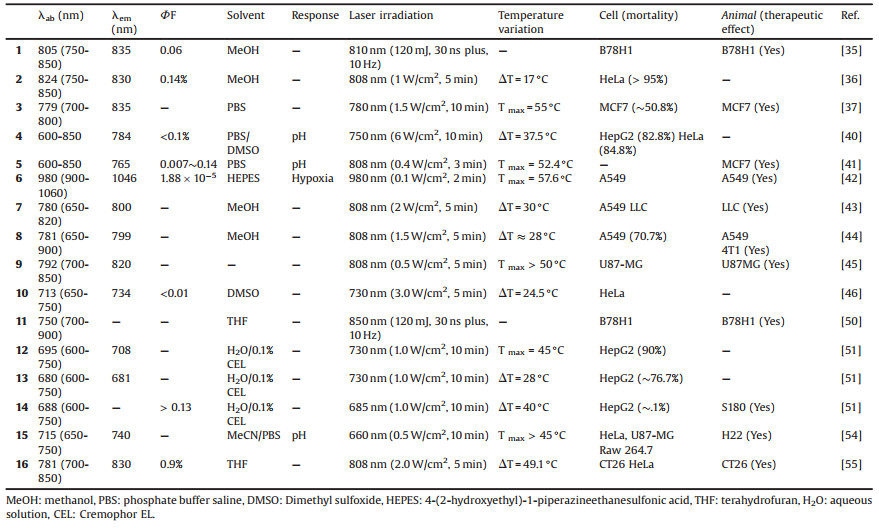
PTT [18-21] is a new treatment strategy for cancer. As we all know, the main methods of clinical therapy are surgery, chemotherapy and radiotherapy. Surgery can remove early solid tumors, which may not be completely ablated, leading to metastasis and recurrence of postoperative cancer. Chemotherapy [22-24] and radiotherapy [25-26] are common treatment strategies, which have an inhibitory effect on tumors. However, the patient's adaptability is poor due to the side effects, drug tolerance and inherent toxicity of radiation. Based on the deficiencies of the above treatment methods, non-invasive PTT has received extensive attention due to its high specificity, short treatment time, significant effects, little damage to surrounding tissues, and good bioavailability. Materials with photothermal efficiency are important for PTT. In the development of PTT materials, the first to third generation photothermal materials are nano-platforms [27-29], including precious metal nanoparticles, carbon nanoparticles and metal complex nanoparticles. Due to the high cost of the nanoparticles and the difficulty in degradation, the fourth generation organic small molecule dyes having photothermal properties avoid the above disadvantages. However, few reviews have been reported from the point of small molecule fluorescent dyes. In this review, we provide a concise overview of small organic molecules used in PTT in recent years. And the structure of the small organic molecules and the main data are shown in Figs. 1-5 and Table 1.

|
Download:
|
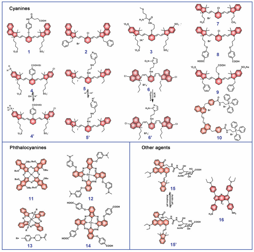
| Fig. 1. The library of small molecule fluorescent dyes for photothermal therapy. | |

|
Download:
|
| Fig. 2. (A) Schematic illustration of 5 as a versatile theranostic probe for pH sensing, tumor targeting and PTT. (B) UV-vis spectra (Abs: absorbance) and fluorescence spectra (Flu) of 5 in PBS, pH 4.4. (C) UV-vis spectra of 5 at various pH values in 20 mmol/L PBS buffer. (D) Infrared thermal images of subcutaneous MCF7 tumor xenograft mice after 3 min NIR laser irradiation (808 nm, 0.4 W/cm2). Reproduced with permission [41]. Copyright 2017, Royal Society of Chemistry. | |

|
Download:
|
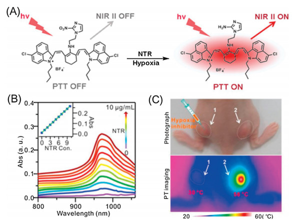
| Fig. 3. (A) Schematic illustration of the NTR-responsive NIR Ⅱ fluorescence/PA probe 6 for visualizing tumors and inducing an NTR-triggered PTT effect. (B) Absorption responses of 5 μg/mL 6 to different concentrations of NTR. Inset: a linear correlation between absorption intensity and concentration of NTR. (C) Photograph and infrared thermal imaging of a mouse with two A549 tumors on the left and right hind legs 14 h after intravenous injection of 6. The room temperature was 23 ℃. Tumor 1 was injected with dicoumarin as an inhibitor of NTR; Tumor 2 was without treatment. Reproduced with permission [42]. Copyright 2018, Ivyspring International Publisher. | |

|
Download:
|
| Fig. 4. (A) Schematic representation of enhanced photothermogenesis by mitochondria-targeting probe 10. (B) Photophysical data for 10 determined in DMSO. (C) Effect of NIR irradiation following 10 administration on the viability of HeLa cells. Confocal fluorescence images of propidium iodide-treated HeLa cells after incubation with various concentration of 10 for 4 h, following by 730 nm irradiation (2.3 W/cm2) for 10 min. The proportion of PI-positive cells was determined from the ratio of PI-positive cells to the total number of cells as determined by Hoechst 33342 staining. Confocal fluorescence images of HeLa cells obtained after incubation with 10 (20 μmol/L) for 4 h following 730 nm laser irradiation (2.3 W/cm2) for 10 min. Dead cells are labeled in red by PI staining, whereas all cells were visualized using Hoechst 33342 (green). (D) Photothermal conversion utility of 10. Infrared thermal images of DMSO solutions of 10. Concentration: 0.5 mmol/L and irradiation intensities of 0, 1.0, 2.3, and 3.0 W/cm2 at 730 nm. Reproduced with permission [46]. Copyright 2017, American Chemical Society. | |

|
Download:
|
| Fig. 5. (A) Schematic of tumor illumination with a targetable sialic acid conjugated NIR profluorophore (15) activatable to lysosomal acidity and nonfluorescent 15 undergoes acidic pH-mediated fluorogenic opening of the intramolecular lactam to give fluorescent and photothermal 15'. (B) UV-vis-NIR absorption spectra of the solutions were collected over the buffer pH range. (C) Fluorescence emission of the solutions was recorded. (D) Acidic pH-dependent photothermal properties of 15. The temperature of sodium phosphate buffer (100 mmol/L, pH 4.5 or 7.5) containing 15 (0 or 0.1 mg/mL) was recorded over the time of irradiation with an NIR laser (660 nm, 0.5 W/cm2). (E) Photothermal effects of 15 on live cells. HeLa, U87-MG and Raw 264.7 cells were cultured for 24 h with 15 (0 or 100 mg/mL) or SA (0 or 100 mg/mL) in DMEM and then either irradiated with an NIR laser (660 nm, 0.5 W/cm2), or not subjected to irradiation in fresh DMEM for 10 min, and then further cultured for 24 h. Cell viability was determined by MTT assay. Reproduced with permission [54]. Copyright 2014, Royal Society of Chemistry. | |
|
|
Table 1 The data of small molecule fluorescent dyes. |
2. Cyanine-based photothermal agents
Heptamethine cyanine dye [30-33] is a commonly used fluorescent molecular skeleton, in which various heterocyclic rings such as benzazole and its derivatives, pyridine, quinoline, thiazole and pyrrole are often used as terminal groups. Nowadays, more and more attention has been focused on cyanine compounds which have been proven their accumulation in the tumor site and the intense absorption bands are in the range of 800-900 nm. Besides, they are superior to other dyes in terms of good solubility and stability. Compared to the similar structure ICG [34], heptamethine cyanine dyes show better photothermal conversion efficiency. At the same time, since heptamethine cyanine dyes have more chemical sites that can be modified, they can be used in the design of responsive or targeted photothermal compounds. In addition, some heptamethine cyanine dyes can also release reactive oxygen species (ROS) under light irradiation for the synergistic treatment of photodynamic therapy (PDT) and PTT.
Early in 2009, Camerin et al. synthesized compound 1, a novel far-red absorbing cyanine, with photothermal sensitization and therapeutic properties [35]. The absorption band of 1 was narrow and the maximum peak was 805 nm. Its maximum emission peak was at 835 nm and fluorescence quantum yield was at 0.06 compared with IR-125 (from Acros Organics). Compound 1 was taken up by cancer cells quickly and the concentration would be at least up to 7.7 mmol/L. The photoinactivation efficiency in HT1080 cells was very low, compared with B78H1 cells whose photoinactivation efficiency increased with prolong incubation (810 nm, 30 ns pulses, 10 Hz, 120 mJ). Most importantly, compound 1-loaded tumors remained tumor-free for about 8 days after PTT and 50% of the mice exhibited no detectable tumor regrowth about 30 days after PTT. In contrast, the tumor volume of untreated control mice reached to 400 mm3 within 28 days. Furthermore, in 2017, the group of Chen developed a mitochondria targeted cyanine dye 2 for both fluorescence imaging and near-infrared PTT owing to its two separated excitation wavelength channels [36]. For fluorescence imaging, 2 can emit red fluorescence under 552 nm excitation in mitochondria. For PTT, it converted optical energy into heat. It exhibited strong NIR absorption at about 824 nm in various organic solvents due to the monomeric form with low fluorescence quantum yield (QY). Upon excitation (5 min, 808 nm laser, 1.0 W/cm2), the temperature of the PBS solution containing 20 μg/mL of 2 increased rapidly for around 17 ℃, whereas a control PBS solution increased only 2 ℃. Otherwise, the photothermal conversion efficiency of 2 was calculated to be 17.4%, which was comparable to that of ICG (18.5%). The cells incubated with 2 (5 μg/mL) showed 70% mortality and 95% at higher concentrations after exposure (5 min, 808 nm laser, 1.0 W/cm2). At the same time, 2 exhibited low cytotoxicity at the concentrations below 20 μg/mL in the nonirradiation group. In 2018, Tang et al. described a new small molecule NIR fluorescent dye-labeled Met, which was an essential amino acid for protein synthesis [37]. In the research, dye 3 was synthesized with absorption peak at 779 nm and emission peak at 835 nm. After being exposed to irradiation of a 60 W light bulb for an hour, the absorption of 3 was higher than that of the intermediate product, indicating that 3 had better photostability. The photothermal conversion efficiency of 3 was measured, which reached 55 ℃ under 780 nm laser irradiation for 10 min at 1.5 W/cm2 in the 2 μg/mL solution of 3. Moreover, it caused the death of 50.8% cells after incubating with compound 3 (20 μg/mL) when irradiated with 2 W/cm2 laser for 10 min. Based on great photothermal properties and photothermal toxicity to cells, 3 was injected via tail vein with 200 mL (200 μg/mL). When irradiated with NIR light (780 nm, 2 W/cm2) for 20 min after injecting, the temperature of tumor reached 43 ℃ and the tumor completely disappeared at the 12th day, which demonstrated good photothermal treatment effect.
In order to compensate for the defect that a single small molecule cannot be accurately located in tumor cells, some cyanine dyes with pH responsibility were designed and reported [38, 39]. Due to the high infiltration, invasion and metastatic characteristics of cancer, tumor cells have more acidic lysosomal microenvironment (pH(lys) 3.8-4.7), which is a potential candidate for accumulation of dyes in tumor. Zhang and coworkers designed and synthesized four heptamethine cyanines with pH switchable NIR fluorescence and photothermal effects [40]. Through the study of photophysical properties, the authors found that all compounds displayed absorption at 600-850 nm and emission at 780 nm. The fluorescence QY of compounds increased up to 1020-fold upon acidification from pH 7.4 to 2.4 in physiologically acidic conditions, but their photothermal efficiencies concomitantly decreased up to 7.1-fold. By precisely regulating its pKa value (4.6), the fluorescent signal of the dye was only turned on in the acidic environment of cancer cell lysosomes, enabling selective tracer of cancer cells. Subsequently, the author used 4, which had the best photophysical properties as a therapeutic dye to study its PTT effect. Besides cancer cells imaging by sensing acidic pH(lys), 4 also showed higher mitochondrial uptake in cancer cells than normal cells. Meanwhile, 4 had better photothermal toxicity. The experimental data displayed that 82.8% HepG2 cells and 84.8% HeLa cells were dead after combined treatment of 4 with laser irradiation (750 nm, 6.0 W/cm2, 10 min) and had lower cytotoxicity of normal cells with 9.4% for dead HL-7702 cells. All in all, these theranostic dyes are applicable for assisting the image-guided tumor photoablation by selective aggregation in tumor cells and reducing damage to normal tissue. Coincidentally, Meng et al. reported a fluorophorespacer-receptor molecular dye 5, which was verified as "All in One" multifunctional agent including pH sensitive tumor targeting, NIR fluorescence imaging, high spatial resolution photoacoustic (PA) imaging and efficient NIR PTT [41] (Fig. 2A). The authors selected IR822 as the basic skeleton and connected a proton receptor N1-(pyridin-4-ylmethyl) ethane-1, 2-diamine (PY) on 5' carbon site to form compound 5. Owing to basic skeleton with similar structure, the dye can selectively accumulate in tumors. It can be observed that 5 had a strong absorption and emission in NIR region and absorption peak shifted from 637 nm to 792 nm with the increase of pH value, especially from pH 5.6 to 7.4 (Figs. 2B and C). In basic buffer solutions (pH 8.0), the fluorescence intensity and QY of 5 was very low because of intramolecular photoinduced electron transfer (PET) effect. These results illustrated that 5 may have good photothermal effect and PA signal. The author also verified the above conjecture through cell experiments. In the study of PTT effects, a temperature increase to 57 ℃ upon irradiation of MCF7 tumor bearing BALB/c nude mice with the treatment of 5 (3 min, 808 nm laser, 0.4 W/cm2) (Fig. 2D). Moreover, mice in the PBS plus laser group and 5 without laser group showed low survival rates of 10% and 20%, while mice treated with 5 plus irradiation survived over 30 days without a single death. These studies showed that 5 was a biocompatible small molecule presenting highly effective PTT without severe adverse effect at tested doses.
The pH regulation is not the only response strategy for NIR dyes. Hypoxia is also a common characteristic of solid tumor, which is related to resistance to radio-chemotherapy. Recently, Cai and coworkers reported a NIR small molecule probe 6 conjugating a nitro imidazole group (2-(2-nitroimidazolyl) ethylamine, MZ) for hypoxia-activated PTT and high-contrast NIR Ⅱ and PA tumor imaging [42] (Fig. 3A). The electron-withdrawing MZ group induced electron-transfer, resulting in fluorescence quenching and absorbance decreasing, and it also improved the aqueous solubility. In the presence of nitroreductase (NTR), the MZ group was reduced to the amine group and the NIR fluorescence was recovered. The maximum excitation and emission wavelengths were 980 nm and 1046 nm after the probe was reduced by NTR, and the fluorescence quantum yield (ΦF) was 0.006. The intensity of absorption peak (980 nm) of 6 increased with increasing NTR concentration from 0 to 10 μg/mL (Fig. 3B). The probe can pinpoint the location of tumor and be used in PTT because of its enhanced absorption and response to NTR. Otherwise, the increases in NIR absorption of the probe after reaction with NTR led to a strong PA signal. After 980 nm NIR laser irradiation (0.1 W/cm2) for 2 min, the temperature of solution increased from 30.4 ℃ to 57.6 ℃ owing to the response of NTR. A remarkable photothermal conversion efficiency of 20.2% was measured according to previous data. In order to confirm the selective photothermal effect caused by hypoxia in vivo, dicoumarin solution (0.2 mmol/L), an inhibitor of endogenous NTR, was injected into tumor 1 on the left hind leg, while tumor 2 on the right hind leg was untreated. After intravenous injection of the A549 tumor-bearing BALB/c nude mice, the temperature of tumor 2 increased from 30 ℃ to 58 ℃ under 980 nm NIR laser (Fig. 3C). In contrast, the temperature of tumor 1 was still at 30 ℃. On the 5th day after the PTT, the tumor disappeared while no obvious side effect of the major blood indexes and liver-function indexes.
In order to improve the therapeutic effect of tumors, PTTand PDT are combined as a new method of therapy. Based on the identification of structure-inherent and multi-functional small molecules, it is worth developing structure-inherent small-molecule photosensitizers (PSs) for targeting, imaging, and therapy. As we all know, mitochondria has been used as ideal tumor target for treatment.Based onthesefindings, Chen et al. developed a lipophilic cationic heptamethine dye, 7, which can further retain in the mitochondria and was excited with laser to generate local heat and ROS [43]. Compound 7 had the maximum absorbance and emission wavelength at near 780 nm and 800 nm. The temperature of 7 irradiatedwith808 nmlaser(2 W/cm2, 5 min)wasfoundtoincrease sharply about 30 ℃. Moreover, the generation of ROS was detected at the same condition. Subsequently, the cell viability of A549 incubated with 7 upon to irradiation exhibited a remarkable decreasewith the increased concentration. The mice bearing tumor were injected with 7 at a dose of 0.3 mg/kg via the tail vein. The real time tumor surface temperature of the mice in irradiation group increased to about 59 ℃, which was enough to lead the death of cancer cells. Finally, large areas of severe tumor tissue damage were observed in tumors of 7 plus irradiation group after hematoxylin and eosin (H&E) staining, which illustrated that 7 was an effective photothermal agent.
Luo et al. synthesized a series of mitochondria-targeted PSs modified with various N-alkyl side chains, of which heptamethine cyanine dye 8 was implied for synchronous cancer PDT and PTT [44]. The maximum absorbance and emission wavelengths of 8 were near 781 nm and 799 nm in MeOH. The temperature of compound 8 rose about 28 ℃ when exposed to the NIR laser irradiation (808 nm, 1.5 W/cm2, 5 min), which had obvious photothermal effect. When the concentration of 8 was 12×10-6 mol/L, there were nearly no living cells detected in both PTT and PDT, nevertheless cell viability was 29.3% in PTT alone and 67.4% in PDT alone. Importantly, effective accumulation in tumor site at 24 h post-injection and tumor reduction was seen in subcutaneous tumor xenograft models bearing A549 or 4T1 cancer cells. The real-time surface temperature of tumors could increase to near 60 ℃ within 5 min irradiation. Tumor growth in the irradiation groups was completely inhibited and the survival rate was even up to 100% during the observation period. Furthermore, targeted imaging and phototherapy in patient-derived Xenograft model were verified as well. Moreover, Gu and her coworkers introduced a new multifunctional small molecule fluorescent dye 9, which was used for synchronous long-duration cancer imaging, PDT and PTT [45]. Under the excitation of 765 nm, 9 showed a quite strong fluorescence at 820 nm. As a result, it could be used to optical image in vivo. Under 808 nm laser (0.5 W/cm2) for 5 min, the temperature of 9 increased rapidly above 50 ℃, whereas ICG increased only in the first 3 min but no further temperature elevation afterward. In vitro photothermal effect of 9 was detected in the U87MG cells. In 9-treated groups and in the pure laser group, there was no obvious cell death. However, the cells treated with 9 after laser irradiation were irreversibly damaged by synergistic PTT/PDT effect. After intravenously injecting and under the 808 nm laser irradiation, it exhibited antitumor effect.
Although FDA has agreed on several photosensitizers PSs, there are still many problems. Organic PSs have been also a hot spot of research. Cyanine-based dyes, which exhibited highly efficient photothermal conversion, had emerged as PSs. In addition, some innovations based on basic skeleton were studied. Jung et al. reported a mitochondria-targeted NIR-absorbing cryptocyanine dye 10 for PTT [46] (Fig. 4A). 10 was characterized with efficient light absorption, low fluorescence quantum yield (ΦF < 0.01) and low singlet oxygen (QY < 0.02). The absorption spectra of 10 showed a peak at 713 nm and fluorescent maximum at 734 nm in DMSO (Fig. 4B). The molar extinction coefficients and fluorescence QYs were 15.5×10⁴ L mol-1 cm-1 and 0.007, respectively. It was worth noting that fluorescence QYs of 10 was much lower than ICG (0.078). The photothermal conversion efficiency was tested under conditions of 730 nm laser irradiation in DMSO. Upon irradiation for 5 min (3.0 W/cm2), the temperature of the solution was boosted by 24.5 ℃ (Fig. 4D). The efficiency of 10 was approximately 9.5%, which was approximately 3-fold higher than the value reported for ICG (~3.1%). Moreover, the extent of temperature increase was found to correlate well with the laser power as well as the concentration of 10. The temperature decrease was recorded so as to estimate the heat transfer rate from the solution to the environment after switching the laser off. At the same time, ROS coproduction during PTT could further improve the efficiency of tumor treatment. The authors speculated that ROS generation mediated by PTT in mitochondria was due to an electron transport chain (ETC) that functions in whole or in part of 10 originated from its photothermal effect upon to photo-irradiation. The singlet oxygen QYs (ΦΔ) of 10 was 0.015, which was 4.3 times lower than that of ICG (0.065). Fluorescence images of 10 in HeLa cell showed that 10 localized in mitochondria. Photothermal cytotoxicity of 10 in cells was measured by propidium iodide (PI staining), the result showed that 10 had no significant cytotoxicity in the dark but gave to rise cytotoxicity upon photoirradiation (Fig. 4C).
3. Phthalocyanine-based photothermal agentsPhthalocyanine [47-49] as a small molecule fluorescent dye is always used in PDT because of their long absorbing wavelengths (λmax >660 nm) and tunable photophysical properties via facile chemical modifications. Therefore, the application of phthalocyanine derivatives in PTT is described.
Recently, it is found that photothermally sensitized process leads to fast and irreversible cell death. In some types of skin and tissues, photothermal sensitization would induce extensive chemical and mechanical damage. It has been reported that some dyes like azo-dyes and cyanines decorated with transition metal ions could be potential alternative for photothermal sensitization. Camerin et al. reported a photothermal sensitizer named as Ni(Ⅱ)-octabutoxy-naphthalocyanine 11, which could generate photothermal inactivation of tumor cell in vitro and in vivo [50]. The features of compound 11 incorporated large molar extinction coefficient, high tumor cells affinity, efficient conversion of electronic energy into thermal energy and formation of aggregated naphthalocyanine clusters in specific subcellular areas. The absorption spectrum of 11 after 48 h incubation in C32 cells and HT1080 cells had a wide band of 800-900 nm, with the maximum absorption peak at 850 nm. The C32 cells formed a deep hole and reached the cell nucleus incubating with 11 for 18 h and 1 min of irradiation (850 nm), which indicated excellent cytotoxic effects. The intratumoral temperature after injection of 11 (1.8 mg/kg) rapidly increased within 140 s when exposed to the pulsed Ti: sapphire laser. The tumor of 11-sensitized mice effectively displayed an apparently complete disappearance of the lesions with initial eschar formation indicative of massive hemorrhagic necrosis, demonstrating high curative efficiency.
Huang et al. published 12 via PET-induced route by tuning peripheral substituents, 13 via PET-induced route by altering axial ligands and 14 via paramagnetic metal-induced pathway by changing central metal ions [51]. 12 showed very weak fluorescence which was possibly quenched by the PET effect induced by its aniline groups. Upon irradiation at 685 nm or 730 nm, 12 exhibited a strong photothermal conversion. To evaluate phototherapeutic effect of 12, human hepatocarcinoma (HepG2) cells were incubated with 10 μmol/L of 12 and exposed to a laser irradiation (730 nm, 1.0 W/cm2) for 10 min. The cell death rate reached up to 90%. Compound 13 axially substituted with piperazine groups had low fluorescence quantum yield. The temperature of 13 aqueous solution (containing 0.1% CEL) increased 28 ℃ and the cell viability decreased to about 32% at 5 μmol/L of 13 under laser irradiation. 14 was designed by changing the metal ion Zn2+ to Cu2+, which displayed strong heating effect. Under a laser irradiation (685 nm, 1.0 W/cm2, 10 min), 10 μmol/L of 14 in water with 0.1% CEL can induce a significant temperature increase of about 40 ℃. Moreover, the inhibition of 14 (5 μmol/L) against HepG2 cell was around 92.1% while no significant cytotoxicity was observed under dark conditions. Among these molecules, 14 was injected intratumorally to mice bearing S180 tumors. The mean temperature of tumors increased to about 53 ℃ under laser irradiation (685 nm, 0.2 W/cm2) for 10 min.
4. Other photothermal agentsIn addition to cyanine and phthalocyanine, there are other commonly used structures in fluorescence imaging, including BODIPYs [52] and rhodamine [53]. Based on the imaging-induced PTT strategy, the photothermal properties of these structures have also been developed. In this section, we summarized these NIR dyes for PTT.
Currently, the group of Wu described a rhodamine derivative 15 with linking the targeting molecule sialic acid (SA), which was identified in a broad spectrum of cancers [54] (Fig. 5A). Optical properties research findings of 15 showed the peaks of absorption and emission were 715 nm and 740 nm. In the meanwhile, in acidmediated environment, fluorogenic isomerization of 15 gave rise to fluorescence intensity changes in the environment of pH 4.5-6.0 compared to pH 7.2, indicating that 15 can respond to the acidic environment of lysosomes in cancer cells (Figs. 5B and C). Cell experiments also demonstrated that the dye was localized to the lysosomes of tumor cells. Meanwhile, 15 can be aggregated at the tumor site for fluorescence imaging of the tumor site. Subsequently, HeLa, U87-MG and Raw 264.7 cells co-incubated with 15 and SA respectively were either irradiated with an NIR laser or not. With excitation light of 660 nm and a power density of 0.5 W/cm2 to irradiate cells, it displayed further decreased viability relatively compared that treated with 15 alone. There was no doubt that 15 showed excellent PTT efficiency (Figs. 5D and E). In addition, the author detected that the efficacy of photothermal killing to tumor cells could be further increased with an appropriate instrumental laser (715 nm) and had very low biotoxicity at cellular levels. Therefore, 15 was an excellent small molecule for fluorescenceinduced tumor imaging as well as PTT. Y. Liu et al. designed a multifunctional NIR probe 16, based on BODIPY structure for PTT and self-monitoring treatment [55]. Through a series of in vitro experiments, the authors found that 16 exhibited an intense absorption peak at 781 nm and the maximum emission wavelength at 830 nm in THF, and the QY in DMF was 0.9%. Upon photoirradiation (808 nm laser, 2 W/cm2, 5 min), compound 16 produced a temperature increase of 52 ℃ (from 22.5 ℃). A very high photothermal conversion efficiency of 50% was found. The 3-(4, 5-dimethyl-2-thiazolyl)-2, 5-diphenyl-2H-tetrazolium bromide (MTT) experiment revealed that the cell survival rate was 80% in the absence of light, which indicated the probe had excellent bioavailability. In the mouse tumor model, the tumor inhibition rate was 96% after intratumoral injection and the tumor inhibition rate was 86% after tail-vein injection. There was nearly no change in weight by PTT. Based on the above results, 16 was a promising imaging-guided photothermal agent for PTT.
Currently, the group of Wu described a rhodamine derivative 15 with linking the targeting molecule sialic acid (SA), which was identified in a broad spectrum of cancers [54] (Fig. 5A). Optical properties research findings of 15 showed the peaks of absorption and emission were 715 nm and 740 nm. In the meanwhile, in acidmediated environment, fluorogenic isomerization of 15 gave rise to fluorescence intensity changes in the environment of pH 4.5-6.0 compared to pH 7.2, indicating that 15 can respond to the acidic environment of lysosomes in cancer cells (Figs. 5B and C). Cell experiments also demonstrated that the dye was localized to the lysosomes of tumor cells. Meanwhile, 15 can be aggregated at the tumor site for fluorescence imaging of the tumor site. Subsequently, HeLa, U87-MG and Raw 264.7 cells co-incubated with 15 and SA respectively were either irradiated with an NIR laser or not. With excitation light of 660 nm and a power density of 0.5 W/cm2 to irradiate cells, it displayed further decreased viability relatively compared that treated with 15 alone. There was no doubt that 15 showed excellent PTT efficiency (Figs. 5D and E). In addition, the author detected that the efficacy of photothermal killing to tumor cells could be further increased with an appropriate instrumental laser (715 nm) and had very low biotoxicity at cellular levels. Therefore, 15 was an excellent small molecule for fluorescenceinduced tumor imaging as well as PTT. Y. Liu et al. designed a multifunctional NIR probe 16, based on BODIPY structure for PTT and self-monitoring treatment [55]. Through a series of in vitro experiments, the authors found that 16 exhibited an intense absorption peak at 781 nm and the maximum emission wavelength at 830 nm in THF, and the QY in DMF was 0.9%. Upon photoirradiation (808 nm laser, 2 W/cm2, 5 min), compound 16 produced a temperature increase of 52 ℃ (from 22.5 ℃). A very high photothermal conversion efficiency of 50% was found. The 3-(4, 5-dimethyl-2-thiazolyl)-2, 5-diphenyl-2H-tetrazolium bromide (MTT) experiment revealed that the cell survival rate was 80% in the absence of light, which indicated the probe had excellent bioavailability. In the mouse tumor model, the tumor inhibition rate was 96% after intratumoral injection and the tumor inhibition rate was 86% after tail-vein injection. There was nearly no change in weight by PTT. Based on the above results, 16 was a promising imaging-guided photothermal agent for PTT.
Currently, the group of Wu described a rhodamine derivative 15 with linking the targeting molecule sialic acid (SA), which was identified in a broad spectrum of cancers [54] (Fig. 5A). Optical properties research findings of 15 showed the peaks of absorption and emission were 715 nm and 740 nm. In the meanwhile, in acidmediated environment, fluorogenic isomerization of 15 gave rise to fluorescence intensity changes in the environment of pH 4.5-6.0 compared to pH 7.2, indicating that 15 can respond to the acidic environment of lysosomes in cancer cells (Figs. 5B and C). Cell experiments also demonstrated that the dye was localized to the lysosomes of tumor cells. Meanwhile, 15 can be aggregated at the tumor site for fluorescence imaging of the tumor site. Subsequently, HeLa, U87-MG and Raw 264.7 cells co-incubated with 15 and SA respectively were either irradiated with an NIR laser or not. With excitation light of 660 nm and a power density of 0.5 W/cm2 to irradiate cells, it displayed further decreased viability relatively compared that treated with 15 alone. There was no doubt that 15 showed excellent PTT efficiency (Figs. 5D and E). In addition, the author detected that the efficacy of photothermal killing to tumor cells could be further increased with an appropriate instrumental laser (715 nm) and had very low biotoxicity at cellular levels. Therefore, 15 was an excellent small molecule for fluorescenceinduced tumor imaging as well as PTT. Y. Liu et al. designed a multifunctional NIR probe 16, based on BODIPY structure for PTT and self-monitoring treatment [55]. Through a series of in vitro experiments, the authors found that 16 exhibited an intense absorption peak at 781 nm and the maximum emission wavelength at 830 nm in THF, and the QY in DMF was 0.9%. Upon photoirradiation (808 nm laser, 2 W/cm2, 5 min), compound 16 produced a temperature increase of 52 ℃ (from 22.5 ℃). A very high photothermal conversion efficiency of 50% was found. The 3-(4, 5-dimethyl-2-thiazolyl)-2, 5-diphenyl-2H-tetrazolium bromide (MTT) experiment revealed that the cell survival rate was 80% in the absence of light, which indicated the probe had excellent bioavailability. In the mouse tumor model, the tumor inhibition rate was 96% after intratumoral injection and the tumor inhibition rate was 86% after tail-vein injection. There was nearly no change in weight by PTT. Based on the above results, 16 was a promising imaging-guided photothermal agent for PTT.
5. ConclusionIn this review, we have summarized small molecule fluorescent dyes that can be used in PTT. We have found that these dyes have near-infrared UV-vis absorption bands or a wide absorption range. We speculate that small molecules harvest photons under the illumination of NIR excitation light, which leads to the changes of energy in the intramolecular structure. When the fluorescence is released, the excess energy is converted into heat for PTT. Hence, based on the basic fluorescence skeleton such as cyanines, phthalocyanines, rhodamine analogues and BODIPYs, the ingenious modifications to make the absorption strongly in the NIR region seem to be a great strategy to synthetize new small molecular fluorescent dyes. However, there are no exact rules to design the perfect structure until now.
Although organic small molecule fluorescent dyes have made some progress these years, it is still necessary to overcome certain obstacles in order to achieve clinical application. It is vital to figure out the photothermal conversion mechanism of organic small molecule fluorescent dyes more clearly. What is more, photothermal conversion in small molecule NIR dyes shows the tendency of negative correlation with quantum yield, therefore, under the premise of imaging, it tends to design fluorescent dyes with low quantum yield to get the excellent PTT efficiency. In the future, to construct photothermal agents integrating diagnosis and treatment, the researchers can study some photothermal agents which have the absorption in the range of NIR area with special luminous properties like aggregation-induced emission. And it has become the focus of future research to maximize the efficiency of PTT. At the same time, it is also a potential hot of research to develop photothermal agents based on fluorescent dyes which can accurately target tumor subcellular organelles and achieve multimodal imaging and treatments of NIR small molecule fluorescent dyes through appropriate strategies. We expect that there will be a greater breakthrough in the field of PTT based on small molecule fluorescent dyes in the near future.
AcknowledgmentsWe are grateful to the Natural Science Foundation Committee of China (NSFC, No. 81671803), the National Key Research and Development Program (No. 2017YFC0107700), the Outstanding Youth Foundation of Jiangsu Province (Nos. GX20171114003, BK20170030), Fok Ying Tung Education Foundation (No. 161033), "Double First-Class" University project (Nos. CPU2018GY06 and CPU2018GY24) and the Priority Academic Program Development of Jiangsu Higher Education Institutions, for their financial support. Yin acknowledges the National Natural Science Foundation of China (Nos. 21676113, 21402057, 21772054, 21472059), Distinguished Young Scholar of Hubei Province (No. 2018CFA079) for the financial support. This work is also supported by the 111 Project (No. B17019).
| [1] |
R.L. Siegel, S.A. Fedewa, K.D. Miller, et al., CA:Cancer J. Clin. 65 (2015) 457-480. DOI:10.3322/caac.21314 |
| [2] |
M.R. Hansen, R. Graf, H.W. Spiess, Chem. Rev. 116 (2016) 1272-1308. DOI:10.1021/acs.chemrev.5b00258 |
| [3] |
H.S. Krishnan, L. Ma, N. Vasdev, et al., Chem.-Eur. J. 23 (2017) 15553-15577. |
| [4] |
S.M. Ametamey, M. Honer, P.A. Schubiger, et al., Chem. Rev. 108 (2008) 1501-1516. DOI:10.1021/cr0782426 |
| [5] |
Y.H. Chen, T.W. Wei, Z.J. Zhang, et al., Chin. Chem. Lett. 28 (2017) 1957-1960. DOI:10.1016/j.cclet.2017.05.010 |
| [6] |
X.X. Ma, L.L. Hu, X. Han, et al., Chin. Chem. Lett. 29 (2018) 1489-1492. DOI:10.1016/j.cclet.2018.06.022 |
| [7] |
W.J. Chen, Y.L. Pan, J.H. Chen, et al., Chin. Chem. Lett. 29 (2018) 1439-1435. |
| [8] |
P.Z. Zhang, Z.Q. Guo, C.X. Yan, et al., Chin. Chem. Lett. 28 (2017) 1952-1956. DOI:10.1016/j.cclet.2017.08.038 |
| [9] |
S.L. Leng, Q.L. Qiao, Y. Gao, et al., Chin. Chem. Lett. 28 (2017) 1911-1915. DOI:10.1016/j.cclet.2017.03.034 |
| [10] |
Y. Kang, J.L. Fan, Q. Jin, et al., Chin. Chem. Lett. 28 (2017) 1991-1993. DOI:10.1016/j.cclet.2017.08.054 |
| [11] |
Z.W. Yuan, L.J. Gui, H.Y. Chen, et al., ACS Appl. Mater. Interfaces 10 (2018) 30994-31007. DOI:10.1021/acsami.8b09841 |
| [12] |
Z.Q. Xu, X.T. Huang, X. Han, et al., Chem 7 (2018) 1609-1628. |
| [13] |
S.Y. Lee, J. Li, X. Zhou, et al., Coord. Chem. Rev. 366 (2018) 29-68. DOI:10.1016/j.ccr.2018.03.021 |
| [14] |
D. Wu, L.Y. Chen, W. Lee, et al., Coord. Chem. Rev. 354 (2018) 74-97. DOI:10.1016/j.ccr.2017.06.011 |
| [15] |
X. Han, F. Hu, S.H. Liu, et al., Sens. Actuators B:Chem. 277 (2018) 55-61. DOI:10.1016/j.snb.2018.08.109 |
| [16] |
H.J. Yoon, H.S. Lee, J.Y. Lim, et al., ACS Appl. Mater. Interfaces 9 (2017) 5683-5697. DOI:10.1021/acsami.6b16801 |
| [17] |
X. Zheng, D. Xing, F. Zhou, et al., Mol. Pharmaceutics 8 (2011) 447-456. DOI:10.1021/mp100301t |
| [18] |
K. Zhang, X.D. Meng, Y. Cao, et al., Adv. Funct. Mater. 28 (2018) 1804634. DOI:10.1002/adfm.201804634 |
| [19] |
F.P. Gao, Y.X. Lin, L.L. Li, et al., Biomaterials 35 (2014) 1004-1014. DOI:10.1016/j.biomaterials.2013.10.039 |
| [20] |
K.K. Ng, G. Zheng, Chem. Rev. 115 (2015) 11012-11042. DOI:10.1021/acs.chemrev.5b00140 |
| [21] |
H.S. Jung, P. Verwilst, A. Sharma, et al., Chem. Soc. Rev. 47 (2018) 2280-2297. DOI:10.1039/C7CS00522A |
| [22] |
K.R. Jordan, B.R. Loman, M.T. Bailey, L.M. Pyter, Cancer 124 (2018) 3990-3999. DOI:10.1002/cncr.31584 |
| [23] |
M.M. Acharya, N.N. Chmielewski, A.C. Liao, et al., Cancer Res. 75 (2015) 676-686. DOI:10.1158/0008-5472.CAN-14-2237 |
| [24] |
L. Liu, Z.H. Fei, M.F. Chen, et al., Radiation Oncology 13 (2018) 148. DOI:10.1186/s13014-018-1092-0 |
| [25] |
Q. Zhu, Y.L.M. Kirova, L. Cao, et al., Cancer Treatment Reviews 68 (2018) 9-15. DOI:10.1016/j.ctrv.2018.03.008 |
| [26] |
R.H. Press, C. Zhang, R.J. Cassidy, et al., Cancer 124 (2018) 3586-3595. DOI:10.1002/cncr.31589 |
| [27] |
R. Vankayala, K.C. Hwang, Adv. Mater. 30 (2018) 1706320. DOI:10.1002/adma.201706320 |
| [28] |
A.D. Shao, Y.S. Xie, S.J. Zhu, et al., Angew. Chem. Int. Ed. 54 (2015) 7275-7280. DOI:1002/anie.201501478 |
| [29] |
Z.M. Tang, H.L. Zhang, Y.Y. Liu, et al., Adv. Mater. 29 (2017) 1701683. DOI:10.1002/adma.201701683 |
| [30] |
D. Wu, Y.Z. Shen, J.H. Chen, et al., Chin. Chem. Lett. 28 (2017) 1979-1982. DOI:10.1016/j.cclet.2017.07.004 |
| [31] |
X.Q. Chen, D. Lee, G. Kim, et al., Biomaterials 122 (2017) 130-140. DOI:10.1016/j.biomaterials.2017.01.020 |
| [32] |
Z.W. Gao, J.Y. Sun, M. Gao, et al., Sens. Actuators B:Chem. 265 (2018) 565-574. DOI:10.1016/j.snb.2018.03.078 |
| [33] |
X.M. Wu, X.R. Sun, Z.Q. Guo, et al., J. Am. Chem. Soc. 136 (2014) 3579-3588. DOI:10.1021/ja412380j |
| [34] |
X.C. Cai, B. Liu, M.L. Pang, et al., Dalton Trans. 17 (2018) 16329-16336. |
| [35] |
M. Camerin, G. Jori, L.D. Ciana, et al., Photochem. Photobiol. Sci. 8 (2009) 1422-1431. DOI:10.1039/b908495a |
| [36] |
G.Y. Pan, H.R. Jia, Y.X. Zhu, et al., ACS Biomater. Sci. Eng. 3 (2017) 3596-3606. DOI:10.1021/acsbiomaterials.7b00480 |
| [37] |
Y. Wu, W. Zhang, D. Xu, et al., Lasers Med. Sci. 33 (2018) 1601-1608. DOI:10.1007/s10103-018-2532-7 |
| [38] |
L.J. Gui, Z.W. Yuan, H.Y. Chen, et al., Chem. Commun. 54 (2018) 9675-9678. DOI:10.1039/C8CC04246B |
| [39] |
Z.Q. Xu, J.H. Chen, L.L. Hu, et al., Chin. Chem. Lett. 28 (2017) 1935-1942. DOI:10.1016/j.cclet.2017.07.018 |
| [40] |
J.Y. Zhang, Z.I. Liu, P. Lian, et al., Chem. Sci. 7 (2016) 5995-6005. DOI:10.1039/C6SC00221H |
| [41] |
X.Q. Meng, W.J. Li, Z.H. Sun, et al., J. Mater. Chem. B 5 (2017) 9405-9411. DOI:10.1039/C7TB02496G |
| [42] |
X.Q. Meng, J.L. Zhang, Z.H. Sun, et al., Theranostics 8 (2018) 6025-6034. DOI:10.7150/thno.26607 |
| [43] |
J. Chen, X. Tan, S.L. Luo, et al., J. Innov. Opt. Heal. Sci. 4 (2018) 1850016. |
| [44] |
S.L. Luo, X. Tan, S.T. Fang, et al., Adv. Funct. Mater. 26 (2016) 2826-2835. |
| [45] |
Y. Ma, M. Zhang, P. Li, et al., Part. Part. Syst. Charact. 34 (2017) 170076. |
| [46] |
H.S. Jung, J.H. Lee, K. Kim, et al., J. Am. Chem. Soc. 139 (2017) 9972-9978. DOI:10.1021/jacs.7b04263 |
| [47] |
N. Manjunatha, M. Imadadulla, K.S. Lokesh, et al., Dyes Pigments 153 (2018) 213-224. DOI:10.1016/j.dyepig.2018.01.042 |
| [48] |
X. Li, D. Lee, J.D. Huang, J. Yoon, Angew. Chem. Int. Ed. 57 (2018) 9885-9890. |
| [49] |
G. Mattioli, R. Larciprete, P. Alippi, et al., Chem.-Eur. J. 23 (2017) 16319-16327. DOI:10.1002/chem.201703255 |
| [50] |
M. Camerin, S. Rello, A. Villanueva, et al., Eur. J. Cancer 41 (2005) 1203-1212. DOI:10.1016/j.ejca.2005.02.021 |
| [51] |
X.S. Li, X.H. Peng, B.D. Zheng, et al., Chem. Sci. 9 (2018) 2098-2104. DOI:10.1039/C7SC05115H |
| [52] |
A. Turksoy, D. Yildiz, E.U. Akkaya, Coordin. Chem. Rev. 379 (2019) 47-64. DOI:10.1016/j.ccr.2017.09.029 |
| [53] |
D. Lee, K.M.K. Swamy, J. Hong, et al., Sens. Actuators B:Chem. 266 (2018) 416-421. DOI:10.1016/j.snb.2018.03.133 |
| [54] |
X.J. Wu, M.Z. Yu, B.J. Lin, et al., Chem. Sci. 6 (2015) 798-803. DOI:10.1039/C4SC02248C |
| [55] |
Y. Liu, N. Song, L. Chen, et al., Chem.-Asian J. 13 (2018) 989-995. |