 2019, Vol. 30


b Department of Quality Assurance, Jiangsu Yangnong Chemical Group Co., Ltd., Yangzhou 225009, China
Surface-enhanced Raman spectroscopy (SERS), a sensitive and rapid detection method, could offer abundant molecular vibration information. So it has been applied in many fields, such as environmental chemistry [1], hazardous chemical [2-4], food safety [5-7], chemical contaminants [8-10], biomolecules [11, 12] as well as forensic science [13, 14]. Enhancement of Raman signals upon deposition of analyst onto metallic substrate with nanoscale surface roughness (SERS substrate) is accomplished through electromagnetic and chemical mechanisms. Electromagnetic enhancement originates through strong electromagnetic field generated by localized surface plasmons excited on metallic nanostructure by incident light beam while chemical enhancement arises from increased polarizability of molecule absorbed onto SERS substrate. Combination of these mechanisms generates Raman enhancement factor up to order of 1014 enabling detection of extremely low concentration of analyte, in some cases down to single molecule level [15]. In SERS substrate plays an essential role, therefore studies on substrate synthesis or preparation has been growing during the last few decades. When SERS is applied in practice, people often complain the poor stability and short life of the substrate. In the present study, a new strategy of using substrate is proposed to overcome this problem, saying that the substrate is synthesized in the field and the SERS detection uses fresh substrate. For this purpose, the synthesis must be simple, rapid and easy to operate.
Among the many SERS substrates developed, the most common used ones were silver or gold colloids, or those prepared based on metallic silver or gold (and occasionally copper) nanoparticles [14, 16-20]. Silver colloid or silver nanoparticles (AgNPs) have higher SPR refractive index sensitivity and SERS activity. The simplest and most commonly used synthetic method is the chemical reduction of silver salt that usually makes use of a soluble silver salt, a reducing agent and a stabilizing agent. Sodium borohydride [21], ascorbic acid [22, 23] and trisodium citrate [24, 25] are three typical reductants. Borohydride and ascorbic acid usually react with metal salt at room temperature, but borohydride is used to prepared small spherical nanoparticles (< 10 nm) [26, 27]. Trisodium citrate can only react at high temperatures while the stabilizing agent caps the particles and prevents further growth or aggregation. Polymers and organic molecules bind to the particle surface and thus serve the role of a stabilizer. Trisodium citrate can also serve as the stabilizer which has been reported in other works [27-29]. However, it is difficult to control the preparation of silver nanoparticles because of their active chemical properties, which lead to their being easily oxidized in air and the difficulty to maintain the long-term stability. It is of great significance to develop rapid and stable preparation method for silver nanostructure as SERS substrate.
In this study, ascorbic acid and trisodium citrate were chosen as the reducing agent and the stabilizing agent separately. They were mixed to reduce Ag ions, which can accomplish the one step synthesis (one-pot synthesis) and rapid preparation of silver colloid. Human body is just a natural thermostat, which can provide free energy and stable environment for the reaction. This study aims at developing a method to prepare silver colloid in the field with one-pot synthesis under body-heat. And it is essential for a substrate to supply good stability, good SERS performance and experimental repeatability. The synthesis conditions were optimized, and then stability of this substrate was investigated, results showed that this synthetic method of silver colloid is convenient, rapid and stable. Finally, the developed SERS substrate was used to detect several potential contaminants.
For preparation of silver colloid, the syntheses were carried out by using ascorbic acid as a reductant and trisodium citrate as a stabilizer. 5.0 mL 0.6 mmol/L ascorbic acid aqueous solution containing a certain concentration of trisodium citrate was transferred to a 10 mL vial (18.4 mm I.D. and 65 mm H.) and its pH was adjusted with NaOH/HNO3 solutions. And 0.05 mL 0.1 mol/L AgNO3 aqueous solution was added quickly. The vial was then grasped with the palm of the experimenter for several minutes without shaking, after the reaction was accomplished by body-heat, a color change from achromatic color to greyish-brown of the solution can be observed. The prepared silver colloid can be used immediately for SERS applications.
As the SERS substrate, when the SERS spectrum were measured, 200 μL silver colloid prepared were transferred into a 2 mL centrifuge tube, mixed with 200 μL sample solution and 100 μL 1% (w/v) NaCl solution. After several times' shaking, the materials were taken out and put on a quartz plate for a SERS detection. A SERS spectrum was measured using a portable Raman instrument (i-Raman, B & W Tek Inc., USA) at 785 nm excitation energy attached with a microscope (20× objective). The integration time for each measurement was 10 s A clear scheme that clearly exhibits the SERS detection workflow was shown in Scheme S1 (Supporting information). And SERS intensity was used to optimize synthesis conditions.
The reduction of silver ion by ascorbic acid (C6H8O6) has been proposed to occur according to the following reaction [30]:

|
The reaction stoichiometry suggests that for reduction of 5.0 μmol AgNO3, 2.5 μmol ascorbic acid is required. In the reactions of this study, the dosages of silver nitrate and ascorbic acid were set as 0.05 mL, 0.1 mol/L and 5 mL, 0.6 mmol/L, respectively. In order to ensure abundant reduction, we adjusted the amount of ascorbic acid up to 3.0 μmol (the stoichiometric request is 2.5 μmol). The concentration of trisodium citrate was set as 3.0 mmol/L in ascorbic acid solution, which was added to control the size of the nanoparticles. The silver colloid substrates were obtained by holding in hand for 15 min.
Qin et al. [27] reported that the reaction rate of Ag+ → Ag0 was accelerated with elevated pH, which was attributed to the different reactivity of ascorbic acid under different pH values. The pH of the reactions was adjusted to 5, 6, 7, 8, 9 and 10 by addition of 1.0 mol/L HNO3 or NaOH solution. The pH was effective to the mediate number of the nucleus and thus the size of the resulting silver nanoparticles [31, 32]. Different particle size distribution will generate different SERS enhancement effect. Fig. 1A gives SERS intensity of 1.0 × 10⁻⁷ mol/L rhodamine 6 G (R6 G) at different pH. It can be found that under neutral conditions R6 G had the highest SERS signal, which could reach about 3.1 ×10⁴. When pH decreased to 8, 9 and 10, the corresponding intensity decreased by about 19% (2.5 ×10⁴), 26% (2.3 × 10⁴) and 58% (1.3 × 10⁴), respectively. Fig. 1B shows the trend of maximum absorbance within 15 min reaction, when pH was 7, 8 and 9. In a neutral environment, the reaction equilibrium timewas approximately11 min, but whenpH valuewas 9, the equilibrium time was only 5 min, greatly shortening the reaction time. Therefore, we chose the pH of the reaction tobe 9, and the reaction time was decreased by about 67%, although the SERS intensity was decreased about 26% comparing with those at pH 7.

|
Download:
|
| Fig. 1. (A) SERS intensity (1.0 × 10⁻⁷ mol/L R6 G) of the silver colloid synthesized under different pH values; (B) The relationship between maximum absorbance values of different pH values with reaction time. | |
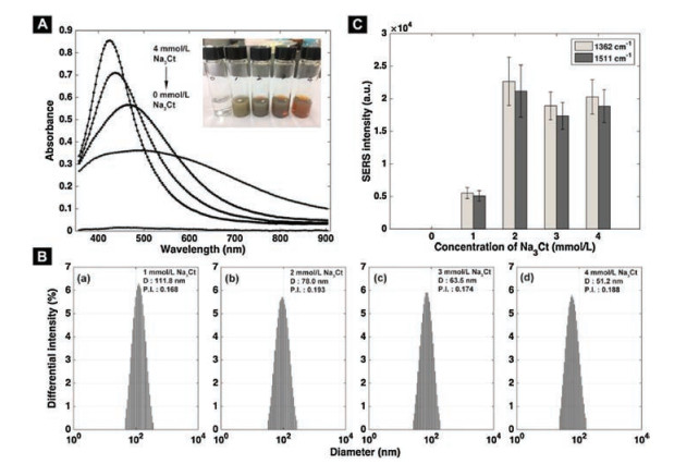
It is known that the stabilizing agent caps the particle and prevents further growth or aggregation. In this case, citrate ions serve as the role of a stabilizer. At pH 9 the silver colloid was synthesized by holding in hand for 5 min. Dynamic light scattering (DLS) (Delsa Nano C, Beckman Coulter, USA) and transmission electron microscopy (TEM) (JEM-2100, JEOL, Japan) results measuring the silver colloids obtained by different concentrations of trisodium citrate were shown in Fig. 2C and Fig. S1 (Supporting information). The DLS results suggested that as the concentration increased the particle average sizes of colloid decreased. The trend was similar to that reported by Arnim and Michael [33]. At high citrate concentrations large lumps of coalesced silver particles are present, due to destabilization by the high ionic strength of the solution [33]. The polydispersity index (P.I.) at different concentrations were 0.168, 0.193, 0.174 and 0.188, which indicated that the uniformity of colloid was well (P.I. < 0.2, [34]). From the results of electron microscopy (Fig. S1), it can be seen that the particles were almost spherical, although some were multiple twinning, staple faults and partial dislocations. Therefore 2 mmol/L of trisodium citrate was chosen for subsequent works.

|
Download:
|
| Fig. 2. (A) Absorption spectra and (B) intensity distribution results obtained by DLS at four concentrations of trisodium citrate: (a) 1 mmol/L (111.8 nm), (b) 2 mmol/L (78.0 nm), (c) 3 mmol/L (63.5 nm), (d) 4 mmol/L (51.2 nm). The horizontal ordinate is expressed in log form; 'D' denotes the average particle size. (C) SERS intensity of 1.0 × 10⁻⁷ mol/L R6 G at different concentrations of trisodium citrate. | |
The digital photograph of silver colloids and their absorption spectra (UV–vis spectrometer, INSION, Germany) at different concentrations of citrate were shown in Fig. 2A. When trisodium citrate is not added, the solution was transparent. As the concentration increased, the color of the colloid turned from grayish green to reddish. The overall spectral shape of the plasmon absorption remains different, showing that the overall distribution of particles is affected by citrate concentration. The difference absorption spectra of colloids in the presence of citrate ions showed an increase in the absorption at maximum absorbance with increase in concentration of citrate, possibly due to the decreased size and/or anisotropy degree of the silver particles [27]. At the same time, the absorption peak was obviously blue-shifted due to a decrease in particle size, which was supported by the DLS results and TEM images (Fig. 2B and Fig. S1). Many authors had associated the blue-shift of the surface plasmon resonance of the silver nanoparticles with their size decrease, which may be the universal case [21, 35-37].
Different concentrations of trisodium citrate would lead to a variety of particle distribution of synthesized colloid. And the SERS enhancement strongly depends on the size, shape and composition of the SERS substrate. Using R6 G (1.0 × 10⁻⁷ mol/L) as the probe molecule, the SERS effect of those silver colloid under different concentrations of trisodium citrate were investigated. The results were shown in Fig. 2C. As the concentration increased, the SERS intensity increased first and then decreased. When the concentration was 2 mmol/L, SERS signal of 1.0 × 10⁻⁷ mol/L R6 G was the highest both at 1362 cm-1 and 1511 cm-1. In this case, the particle size was 76.9 ± 6.2 nm (DLS). Therefore, the optimum concentration of trisodium citrate was 2 mmol/L.
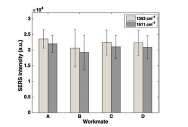
To confirm the reproducibility of the SERS substrates these syntheses were repeated several times by same and different workmates. Under the conditions optimized, i.e., pH was finally selected to be9, the concentration of trisodium citrate was 2 mmol/L, and the reaction time was 5 min, the reproducibility of this method between different production batches and four different workmates was investigated with 1 ×10⁻⁷ mol/L R6 G as the molecular probe. The average Raman intensities of corresponding bands were given in Fig. 3, from which it can be seen that the relative standard deviation (RSD) values of Raman intensities at 1362 cm-1 and 1511 cm-1 obtained by four workmates with three repetitions were 10%–19%. Moreover, Fig. 3 showed very small difference in the outcome of these independent experiments. The RSD between columns estimated by the intensities at 1362 cm-1 and 1511 cm-1 were 5.51% and 5.49% respectively, proved that this new method of synthesis of silver colloid has good reproducibility between different manufacturers.And when the confidence interval was 0.05, thet-test results (analysized by Matlab 8.5.0) showed no significant differences among the four groups. So, those results proved that this new type of substrate has acceptable reproducibility between different batches and manufacturers.

|
Download:
|
| Fig. 3. Average SRES intensities of 1 ×10⁻⁷ mol/L R6 G collected by four different workmates. | |
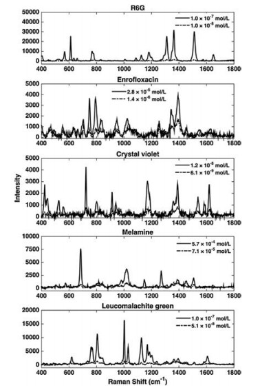
In order to advance the applications of this SERS substrate synthesized by proposed method, it was used for the detections of antibiotic, dye and forbidden feed additives. Solutions of R6 G, enrofloxacin, crystal violet, melamine and leucomalachite green were prepared for the SERS detections. The SERS spectra of these food harmful substances in different concentrations were shown in Fig. 4. R6 G is a conventional SERS probe molecule, and its lowest detection concentration (LDC) was 1.0 × 10⁻⁸ mol/L in this study. It was found that there is a good linear relationship between the Raman intensities and R6 G concentration in the concentration range of 1.0 × 10⁻⁸ – 1.8 × 10⁻⁷ mol/L with a squared correlation coefficient (R2) of 0.992 (Fig. S2 and Table S1 in Supporting information). Compared with other reported LDCs of R6 G being 5.45 ×10⁻⁷ – 1.0 ×10⁻⁹ mol/L [14, 38, 39], our substance has an acceptable sensitivity. Crystal violet can be commonly used as disinfectant for water bodies, and its LDC was 6.1 ×10⁻⁸ mol/L, which is slightly better than the result reported by C.R. Rekha et al. [40]. The LDCs reached with the SERS substrate of enrofloxacin, melamine and leucomalachite green were 1.4 ×10⁻⁶ mol/L, 7.1 × 10⁻⁵ mol/L and 5.1 ×10⁻⁸ mol/L, respectively. According to Chinese standards, enrofloxacin has a limit of 100–300 μg/kg in food, which is much higher than the concentration reached by using our substrate. For melamine and leucomalachite green, although their lowest detection concentrations are slightly higher than that reported in other literatures [5, 39], considering with convenience and applicability in field, our synthesis method has high efficiency. In a word, these applications indicate that the SERS substrate synthesized by the proposed method could serve as a great potential tool in rapid and effective detections of harmful substances.

|
Download:
|
| Fig. 4. The SERS spectra of several harmful substances (line: high concentration; dotted line: the lowest detection concentration). | |
In conclusion, based on one-step reaction and the constant temperature of human body, we have developed a simple, rapid, green energy and low-cost synthesis method of silver colloid, and the homogeneity of nanoparticles is well. And the particle size was well controlled by adjusting the concentration of trisodium citrate. As a SERS substrate, the silver colloid has good enhancement activity and batch reproducibility. The developed substrate was used to detect rhodamine 6 G, enrofloxacin, crystal violet, melamine and leucomalachite green, and showed good performance with lowest detection concentrations of 1.0 ×10⁻⁸ mol/L, 6.1 ×10⁻⁸ mol/L, 1.4 ×10⁻⁶ mol/L, 7.1 ×10⁻⁵ mol/L and 5.1 ×10⁻⁸ mol/L, respectively. The results obtained demonstrate that the developed synthesis method may become an efficient way to supply fresh SERS substrate overcoming the problem of short life of commercial substrates, thus has great potential in various, especially in field determinations.
AcknowledgmentWe thank Prof. Sugiyama from Hokaido University for his help on the operation of the Soft X-ray machine.
Appendix A. Supplementary dataSupplementary material related to this article can befound, in the online version, at doi:https://doi.org/10.1016/j.cclet.2019.02.004.
| [1] |
K. Haruna, T.A. Saleh, M.K. Hossain, A.A. Al-Saadi, Chem. Eng. J. 304 (2016) 141-148. DOI:10.1016/j.cej.2016.06.050 |
| [2] |
C. Zhang, K. Wang, D. Han, Q. Pang, Spectrochim. Acta Part A 122 (2014) 387-391. DOI:10.1016/j.saa.2013.11.066 |
| [3] |
S. Yu, Z. Liu, W. Wang, et al., Talanta 178 (2018) 498-506. DOI:10.1016/j.talanta.2017.09.054 |
| [4] |
H. Zhang, L. Sun, Y. Zhang, et al., Chin. Chem. Lett. 29 (2018) 981-984. DOI:10.1016/j.cclet.2017.10.017 |
| [5] |
Y.Y. Zhang, W.S. Yu, L. Pei, et al., Food Chem. 169 (2015) 80-84. DOI:10.1016/j.foodchem.2014.07.129 |
| [6] |
Y.F. Jiang, D.W. Sun, H.B. Pu, Q.Y. Wei, Trends Food Sci. Technol. 75 (2018) 10-22. DOI:10.1016/j.tifs.2018.02.020 |
| [7] |
F.K. Alsammarraie, M. Lin, A. Mustapha, et al., Food Chem. 259 (2018) 219-225. DOI:10.1016/j.foodchem.2018.03.105 |
| [8] |
F. Liu, H. Gu, Y. Lin, et al., Spectrochim. Acta Part A 85 (2012) 111-119. |
| [9] |
T. Janci, D. Valinger, J.G. Kljusuric, et al., Food Chem. 224 (2017) 48-54. DOI:10.1016/j.foodchem.2016.12.032 |
| [10] |
Y. Kang, W. Chen, H. Zhang, et al., Microchem. J. 137 (2018) 15-21. |
| [11] |
B. Han, Y.L. Zhang, L. Zhu, et al., Sens. Actuator. B:Chem. 270 (2018) 500-507. DOI:10.1016/j.snb.2018.05.043 |
| [12] |
T. Wu, H.T. Wang, B. Shen, et al., Chin. Chem. Lett. 27 (2016) 745-748. DOI:10.1016/j.cclet.2016.01.059 |
| [13] |
C. Muehlethaler, M. Leona, J.R. Lombardi, Forensic Sci. Int. 268 (2016) 1-13. DOI:10.1016/j.forsciint.2016.09.005 |
| [14] |
M.A. Fikiet, S.R. Khandasammy, E. Mistek, et al., Spectrochim. Acta Part A 197 (2018) 255-260. DOI:10.1016/j.saa.2018.02.046 |
| [15] |
S.C. Luo, K. Sivashanmugan, J.D. Liao, et al., Biosens. Bioelectron. 61 (2014) 232-240. DOI:10.1016/j.bios.2014.05.013 |
| [16] |
R. Gillibert, J.Q. Huang, Y. Zhang, et al., Trac-Trend. Anal. Chem. 105 (2018) 185-190. DOI:10.1016/j.trac.2018.05.009 |
| [17] |
F.Y. Diao, X.X. Xiao, B. Luo, et al., Appl. Surf. Sci. 427 (2018) 1271-1279. DOI:10.1016/j.apsusc.2017.08.117 |
| [18] |
M. Ratkaj, S. Miljanic, Vib. Spectrosc. 74 (2014) 104-109. DOI:10.1016/j.vibspec.2014.08.004 |
| [19] |
L.Y. Chen, J.S. Yu, T. Fujita, M.W. Chen, Adv. Funct. Mater. 19 (2009) 1221-1226. DOI:10.1002/adfm.v19:8 |
| [20] |
H. Zhang, L. Sun, Y. Zhang, et al., Talanta 174 (2017) 301-306. DOI:10.1016/j.talanta.2017.06.025 |
| [21] |
L. Xu, J. Peng, M. Yan, et al., Chem. Eng. Process. 102 (2016) 186-193. DOI:10.1016/j.cep.2016.01.017 |
| [22] |
D. Singha, N. Barman, K. Sahu, J. Colloid Interface Sci. 413 (2014) 37-42. DOI:10.1016/j.jcis.2013.09.009 |
| [23] |
Y. Lu, C.Y. Zhang, D.J. Zhang, et al., Chin. Chem. Lett. 27 (2016) 689-692. DOI:10.1016/j.cclet.2016.01.032 |
| [24] |
P.C. Lee, D. Meisel, J. Phys. Chem. 86 (1982) 3391-3395. DOI:10.1021/j100214a025 |
| [25] |
C.H. Ma, J. Zhang, Y.C. Hong, et al., Chin. Chem. Lett. 26 (2015) 1455-1459. DOI:10.1016/j.cclet.2015.10.015 |
| [26] |
R. Baber, L. Mazzei, N.T.K. Thanh, A. Gavriilidis, RSC Adv. 5 (2015) 95585-95591. DOI:10.1039/C5RA17466J |
| [27] |
Y. Qin, X. Ji, J. Jing, et al., Colloid Surf. A 372 (2010) 172-176. DOI:10.1016/j.colsurfa.2010.10.013 |
| [28] |
J. Haber, K. Sokolov, Langmuir 33 (2017) 10525-10530. DOI:10.1021/acs.langmuir.7b01362 |
| [29] |
P. Wagener, A. Schwenke, S. Barcikowski, Langmuir 28 (2012) 6132-6140. DOI:10.1021/la204839m |
| [30] |
K.P. Velikov, G.E. Zegers, A. van Blaaderen, Langmuir 19 (2003) 1384-1389. DOI:10.1021/la026610p |
| [31] |
V.K. LaMer, R.H. Dinegar, JACS 72 (1950) 4847-4854. DOI:10.1021/ja01167a001 |
| [32] |
D.V. Goia, J. Mater. Chem. 14 (2004) 451-458. DOI:10.1039/b311076a |
| [33] |
A. Henglein, M. Giersig, J. Phys. Chem. B 103 (1999) 9533-9539. DOI:10.1021/jp9925334 |
| [34] |
N. Sawtarie, Y.H. Cai, Y. Lapitsky, Colloid Surf. B 157 (2017) 110-117. DOI:10.1016/j.colsurfb.2017.05.055 |
| [35] |
K. Tokarek, J.L. Hueso, P. Kustrowski, et al., Eur. J. Inorg. Chem. 2013 (2013) 4940-4947. |
| [36] |
A.M. Schwartzberg, T.Y. Olson, C.E. Talley, J.Z. Zhang, J. Phys. Chem. B 110 (2006) 19935-19944. DOI:10.1021/jp062136a |
| [37] |
A. Moores, F. Goettmann, New J. Chem. 30 (2006) 1121-1132. DOI:10.1039/b604038c |
| [38] |
Y.X. Wu, P. Liang, Q.M. Dong, et al., Food Chem. 237 (2017) 974-980. DOI:10.1016/j.foodchem.2017.06.057 |
| [39] |
H.S.S. Sharma, E. Carmichael, D. McCall, Vib. Spectrosc. 83 (2016) 159-169. DOI:10.1016/j.vibspec.2016.01.011 |
| [40] |
C.R. Rekha, V.U. Nayar, K.G. Gopchandran, J. Sci.:Adv. Mater. Dev. 3 (2018) 196-205. |