 2019, Vol. 30


b School of Chemistry and Chemical Engineering, Shandong University, Ji'nan 250100, China;
c School of Transportation and Civil Engineering, Shandong Jiaotong University, Ji'nan 250037, China
Halogenated solvents, especially chlorinated solvents have been widely used in both industrial and scientific fields [1, 2]. However, the mass use of these chemicals is imposing many environmental problems (e.g., the contamination of the air and water resources) [3-5], and threatening to our health within longterm exposure (e.g., probable human carcinogen and central nervous system effector) [6-10]. Therefore, the organic solvents waste disposal is classified into two separate categories: halogenated and non-halogenated. This segregation is necessary because halogenated substances require more extensive treatment during the waste disposal in order to minimize environmental pollution. So far, the detection and discrimination of the halogenated solvents among other solvents remains an area of intense interest but of great challenge.
Sensors are important scaffolds for detecting contaminants and monitoring environmental conditions. Fluorescent sensors that can reveal the presence of a contaminant are attractive because they can display changes in emission colors, especially when such changes can be readily discernible by naked eye [11-25].Traditionally, the methods for detection of organic solvents mostly relied on analytical techniques [6, 26-34] (such as gas chromatography, high-performance liquid chromatography and mass spectrometry) which require specially trained personnel and consume lots of money and time. Development of new sensors and methods for chemical detection is now gaining increasing attention. Ooyama et al. [35, 36] designed several D-π-A type pyridinium dyes which show the larger bathochromic shift in halogenated solvents than those in non-halogenated solvents. Ripp et al. [6] created a bioreporter for self-directed detection of CH2Cl2 by taking advantage of a bioluminescent gene. Murai et al. [37] also synthesized several pyridinium 5-aminothiazoles that can be applied to the detection of halogenated solvents, especially CH2Cl2. Xu et al. [1] prepared a naphthalimide-based fluorescent sensor molecule which can efficiently distinguish halogenated solvents from non-halogenated solvents. Xue et al. [38] fabricated an optical sensor film encapsulating modified Fujiwara reagents for detection of CHCl3. Zang et al. [39] reported a fluorescence sensor molecule which enabled instant detection of γ radiation in halogenated solvents (e.g., CHCl3, CH2Cl2). Kim et al. [40] devised a solvatochromic sensor system that can differentiate chloroform and dichloromethane colorimetrically. Che et al. [41] prepared a Platinum (Ⅱ) complex which gave most intense luminescence with volatile halogenated solvents. Yi et al. [42] constructed a multi-responsive MOF that can recognize CCl4 vapor with high selectivity. Despite those promising chemosensors have been reported, the studies on simple, low-cost and efficient sensors that can distinguish halogenated solvents from halogen-free solvents are still relatively rare and the sensoring mechanism is not clear until now.
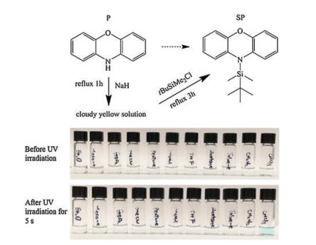
Here, we have developed an effective sensor molecule SP derived from phenoxazine P (Scheme 1) that can discriminate most commonly-used halogen solvents among other solvents with solution color changing from colorless to blue under UV light irradiation (λ = 365 nm). The detection process can be realized by the visual color change and fluorescence change.

|
Download:
|
| Scheme 1. The synthesis route for SP molecule (top) and the photos of SP molecule dissolved in different solvents (bottom) before and after UV light irradiation (λ = 365 nm) for 5 s. | |
The synthetic route for the sensor molecule SP is shown in Scheme 1 (the details of the experiment deposited in the Supporting information). The pure product was obtained through recrystallization in methanol in 77.9% yield. The product was characterized by 1H NMR, 13C NMR, 29Si NMR, IR and EA (Figs. S1-S4 in Supporting information). In CDCl3, the single peak at δ 6.9 is corresponding to eight aromatic hydrogens, while these hydrogens can be separated into three multi-peaks in C6D6 (Fig. S5 in Supporting information). As is clearly shown in Scheme 1, after 5 s irradiation by UV light (λ = 365 nm, power: 10 W, vertical distance: 5 cm) at ambient temperature, the color of SP solution in CDCl3 turns slightly blue. Notably, this color change has also been found in other halogenated solvents, such asCH2Cl2, C2H2Cl4, Me2CHBr, PhCl and soon (Fig. S6 in Supporting information), whereas in non-halogenated solvents such as toluene, acetone, THF, methanol, pentane, hexane, acetonitrile, ethyl acetate, and diethyl ether and so on, there is no color change even after irradiation for several hours or with shorter wavelength irradiation. This promoted us to explore the chromism mechanism of this fast response towards halogenated solvents. We initially employed the NMR technique to determine this color change with CDCl3 as an example. As shown in Fig. S7 in Supporting information, there is no obvious change after 5 s irradiation. But if we zoom in this spectrum, we can see the slightlydifference. A small new peak at δ 0.37 and a shoulder peak at δ 0.99 appear. Then we trace this dynamic process by means of theNMR technique. After1 h irradiation by 365 nm UV light at ambient temperature, both 1H NMR (Fig. 1) and 13C NMR (Fig. S6) showed clear difference. As we can see from Fig. 1, the solution color turnedbluefrom colorlessafter irradiation, and those two new peaks grew obviously at high field area (δ 0.37 and 0.99). With longer irradiation time, these two peaks get stronger whereas the original two peaks at δ 0.27 and δ 0.97 become weaker. At the same time, the multiple peak at δ6.90 becomes weaker as well, although the position did not change. These results indicate the decomposition of the SP molecule with the departure of the silane group. After 6 h irradiation, the two new peaks became dominant and the solution color turned to dark blue, which suggests most of the SP molecules decompose. Moreover, the appearance of the multipeak at about δ 6.70 possibly indicates the formation of new species. In comparison with the pure ClSi(CH3)2(tBu) molecule, we infer that these two new peaks are attributed to the product ClSi(CH3)2(tBu) molecule from the reaction of the SP molecule and chloroform. The 13C NMR (Fig. S8 in Supporting information) and 29Si NMR (Fig. S9 in Supporting information) also confirmed our speculation. Moreover, we found that some precipitates are formed in the solution after longtime exposure from 365 nm UV light, which is responsible for the decreased intensity of the multi-peaks corresponding to the aromatic hydrogen at low field area (δ6.90). Only trace of the startingmaterials can be found after 6 h irradiation, which indicates most of the SP molecules have decomposed and the reaction is nearly complete. Interestingly, the precipitates can dissolve in DMSO. The 1H NMR spectra (Fig. S10 in Supporting information) shows clear multi-peaks around δ6.6 which can be ascribed to the phenylhydrogen.According tothedetailedNMR study, weinfer that the polymer or oligomer which precipitated out from the solution was produced via the photo-induced reaction between chloroform and SP molecules.

|
Download:
|
| Fig. 1. 1H NMR spectra of ClSi(CH3)2(tBu) molecule (bottom) and the SP solution in CDCl3 (0.01 mmol/mL) before and after 365 nm UV irradiation (0–6 h). (insets: the pictures of the solution under ambient light). | |
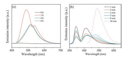
Based on the fast color response of the solution to the 365 nm UV light and the NMR evolutions in chloroform, the irradiation time-dependent fluorescence property was further investigated under the same conditions. Before UV irradiation, the fluorescence spectra showed a broad emission band from 350 nm to 550 nm with the maximum at about 450 nm (Fig. 2a, λex = 326 nm). Here, we should note that, if we use 398 nm as the excitation wavelength before UV irradiation, there is no any emission at all. After 0.5 h irradiation, not only the clear red-shift to 488 nm (λex = 398 nm) for the maximum emission peak but also the much stronger intensity (about 15 times than before) can be detected. Then, we further prolonged the irradiation time to 1 h. Again, 4-fold emission enhancement and a small red-shift to 490 nm (λex = 398 nm) in comparison with 0.5 h irradiation were observed. A shoulder peak at 538 nm was probably due to the formation of some unknown new species. After total 4 h irradiation, the maximum emission peak shifted to 520 nm (λex = 398 nm), whereas the intensity turned half. When prolonging the irradiation time to 6 h, there was no obvious change for the maximum emission peak including its intensity and the profile. Since the solution color changed very quickly from colorless to blue upon exposure to 365 nm UV light, further efforts were paid to investigate the fluorescence properties of SP in CHCl3 at the initial stage (Fig. 2b). As we can see, from 1 min to 9 min' irradiation, the emission intensity at about 412 nm (λex = 346 nm) reduced gradually as irradiation time went on Conversely, a new peak at about 480 nm appeared and get stronger and stronger. Moreover, a slight red-shift was also found for this new peak from 472 nm for 5 min to 480 nm for 7 min and to 486 nm for 9 min.

|
Download:
|
| Fig. 2. (a) Emission spectra of SP solution in CHCl3 (0.01 mmol/mL) after UV 365 nm irradiation (0–6 h). (b) Excitation and emission spectra after short-time 365 nm UV irradiation. | |
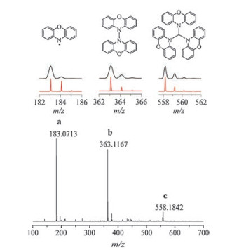
Observation from the NMR and fluorescence spectra suggests that the SP molecule must react with chloroform to form a new compound under UV light. To disclose more about the possible mechanism, the positive-ion ESI-MS of SP in CHCl3 after 1 h UV irradiation was measured. As shown in Fig. 3, the most dominant peak at m/z 183.07 corresponds to C12H9NO (a, calcd. m/z 183.07), which might be the SP radical. Moreover, another two species with lower abundance at 363.12 (b) and 558.18 (c) were also detected and assigned to C24H16N2O2 (calcd. m/z 263.12) and C37H25N3O3 (calcd. m/z 558.19), respectively, by matching the experimental and simulated isotope distributions. They should be the dimer and trimer of the SP molecule, respectively (shown in Scheme S1 in Supporting information).

|
Download:
|
| Fig. 3. High resolution mass spectrum of SP derivatives (middle: corresponding close-up simulated (uper line) and experimental (lower line) spectrum, top: proposed corresponding chemical structure). | |
To further elucidate the mechanism, we also performed the experiments in different solvents, i.e., C6D6 and CD2Cl2, as well as 1 equiv. of CHCl3 in C6D6, the results of which were analyzed by using NMR (1H, 13C and 29Si) and fluorescence spectra (Fig. S11-S20 in Supporting information). In benzene, there's no change even after 48 h 365 nm UV light irradiation. In CD2Cl2, the changing trend is very similar to that in CDCl3 but more complicated. As we mentioned above, 0.01 mmol/mL SP solution in CDCl3 allows fast response to 365 nm UV light within 5 s. We also tried low concentration of CHCl3 in C6D6 with 1 equiv. CHCl3 (10-3 mmol/mL). After 365 nm UV light irradiation, we can also see the color change but with longer time (about 2 min). The changing trend of the NMR spectra is the same as that in CDCl3 but need more time irradiation. Thus, the sensitivity for chloroform is at least 10-3 mol/L. In light of these experiments, we proposed the tentative mechanism of the reaction between the SP molecule and chloroform (Scheme S1 in Supporting information). In the first step, under 365 nm UV light, the ·Cl radical and ·CHCl2 radical were generated by the decompose of chloroform. The ·Cl then triggered the departure of silane group from SP molecule giving the ClSi (CH3)2(tBu) molecule and a species immediately. The ClSi (CH3)2(tBu) molecule has already been proved through NMR spectra and a species (a colored radical) can give stronger luminescence than SP molecule. In the third step, the a can dimerize to form b. At the same time, the a species can also react with ·CHCl2 to produce c1 which then lost the rest of two ·Cl stepwise to form c2 and c species, respectively.
In summary, a phenoxazine-based sensor was developed, which can effectively distinguish the halogenated solvents from non-halogenated solvents. The dynamic NMR spectra and fluorescence spectra as well as the ESI-MS spectra reveal a radicalinvolved sensoring mechanism that triggered by photo. This work not only provide a new avenue to design new sensor towards the halogenated solvents but also give a deep insight to understand the underlying sensoring mechanism.
AcknowledgmentsWe are grateful to the Natural Science Foundation of Shandong Province (Nos. BS2014CL042, ZR2014EEP021, ZR2017MB033), the Key Research and Development Program of Shandong Province, China (Nos. 2016GSF116014 and 2017CXGC1113), the Start-up Grant of Shandong Jiaotong University and the National Undergraduate Innovation Training Program (No. 201611510109), we also acknowledge the financial support from the government of Shandong Province for visiting abroad. We appreciate for Prof. Zhenghu Xu and Prof. Wenguang Wang for their constructive comments on the mechanism in this paper.
Appendix A. Supplementary dataSupplementary material related to this article canbefound, in the online version, at doi:https://doi.org/10.1016/j.cclet.2019.01.027
| [1] |
L. Dai, D. Wu, Q. Qiao, et al., Chem. Commun. (Camb.) 52 (2016) 2095-2098. DOI:10.1039/C5CC09403H |
| [2] |
T. Permpool, A. Sirivat, D. Aussawasathien, L. Wannatong, Polym-Plast. Technol. 52 (2013) 907-920. DOI:10.1080/03602559.2013.763371 |
| [3] |
R.E. Jackson, Technol. Cult. 45 (2004) 55-79. DOI:10.1353/tech.2004.0022 |
| [4] |
U.S. Environmental Protection Agency, Chemical Summary for Methylene Chloride (dichloromethane), Office of Pollution Prevention and Toxics, 1994 Report number 749-F-94-018a.
|
| [5] |
Agency for Toxic Substances, Disease Registry (ATSDR), Toxicological Profile for Methylene Chloride, Public Health Services, U.S. Department of Health and Human Services, Atlanta, 2005.
|
| [6] |
N. Lopes, S.A. Hawkins, P. Jegier, et al., J. Ind. Microbiol. Biotechnol. 39 (2012) 45-53. DOI:10.1007/s10295-011-0997-5 |
| [7] |
C. Barragán-Martínez, C.A. Speck-Hernández, G. Montoya-Ortiz, et al., PLoS One 7 (2012) e51506. DOI:10.1371/journal.pone.0051506 |
| [8] |
Y.L. Chang, C.C. Yang, J.F. Deng, et al., J. Toxicol. Clin. Toxicol. 37 (1999) 497-504. DOI:10.1081/CLT-100102442 |
| [9] |
N.Y. Kim, S.W. Park, J.K. Suh, J. Forensic Sci. 41 (1996) 527-529. |
| [10] |
M. Mahmud, S.N. Kales, Health Perspect. 107 (1999) 769-772. DOI:10.1289/ehp.99107769 |
| [11] |
O.S. Wenger, Chem. Rev. 113 (2013) 3686-3733. DOI:10.1021/cr300396p |
| [12] |
C. Reus, T. Baumgartner, Dalton Trans. 45 (2016) 1850-1855. DOI:10.1039/C5DT02758F |
| [13] |
Y. Ren, A. Orthaber, R. Pietschnig, T. Baumgartner, Dalton Trans. 42 (2013) 5314-5321. DOI:10.1039/c3dt33058c |
| [14] |
J.H. Wang, M. Li, D. Li, Chem. Sci. 4 (2013) 1793-1801. DOI:10.1039/c3sc00016h |
| [15] |
R.F. Landis, M. Yazdani, B. Creran, et al., Chem. Commun. (Camb.) 50 (2014) 4579-4581. DOI:10.1039/c4cc00805g |
| [16] |
X.M. He, J. Borau-Garcia, A.Y.Y. Woo, S. Trudel, T. Baumgartner, J. Am. Chem. Soc. 135 (2013) 1137-1147. DOI:10.1021/ja310680x |
| [17] |
E. Yamaguchi, C. Wang, A. Fukazawa, et al., Angew. Chem. Int. Ed. 54 (2015) 4539-4543. DOI:10.1002/anie.201500229 |
| [18] |
Z.W. Chen, X.N. Mi, J. Lu, et al., Dalton Trans. 47 (2018) 6240-6249. DOI:10.1039/C8DT00909K |
| [19] |
S.N. Wang, T.T. Cao, H. Yang, et al., Inorg. Chem. 55 (2016) 5139-5515. DOI:10.1021/acs.inorgchem.5b02801 |
| [20] |
J.H. Wang, M. Li, D. Li, Chem. Sci. 4 (2013) 1793-1801. DOI:10.1039/c3sc00016h |
| [21] |
Z.W. Chen, X.N. Mi, S.N. Wang, et al., J. Solid State Chem. 261 (2018) 75-78. DOI:10.1016/j.jssc.2018.02.008 |
| [22] |
R.R. Ma, Z.W. Chen, S.N. Wang, et al., J. Solid State Chem. 252 (2017) 142-151. DOI:10.1016/j.jssc.2017.05.018 |
| [23] |
S.N. Wang, R.R. Ma, Z.W. Chen, et al., Sci. China Chem. 59 (2016) 948-958. DOI:10.1007/s11426-015-0537-6 |
| [24] |
L. Yu, Y. Qiao, L. Miao, Y. He, Y. Zhou, Chin. Chem. Lett. 29 (2018) 1545-1559. DOI:10.1016/j.cclet.2018.09.005 |
| [25] |
X.Y. Ren, L.H. Lu, Chin. Chem. Lett. 26 (2015) 1439-1445. DOI:10.1016/j.cclet.2015.10.014 |
| [26] |
W. Buchberger, U. Huebauer, Microchim. Acta 99 (1989) 137-142. DOI:10.1007/BF01242799 |
| [27] |
M.I. Cervera, J. Beltran, F.J. Lopez, F. Hernandez, Anal. Chim. Acta 704 (2011) 87-97. DOI:10.1016/j.aca.2011.08.012 |
| [28] |
M.E. Miller, C.J. Cappon, Clin. Chem. 30 (1984) 781-783. |
| [29] |
B. Mizaikoff, Anal. Chem. 75 (2003) 258-267. |
| [30] |
T. Huybrechts, J. Dewulf, O. Moerman, H.V. Langenhove, J. Chromatogr. A 893 (2000) 367-382. DOI:10.1016/S0021-9673(00)00771-8 |
| [31] |
A. Lara-Gonzalo, J.E. Sanchez-Uría, E. Segovia-García, A. Sanz-Medel, Talanta 74 (2008) 1455-1462. DOI:10.1016/j.talanta.2007.09.036 |
| [32] |
Z. Wang, K. Li, M. Fingas, L. Sigouin, L. Menard, J. Chromatogr. A 971 (2002) 173-184. DOI:10.1016/S0021-9673(02)01003-8 |
| [33] |
A.M.S. Silva, E. de Viana A., M.F. Pimentel, Y.M.B. Almeida, I.M. Raimundo JrⅢ, J. Braz. Chem. Soc. 22 (2011) 1470-1477. DOI:10.1590/S0103-50532011000800010 |
| [34] |
M. Jakusch, B. Mizaikoff, R. Kellner, A. Katzir, Sens. Actuators B Chem. 38 (1997) 83-87. DOI:10.1016/S0925-4005(97)80175-X |
| [35] |
Y. Ooyama, Y. Oda, T. Mizumo, J. Ohshita, Tetrahedron 69 (2013) 5e1760. |
| [36] |
Y. Ooyama, R. Asada, S. Inoue, et al., New J. Chem. 33 (2009) 2311-2316. DOI:10.1039/b9nj00332k |
| [37] |
K. Yamaguchi, T. Murai, Y. Tsuchiya, et al., RSC Adv. 7 (2017) 18132-18135. DOI:10.1039/C7RA01896G |
| [38] |
J.K. Fong, J.K. Pena, Z.L. Xue, et al., Anal. Chem. 87 (2015) 1569-1574. DOI:10.1021/ac503920c |
| [39] |
J.M. Han, M. Xu, B. Wang, et al., J. Am. Chem. Soc. 136 (2014) 5090-5096. DOI:10.1021/ja500262n |
| [40] |
J. Lee, H.T. Chang, H. An, et al., Nat. Commun. 4 (2013) 2461-2469. DOI:10.1038/ncomms3461 |
| [41] |
S.C.F. Kui, S.S.Y. Chui, C.M. Che, N.Y. Zhu, J. Am. Chem. Soc. 128 (2006) 8297-8309. DOI:10.1021/ja061422x |
| [42] |
F.Y. Yi, S.C. Wang, M. Gu, J.Q. Zheng, L. Han, J. Mater. Chem. C 6 (2018) 2010-2018. DOI:10.1039/C7TC05707E |