 2019, Vol. 30


b Department of Chemistry, Southern University of Science and Technology, Shenzhen 518055, China
Urethane, also known as ethyl carbamate, is an ethyl ester of carbamic acid. This compound has been proven to be genotoxic and carcinogenic to many animals, including mice, hamsters and monkeys [1]. It can be absorbed rapidly and nearly completely by the gastro-intestinal tract and the skin [2]. The cellular metabolism of urethane is associated with oxidative stress and DNA damage, which may finally result in lung cancer, skin cancer, or lymph cancer [3]. Meanwhile, urethane is very soluble in water (ca. 2 g/mL) and widely distributed in daily life. It is one of the natural constituents in Tobacco leaves, and can also be found in many alcoholic beverages and fermented food products such as bread, yogurt, soy sauce and so on, threatening people's health [4]. Therefore, urethane has been re-classified as a Group 2A carcinogen (probably carcinogenic to humans) from Group 2B (possibly carcinogenic) by the International Agency for Research on Cancer (IARC) of World Health Organization in March 2007 [5].
Urethane is a small and polar molecule with a molecular weight of only 89 g/mol (EtCO2NH2) and the dipole moment of 0.521 D. Therefore, its detection and analysis are quite difficult especially in aqueous solution [6]. A great deal of effort has been devoted to the quantitative analysis of urethane, and so far different analytical tools for urethane determination have been developed, for example HPLC-FLD, solid phase microextraction coupled with GC–MS, etc. [7]. The standard method for the determination of urethane residues for export in China (SN/T 0285-2012) [8] is as follows: the sample is extracted by dichloromethane and then analyzed by GC–MS. This method requires high operational skill, and is quite expensive and time-consuming and not suitable for the quantification of a large number of samples [9].
Selective recognition of hydrophilic molecules in water is a generally-accepted challenge in supramolecular chemistry [10-12]. This is also the key for molecular sensing [13-16] of these molecules. Recently, we have reported a series of naphtholbased macrocyclic receptors [17-27]. Of them, endo-functionalized molecular tubes 1a and 1b (Fig. 1) are shown to be able to selectively recognize hydrophilic molecules in water. A systematic study on the recognition property of molecular tubes 1a and 1b toward 44 hydrophilic molecules [27] reveals that hydrophobic effect through releasing the "high-energy" cavity water and hydrogen bonding are the driving forces for the binding of hydrophilic molecules in water. In addition, hydrogen bonding makes a decisive role in the high binding selectivity. Molecular tubes 1a and 1b are fluorescent and show fluorescent enhancement upon guest complexation. With these results, we wondered whether molecular tubes 1a and 1b can bind urethane and even act as fluorescent sensors for detecting urethane in aqueous solutions. Herein, we report our findings in this regard.

|
Download:
|
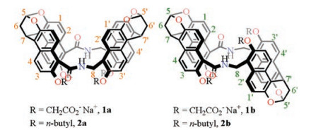
| Fig. 1. Chemical structures of molecular tubes 1a, 1b, 2a, and 2b. The numberings on the structures are used for the assignments of NMR signals. | |
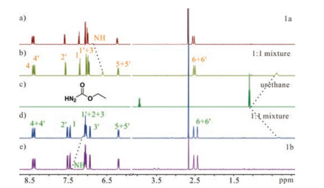
Molecular tubes 1a and 1b can indeed bind urethane in water. In the 1H NMR spectra (H2O:D2O, 9:1) of a 1:1 mixture of urethane with 1a or 1b (Fig. 2), the signals of the methyl group of urethane undergo significant upfield shifts (-0.72 ppm for 1a and -0.76 ppm for 1b) when compared to free guest. This indicates that the guest sits inside the cavities and experiences the shielding effect of four naphthalene panels. In the meantime, the amide NH protons of 1a and 1b, which are not fully exchanged with deuterium in this solvent mixture, are thus visible in the 1H NMR spectra. These signals also undergo upfield shifts (-0.27 ppm for 1a and -0.27 ppm for 1b) when adding one equivalent of urethane into their solutions. This further confirms that urethane is bound inside the cavities because these amide protons are inwardlydirected to the cavity.

|
Download:
|
| Fig. 2. Partial 1H NMR spectra (500 MHz, H2O: D2O = 9: 1, 0.5 mmol/L, 25 ℃) of (a) 1a, (c) urethane, and (e) 1b, (b) 1a and the equimolar mixtures of urethane, (d) 1b and the equimolar mixtures of urethane. | |
Job's plots (Fig. 3 and Fig. S1 in Supporting information) suggest that 1a or 1b binds urethane in a 1:1 binding stoichiometry. In addition, guest exchange rate of these complexes is fast on the NMR timescale. Therefore, NMR titrations were performed to determine the association constants (Ka): 1750 L/mol for 1a and 1210 L/mol for 1b (Figs. S2–S5 in Supporting information). These association constants are consistent with those obtained by ITC titrations and fluorescence titrations (Table 1, Figs. 4 and 5 and Figs. S6–S9 in Supporting information). For such a small and polar molecule, these association constants are rather large. This shows that the cavities of molecular tubes 1a and 1b are suitable to accommodate small hydrophilic molecules in water.

|
Download:
|
| Fig. 3. Job's plot obtained by plotting the chemical shift change (△δ) of the 1a in 1H NMR spectra by varying the ratio of the host and urethane against the mole fraction of Host 1a. The total concentration of the host and the guest is fixed: [Host] + [Guest] = 1.0 mmol/L. This experiment supports the 1:1 binding stoichiometry between urethane and 1a. | |
|
|
Table 1 Association constants Ka (L/mol) of 1a and 1b with urethane. |


|
Download:
|
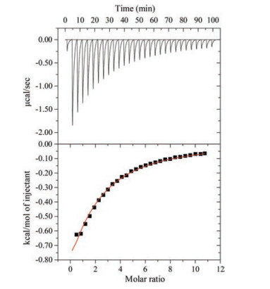
| Fig. 4. ITC titration data for 1a and urethane (H2O, 25 ℃). | |

|
Download:
|
| Fig. 5. Fluorescence spectra of 1a (5.0 × 10⁻⁶ mol/L) when titrated with urethane in deionized H2O at 25 ℃. Inset: curve fit of the titration data according to a 1:1 binding stoichiometry. | |
We would like to understand the driving forces for the binding of highly hydrophilic urethane in the relatively hydrophobic cavities of 1a and 1b. The binding affinities of 2a or 2b to urethane in CDCl3 are negligible (Figs. S10–S113 in Supporting information). This suggests that water and thus hydrophobic effect are very important for the binding of urethane in water. In our recent publication [27], we indicated that the encapsulated water in the cavity forms fewer hydrogen bonds than the bulk water. Upon guest complexation, the "high-energy" cavity waters would be released and more hydrogen bonds are formed. We proposed that this is the driving force for the binding of hydrophilic molecules in rather hydrophobic cavity through the hydrophobic effect. We believe this is also true for the binding of urethane in the cavities of these molecular tubes. Indeed, ITC experiments (Fig. 4) indicate that the binding of urethane in water is mainly driven by enthalpy with negligible entropic contributions (for 1a: △H = -21.3 kJ/mol, -T△S = 0.2 kJ/mol; for 1b: △H = -20.6 kJ/mol, -T△S = 0.3 kJ/mol; T = 298 K).
Hydrogen bonding is also important for the binding of urethane. Slow evaporation of the solution containing urethane and 2a in the mixture of CH2Cl2 and EtOH afford a single crystal, which is suitable for X-ray single crystallography. The structure is shown in Fig. 6. Although this single crystal was not grown from water, solvent molecules are anyway expelled from the cavity once the guest is bound inside the cavity. Even if the crystal was grown from water, the water molecules would be expelled anyway and the environment would be totally different from that in the aqueous solution. Therefore, this single crystal structure can still provide some information on the binding mode, binding stoichiometry, and noncovalent interactions.

|
Download:
|
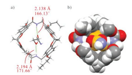
| Fig. 6. (a) Stick and (b) space-filling representation of the X-ray single crystal structure of urethane@2a. | |
As shown in Fig. 6, urethane is indeed inside the cavity of molecular tube 2a. 1:1 binding stoichiometry was observed, which is in line with the results from Job's plots. Two hydrogen bonds are involved between urethane's oxygen atoms and the host's amide protons. The distance between two amide NH of 2a and two oxygen atoms of urethane are 2.194 Å and 2.138 Å, respectively. The bond angles are 171.66° and 166.13°, respectively. This shows that N─H…O hydrogen bonds also play an important role in the host-guest complexes. However, we think the hydrogen bonds are not the main contributor to the binding, because the amide protons undergo upfield shifts when adding urethane in the solution of hosts (Fig. 2) which suggests urethane forms weaker hydrogen bonds with the amide protons than do the encapsulated water molecules [27]. That is, hydrophobic effect through releasing the "high-energy" cavity water is the major contributor. Nevertheless, once the hydrophobic effect brings the guest into the cavity, hydrogen bonding would be involved anyway.
Molecular tubes 1a and 1b show fluorescent enhancement in the presence of urethane. Therefore, they can be used as fluorescent sensors for urethane in water. The limit of detection (LOD, 3δ/slope) and the upper concentration limit were then determined: for 1a, 6.2 – 60 μmol/L; while for 1b, 29.8 – 35 μmol/L (Fig. 7 and Fig. S14 in Supporting information). Obviously, 1a has a wider applicable concentration range for the detection of urethane in water. This limit of detection can meet the requirements for the control of urethane in some distilled wines (such as Brazilian sugar cane spirits, Stone fruit spirits, etc.) and beers [15]. In addition, ethanol would not interfere with the detection of urethane in the alcoholic drinks, because 1a binds very weakly to ethanol (9 L/mol) and shows no obvious fluorescent response [27]. Furthermore, fluorescent sensing of urethane in beer (Tsingtao Beer) by 1a was performed. The intrinsic fluorescence of 1a was quenched to some extent. Nevertheless, fluorescent enhancement was still observed with the addition of urethane. Consequently, 1a can work as a fluorescent sensor to urethane even in beer (Figs. S15 and S16 in Supporting information). The limit of detection (LOD, 3δ/slope) and the upper concentration limit are 22.9 μmol/L and 60 μmol/L, respectively. As for other alcoholic drinks and fermented food products, urethane can be detected after concentrating the samples with extraction.

|
Download:
|
| Fig. 7. Fluorescence change (403 nm) of 1a with the addition of urethane in deionized H2O, and the LOD and the detection range of urethane. | |
In summary, we reported a detailed study on the molecular recognition and fluorescent sensing of urethane, which is a Group 2A carcinogen. The endo-functionalized molecular tubes are the effective receptors for this small and polar molecule in water. 1H NMR, ITC and fluorescence titrations and single crystal X-ray crystallography reveal that the hydrophobic effect through releasing the "high-energy" cavity water is the major driving force. Hydrogen bonding is involved as well, but should play a minor role. Finally, fluorescent detection of urethane in water and beer was performed. With the syn-configured molecular tube, a wide detection concentration range (6.2 – 60 μmol/L in water, and 22.9 – 60 μmol/L in beer) was achieved, which may be used in the detection of urethane in other alcoholic drinks. We think this method should be amenable to high-throughput detection of urethane in alcoholic beverages industry and fermented food industry.
AcknowledgmentsThis research was financially supported by the National Natural Science Foundation of China (Nos. 21572097, 21772083 and 21822104), and the SZSTI (Nos. JCYJ20170307105848463 and KQJSCX20170728162528382). We are grateful to SUSTech-MCPC for instrumental support.
Appendix A. Supplementary dataSupplementary material related to this article canbefound, in the online version, at doi:https://doi.org/10.1016/j.cclet.2018.11.033.
| [1] |
F.A. Beland, W.R. Benson, P.W. Mellick, FoodChem.Toxicol. 43 (2005) 1-19. DOI:10.1016/j.fct.2004.07.018 |
| [2] |
S.W. Cha, H.K. Gu, K.P. Lee, et al., Toxicol. Lett. 115 (2000) 173-181. DOI:10.1016/S0378-4274(00)00176-4 |
| [3] |
X. Zhao, G. Du, H. Zou, et al., Trends Food Sci. Technol. 32 (2013) 97-107. DOI:10.1016/j.tifs.2013.05.009 |
| [4] |
Z. Ajtony, N. Szoboszlai, L. Bencs, et al., Food Chem. 141 (2013) 1301-1305. DOI:10.1016/j.foodchem.2013.04.011 |
| [5] |
R. Baan, K. Straif, Y. Grosse, et al., Lancet Oncol. 8 (2007) 292-293. DOI:10.1016/S1470-2045(07)70099-2 |
| [6] |
J.V. Weber, V.I. Sharypov, Environ. Chem. Lett. 7 (2009) 233-247. DOI:10.1007/s10311-008-0168-8 |
| [7] |
A. Armnda, O. Beatriz, H. Paulo, Anal. Bioanal. Chem. 382 (2005) 498-503. DOI:10.1007/s00216-005-3061-3 |
| [8] | |
| [9] |
Z.H. Jiao, Y.C. Dong, Q.H. Chen, Compr. Rev. Food Sci. Food 13 (2014) 611-626. DOI:10.1111/1541-4337.12084 |
| [10] |
B.D. Smith, Synthetic Receptors for Biomolecules:Design Principles and Applications. Cambridge, U.K: The Royal Society of Chemistry, 2015.
|
| [11] |
H.H.L. Lee, J.W. Lee, Y. Jang, et al., Angew. Chem. Int. Ed. 55 (2016) 8249-8253. DOI:10.1002/anie.201601320 |
| [12] |
A.P. Davis, S. Kubik, A.D. Cort, Org. Biomol. Chem. 13 (2015) 2499-2500. DOI:10.1039/C5OB90026C |
| [13] |
Y. Zhang, Y. Fang, N.Z. Zhang, et al., Chin. Chem. Lett. 27 (2016) 1673-1678. DOI:10.1016/j.cclet.2016.04.011 |
| [14] |
X. Nie, X. Ning, Y.Y. Zhao, et al., Chin. Chem. Lett. 28 (2017) 619-624. DOI:10.1016/j.cclet.2016.11.013 |
| [15] |
W. Wu, S. Song, X.W. Cui, et al., Chin. Chem. Lett. 29 (2018) 95-98. DOI:10.1016/j.cclet.2017.08.049 |
| [16] |
M.G. Zhou, S.Y. Qin, Z.Z. Feng, et al., Chin. Chem. Lett. 29 (2018) 973-976. DOI:10.1016/j.cclet.2017.10.010 |
| [17] |
F. Jia, Z.F. He, L.P. Yang, et al., Chem. Sci. 6 (2015) 6731-6738. DOI:10.1039/C5SC03251B |
| [18] |
L.P. Yang, S.B. Lu, A. Valkonen, et al., Beilstein J. Org. Chem. 14 (2018) 1570-1577. DOI:10.3762/bjoc.14.134 |
| [19] |
Z.F. He, G. Ye, W. Jiang, Chem. -Eur. J. 21 (2015) 3005-3012. DOI:10.1002/chem.201405912 |
| [20] |
G. Huang, A. Valkonen, K. Rissanen, W. Jiang, Chem. Commun. 52 (2016) 9078-9081. DOI:10.1039/C6CC00349D |
| [21] |
Y.L. Ma, H. Ke, A. Valkonen, et al., Angew. Chem. Int. Ed. 57 (2018) 709-713. DOI:10.1002/anie.201711077 |
| [22] |
J.S. Cui, Q.K. Ba, H. Ke, et al., Angew. Chem. Int. Ed. 57 (2018) 7809-7814. DOI:10.1002/anie.v57.26 |
| [23] |
G.B. Huang, W.E. Liu, A. Valkonen, et al., Chin. Chem. Lett. 29 (2018) 91-94. DOI:10.1016/j.cclet.2017.07.005 |
| [24] |
H.X. Chai, L.P. Yang, H. Ke, et al., Chem. Commun. 54 (2018) 7677-7680. DOI:10.1039/C8CC04195D |
| [25] |
G.B. Huang, S.H. Wang, H. Ke, et al., J. Am. Chem. Soc. 138 (2016) 14550-14553. DOI:10.1021/jacs.6b09472 |
| [26] |
L.L. Wang, Z. Chen, W.E. Liu, et al., J. Am. Chem. Soc. 139 (2017) 8436-8439. DOI:10.1021/jacs.7b05021 |
| [27] |
H. Yao, H. Ke, X.B. Zhang, et al., J. Am. Chem. Soc. 140 (2018) 13466-13477. DOI:10.1021/jacs.8b09157 |