 2019, Vol. 30


Coordination polymers, in particular metal-organic frameworks, represent a rapidly growing class of hybridized materials [1-6]. These materials have been used in many applications, including gas separation and storage, catalysis, drug delivery, sensing and so on [7-26]. With significant developments made in powder and single-crystal state, it becomes rather necessary to integrate the coordination polymers into devices to meet the requirements of future applications in microelectronics, batteries, etc. A key step will be the method to deposit the coordination frameworks films onto substrates preferentially. To date, fabrication of the coordination polymer thin films and patterns has been achieved by many methods including surface-mounted metalorganic framework [27-30], solvothermal method [31], coordination replication [32-34], chemical vapor deposition (CVD) [35-37], atomic layer deposition (ALD) [38-44], and other ways [45-49]. In almost all the cases, chemical pairing between substrates and frameworks has to be considered to ensure strong interactions/ reactions between the two. If we want to deposit the coordination polymers on a non-specified substrate, it becomes difficult. So far, hot pressing developed by Wang's group [50] represents the few successful methods, to the best of our knowledge, which can deposit coordination polymers tightly onto substrates with no need to pair the substrates and frameworks first. The versatility of this method makes deposition of the coordination polymers on substrates with the help of heat and pressure. Because of the heating and pressing steps, this method is better suited for thermally stable coordination polymers and strong substrates.
Recently, we found that Hofmann-type cyano-bridged nanoplates could work as nanoglue by evaporation of the droplets of suspended nanoplates between articles [51]. The non-covalent interaction between the nanoplates and articles can be collected through lamellar stacking to make sure strong and non-specific adhesion. Moreover, because no other additive was used, impurities can be refrained. By taking advantage of this adhesion effect, we report a room-temperature method to deposit prefabricated coordination polymer nanoplates to generate films. Because neither heating nor pressing is required, the thermally unstable nanoplates can be deposited on brittle glass slides. By controlling the deposition conditions, the films can be either a ring-like or disk-like structure. On the basis of the phenomenon, periodic patterns can be realized.
The Hofmann-type cyano-bridged coordination polymer (Ni(H2O)2[Ni(CN)4]·4H2O) used in this work is denoted as NiCN-Ni in the following parts. The Ni-CN-Ni nanoplates were synthesized via a sodium citrate assisted crystallization method [52, 53]. The nanoplates have a lateral size of (170 ± 20) nm as shown in Fig. S1 (Supporting information). For comparison, the NiCN-Ni particles obtained without using sodium citrates are agglomerated particles according to scanning electron microscopy (SEM). Fig. S2 (Supporting information) illustrates that the agglomerated particles are of a size around (130 ± 20) nm. Both the two samples have the same crystal structures as indicated by powder X-ray diffractions (PXRD) (Fig. S3 in Supporting information). The diffraction patterns of both samples are assigned to an orthorhombic system, Pnma group, which is the same as the simulated pattern from single crystals of Ni(H2O)2[Ni(CN)4]·4H2O [54].
By dispersing these obtained Ni-CN-Ni powders into distilled water, we prepared aqueous Ni-CN-Ni suspension which was used for deposition later. Before making film, we tested the adhesion strength of both Ni-CN-Ni powders. The Ni-CN-Ni suspension was sandwiched between the glass slides as shown in Fig. S4a (Supporting information). After drying up, shearing tensile test was carried out. Fig. S4b (Supporting information) illustrates that the Ni-CN-Ni nanoplateshave the abilityto glueglass slides together.The adhesion strength is about 45 N/cm2. In contrast, the agglomerated particles show no adhesive ability. The difference in adhesion strength is due to the different shape of the nanoplates and agglomerated particles. The Ni-CN-Ni nanoplates stack on each other in a lamellar way after the evaporation ofwater [51]. The lamellar stacking cause significant reduction of exposed surfaces, leading to strong van der Waals (vdWs) forces [51]. The vdWs forces result in cohesion and adhesion effects, thereby allowing the nanoplates to bind with glass slides tightly. As for the agglomerated particles, they can not stack on each other in a lamellar way. The contact area between the particles and substrates is very limited, hindering possible contribution from vdWs forces. As a result, the adhesive phenomenon does not emerge in this case. Because only nanoplates can bind on substrates tightly, we use the nanoplates suspension to fabricate robust thin films.
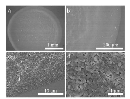
To form Ni-CN-Ni thin film, we dropt 40 mL of the Ni-CN-Ni nanoplates suspension (50 mg/mL) on glass slide following by evaporation in ambient atmosphere (25℃, 60% RH). After drying up, a ring-like film was left on the substrate. Fig. 1a shows SEM images of the film. The surface of the thin film is smooth, suggesting that the film is continuous and high quality. To visualize the difference in the edge and the center parts, enlarged image were taken from the selected areas. Fig. 1b illustrates that the edge of the ring is smooth. Nanoplates were packed densely in the edge area. A clear line is visualized at the interface between the film and substrate, indicating a sharp separation between the coated and un-coated areas. The center part of the film is composed of stacked nanoplates, forming a membrane-type structure (Fig. 1d). To see the surface of deposited film more clearly, we also tested the films by atomic force microscopy (AFM). Fig. S5 (Supporting information) illustrates that the Ni-CN-Ni nanoplates stack together in a lamellar way both in the center and on the edge, similar to the SEM observation. After forming this thin film structure, we measured its N2 adsorption capability. A significant uptake of N2 molecules still could be recorded as shown in Fig. S6 (Supporting information), suggesting that the intrinsic pores in the frameworks were still accessible in this film state. Such a ring-like film is similar to coffee ring which has been seen widely in our daily life. In 1997, Deegan et al. reported that drying up of a drop of coffee could form a non-uniform ring-like pattern on a solid surface [55]. The capillary flow during evaporation of water drives the particles moving from center to the pinned edge, depositing the particles on the edge along the perimeter [56-58]. In recent years, the coffee ring effect has been utilized for building functional films by using many types of materials such as graphene, metallic nanoparticles and micron-sized particles [59-62].

|
Download:
|
| Fig. 1. (a) SEM image of ring-like film formed by Ni-CN-Ni nanoplates in concentration of 50 mg/mL. (b) SEM image of the edge of the ring pattern shown in (a). (c) Enlarged SEM image of edge of the ring pattern. (d) Magnified SEM image of the center part of the ring pattern. | |
To see whether the ring-like film could be alternated, we checked the influence of concentration of the nanoplates in the suspension. The Ni-CN-Ni nanoplates suspension with various concentrations has been prepared first. After drying up a dropt of the suspension on the glass slides, ring-like structures were generated in all the cases although the concentrations were in a range from 20 mg/mL to 200 mg/mL (Fig. S7 in Supporting information). We verified the crystal structure of these films by XRD (Fig. S8 in Supporting information). Comparing with the NiCN-Ni nanoplates powder, the crystal structure has not been changed in a film state. However, diffraction intensity of the peak representing (200) facet was increase obviously, indicating that the films were composed of lamellar stacking layers of the nanoplates. After that, we measure the thickness of the center and edge parts of all the rings (Table S2 in Supporting information). The increase of the thickness correlates to the change of the nanoplates concentration. Table S2 indicates that both the center and edge parts become thicker and thicker with the increase of the nanoplates concentration. The probable reason is that the higher the concentration the more the nanoplates can be deposited in the same area, making the thickness of the thin film increase.
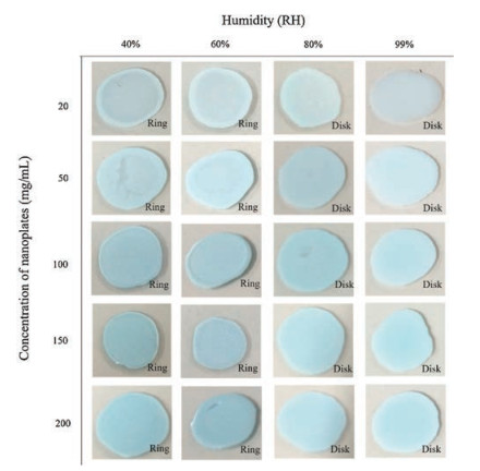
We then investigated the film structure upon changing of the humidity and concentration both. The environmental humidity was set from 40% RH to 99% RH, and the concentration of the nanoplates was tuned from 20 mg/mL to 200 mg/mL. The optical microscopy images of the obtained films were shown in Fig. 2. The patterns are close to ring-like structure when the humidity was 40% RH and 60% RH. However, when the relative humidity was kept at 60% RH, the coffee ring effect was gradually suppressed with the increase of the nanoplates concentration, for example, 200 mg/mL. We further increased the concentration to 300 mg/mL and 500 mg/mL. The photos and thickness statistics of the obtained patterns were shown in Fig. S9 (Supporting information). Arbitrarily, the film changed from ring to disk when the concentration was increased to 500 mg/mL. When the humidity was rose to 80% RH and 99% RH, the difference between the thickness of the edge parts and center parts became neglectable. Even at a very low concentration of 20 mg/mL, the pattern is no more like rings, but like disks. To compare with the ring-like film, we measured SEM (Fig. S10 in Supporting information) and AFM images (Fig. S11 in Supporting information) of the disk-like film. The surface of disk-like thin film and the stacking way of nanoplates are similar to the ring-like pattern. According to the thickness statistics, we demonstrated that the ring effect has been suppressed under high humidity (Tables S3 and S4 in Supporting information).

|
Download:
|
| Fig. 2. Films formed by depositing Ni-CN-Ni nanoplates under different humidity and concentrations. | |
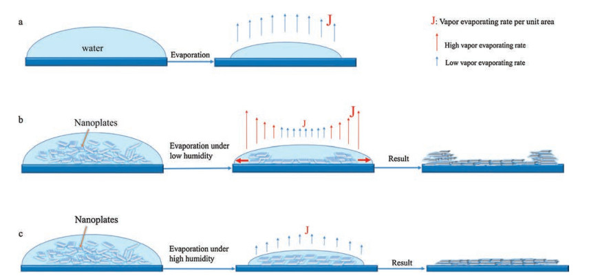
On the basis of the results, a phase diagram was obtained in Fig. 3. A boundary line separates the diagram into two parts. At the low humidity and nanoplates concentration, the films are in ringlike structure. In the other region, the films are flat disks. To explain this phenomenon, we referring the mechanism for formation of coffee rings. If the droplets do not contain the nanoplates, the droplet shrinks in the process of evaporation, eventually disappear, leaving on the substrates (Fig. 4a). However, when there are suspended nanoplates inside the droplet, the situation becomes different. The Ni-CN-Ni nanoplates have the ability to pin to the substrate. As illustrated in Fig. 4b, the liquid on the edge is thinner than that in the center of a droplet. Therefore, the evaporating flux J at the edge is larger than that in the center, which means that the evaporation speed of the water at the edge is faster than at the center. As the water on the edge evaporates, the Marangoni flow pushes the nanoplates to move from center to edge [63]. Eventually, most of the nanoplates are deposited at the edge after the evaporation of the water, forming dense packing along the perimeter. This is how the Ni-CN-Ni nanoplates can form ring-like pattern during its drying process. As for higher humidity mentioned above, the evaporation speed of water becomes extremely slow, making the evaporating speed at the edge similar to the rate near the center of the droplet (Fig. 4c). Therefore, the evaporating flux J is assumed to be close at every parts of a droplet, hindering the Marangoni flow. In this case, the Ni-CN-Ni nanoplates can barely move from center to edge, depositing uniformly as well. As for the effect of nanoplates concentration, we considered the change of viscosity of the suspensions. When the concentration of the Ni-CN-Ni nanoplates becomes high, the viscosity of the suspension is increased. The diffusion of the nanoplates in the droplet can certainly be disturbed. The Marangoni flow during evaporation of water can not move the nanoplates to the edge parts. As a result, a uniform film can be obtained.

|
Download:
|
| Fig. 3. Phase diagram of the structure of Ni-CN-Ni thin films. Region I is ring-like deposition region. Region II is disk-like deposition. | |

|
Download:
|
| Fig. 4. Schematic illustrate for the preferential deposition of the Ni-CN-Ni nanoplates. (a) Evaporation process of water-only droplets. (b) Evaporation process of the droplet of the Ni-CN-Ni nanoplates suspension under a low humidity atmosphere. (c) Evaporation process of the droplet of the Ni-CN-Ni nanoplates suspension under a high humidity atmosphere. | |
Since the Ni-CN-Ni nanoplates can form either ring-like or disklike film we can realize the preferential deposition of Ni-CN-Ni thin films. As shown in Fig. S12 (Supporting information), the Ni-CN-Ni nanoplates can form ring-like pattern arrays, whereas disk-like patterns are generated under higher humidity. This demonstrates that controllable cyanometallate coordination polymer thin films can be realized by evaporation of colloidal droplets onto the substrates with no need to chemically pair the substrates and frameworks at a room temperature. Furthermore, this technique can be utilized to deposit thin films on flexible substrates. Fig. S13 (Supporting information) shows that the ring-like pattern can be formed on Cu and Al foils. The films could attach on the soft substrates tightly even under folding.
In this work, we found that a kind of metal-cyanide coordination polymer, Ni-CN-Ni nanoplates could form coffee-ring during the process of evaporation. Besides, the humidity has an important effect on the formation of coffee-ring pattern. The ring pattern would be changed to disk-like patterns by simply changing the deposition condition. The reason that whether the Ni-CN-Ni nanoplates could form ring-like/disk-like pattern is due to its ability to pin to the substrate. This work demonstrated that preferential deposition of coordination polymers could be realized in such a simple way.
AcknowledgmentThis work was supported by the National Natural Science Foundation of China (No. 21473059).
Appendix A. Supplementary dataSupplementary material related to this article can be found, in the online version, at doi:https://doi.org/10.1016/j.cclet.2018.11.007.
| [1] |
R. Robson, Dalton Trans. (2000) 3735-3744. |
| [2] |
S. Sen, N. Hosono, J.J. Zheng, et al., J. Am. Chem. Soc. 139 (2017) 18313-18321. DOI:10.1021/jacs.7b10110 |
| [3] |
R. Robson, Dalton Trans. (2008) 5113-5131. |
| [4] |
K. Sabyrov, J. Jiang, O.M. Yaghi, G.A. Somorjai, J. Am. Chem. Soc. 139 (2017) 12382-12385. DOI:10.1021/jacs.7b06629 |
| [5] |
H. Furukawa, N. Ko, Y.B. Go, et al., Science 329 (2010) 424-428. DOI:10.1126/science.1192160 |
| [6] |
Y.X. Sun, W.Y. Sun, Chin. Chem. Lett. 25 (2014) 823-828. DOI:10.1016/j.cclet.2014.04.032 |
| [7] |
W. Fan, Y. Wang, Z. Xiao, et al., Chin. Chem. Lett. 29 (2018) 865-868. DOI:10.1016/j.cclet.2017.11.020 |
| [8] |
X.L. Zhao, D. Tian, Q. Gao, et al., Dalton Trans. 45 (2016) 1040-1046. DOI:10.1039/C5DT03283K |
| [9] |
Y.W. Li, K.H. He, X.H. Bu, J. Mater. Chem. A 1 (2013) 4186-4189. DOI:10.1039/c3ta01322g |
| [10] |
Y.W. Li, J.R. Li, L.F. Wang, et al., J. Mater. Chem. A 1 (2013) 495-499. DOI:10.1039/C2TA00635A |
| [11] |
D. Ma, B. Li, Z. Shi, Chin. Chem. Lett. 29 (2018) 827-830. DOI:10.1016/j.cclet.2017.09.028 |
| [12] |
X.Y. Ren, L.H. Lu, Chin. Chem. Lett. 26 (2015) 1439-1445. DOI:10.1016/j.cclet.2015.10.014 |
| [13] |
F.Y. Yi, D. Chen, M.K. Wu, L. Han, H.L. Jiang, ChemPlusChem 81 (2016) 675-690. DOI:10.1002/cplu.201600137 |
| [14] |
Y.Z. Chen, Y.X. Zhou, H. Wang, et al., ACS Catal. 5 (2015) 2062-2069. DOI:10.1021/cs501953d |
| [15] |
Q. Yang, Q. Xu, S.H. Yu, H.L. Jiang, Angew. Chem. Int. Ed. 55 (2016) 3685-3689. DOI:10.1002/anie.201510655 |
| [16] |
C.Y. Sun, C. Qin, C.G. Wang, et al., Adv. Mater. 23 (2011) 5629-5632. DOI:10.1002/adma.v23.47 |
| [17] |
D. Wu, Z. Guo, X. Yin, et al., Adv. Mater. 26 (2014) 3258-3262. DOI:10.1002/adma.v26.20 |
| [18] |
B. Tu, Q. Pang, D. Wu, et al., J. Am. Chem. Soc. 136 (2014) 14465-14471. DOI:10.1021/ja5063423 |
| [19] |
L. Kang, S.X. Sun, L.B. Kong, J.W. Lang, Y.C. Luo, Chin. Chem. Lett. 25 (2014) 957-961. DOI:10.1016/j.cclet.2014.05.032 |
| [20] |
K. Chen, C. Wu, Chin. Chem. Lett. 29 (2018) 823-826. DOI:10.1016/j.cclet.2017.09.040 |
| [21] |
B. Ma, P.Y. Guan, Q.Y. Li, M. Zhang, S.Q. Zang, ACS Appl. Mat. Interfaces 8 (2016) 26794-26800. DOI:10.1021/acsami.6b08740 |
| [22] |
L.H. Cao, F. Shi, W.M. Zhang, S.Q. Zang, T.C.W. Mak, Chem. -Eur. J. 21 (2015) 15705-15712. DOI:10.1002/chem.v21.44 |
| [23] |
H.Y. Li, Y.L. Wei, X.Y. Dong, S.Q. Zang, T.C.W. Mak, Chem. Mater. 27 (2015) 1327-1331. DOI:10.1021/cm504350q |
| [24] |
R.B. Lin, D. Chen, Y.Y. Lin, J.P. Zhang, X.M. Chen, Inorg. Chem. 51 (2012) 9950-9955. DOI:10.1021/ic301463z |
| [25] |
J. Huang, Y. Li, R.K. Huang, et al., Angew. Chem. Int. Ed. 57 (2018) 4632-4636. DOI:10.1002/anie.201801029 |
| [26] |
J.W. Ye, H.L. Zhou, S.Y. Liu, et al., Chem. Mater. 27 (2015) 8255-8260. DOI:10.1021/acs.chemmater.5b03955 |
| [27] |
O. Shekhah, H. Wang, D. Zacher, R.A. Fischer, C. Wöll, Angew. Chem. Int. Ed. 48 (2009) 5038-5041. DOI:10.1002/anie.v48:27 |
| [28] |
D.J. Li, Z.G. Gu, I. Vohra, et al., Small 13 (2017) 1604035. https://www.researchgate.net/publication/314210922_Epitaxial_Growth_of_Oriented_Metalloporphyrin_Network_Thin_Film_for_Improved_Selectivity_of_Volatile_Organic_Compounds
|
| [29] |
Z.G. Gu, J. Zhang, Coord. Chem. Rev. 378 (2019) 513-532. DOI:10.1016/j.ccr.2017.09.028 |
| [30] |
L. Heinke, M. Tu, S. Wannapaiboon, R.A. Fischer, C. Wöll, Microporous Mesoporous Mater. 216 (2015) 200-215. DOI:10.1016/j.micromeso.2015.03.018 |
| [31] |
K.J. Erickson, F. Léonard, V. Stavila, et al., Adv. Mater. 27 (2015) 3453-3459. DOI:10.1002/adma.v27.22 |
| [32] |
K. Liang, C. Carbonell, M.J. Styles, et al., Adv. Mater. 27 (2015) 7293-7298. DOI:10.1002/adma.201503167 |
| [33] |
J. Reboul, S. Furukawa, N. Horike, et al., Nat. Mater. 11 (2012) 717-723. DOI:10.1038/nmat3359 |
| [34] |
M. Nakahama, J. Reboul, K.I. Kamei, S. Kitagawa, S. Furukawa, Chem. Lett. 43 (2014) 1052-1054. DOI:10.1246/cl.140291 |
| [35] |
I. Stassen, M. Styles, G. Grenci, et al., Nat. Mater. 15 (2016) 304-310. DOI:10.1038/nmat4509 |
| [36] |
X. Zhang, W. Wang, Z. Hu, G. Wang, K. Uvdal, Coord. Chem. Rev. 284 (2015) 206-235. DOI:10.1016/j.ccr.2014.10.006 |
| [37] |
S. Hermes, F. Schroder, S. Amirjalayer, R. Schmid, R.A. Fischer, J. Mater. Chem. 16 (2006) 2464-2472. DOI:10.1039/B603664C |
| [38] |
K.B. Klepper, O. Nilsen, P.A. Hansen, H. Fjellvag, Dalton Trans. 40 (2011) 4636-4646. DOI:10.1039/c0dt01716g |
| [39] |
E. Ahvenniemi, M. Karppinen, Chem. Commun. 52 (2016) 1139-1142. DOI:10.1039/C5CC08538A |
| [40] |
A.W. Peters, Z. Li, O.K. Farha, J.T. Hupp, ACS Nano 9 (2015) 8484-8490. DOI:10.1021/acsnano.5b03429 |
| [41] |
K. Khaletskaya, S. Turner, M. Tu, et al., Adv. Funct. Mater. 24 (2014) 4804-4811. DOI:10.1002/adfm.v24.30 |
| [42] |
J. Zhao, B. Gong, W.T. Nunn, et al., J. Mater. Chem. A 3 (2015) 1458-1464. DOI:10.1039/C4TA05501B |
| [43] |
E. Ahvenniemi, M. Karppinen, Chem. Mater. 28 (2016) 6260-6265. DOI:10.1021/acs.chemmater.6b02496 |
| [44] |
C.W. Kung, J.E. Mondloch, T.C. Wang, et al., ACS Appl. Mat. Interfaces 7 (2015) 28223-28230. DOI:10.1021/acsami.5b06901 |
| [45] |
G. Xu, T. Yamada, K. Otsubo, S. Sakaida, H. Kitagawa, J. Am. Chem. Soc. 134 (2012) 16524-16527. DOI:10.1021/ja307953m |
| [46] |
J.W. Xiu, G.E. Wang, M.S. Yao, et al., Chem. Comm. 53 (2017) 2479-2482. DOI:10.1039/C6CC09310H |
| [47] |
M.S. Yao, X.J. Lv, Z.H. Fu, et al., Angew. Chem. Int. Ed. 56 (2017) 16510-16514. DOI:10.1002/anie.201709558 |
| [48] |
M.S. Yao, W.X. Tang, G.E. Wang, B. Nath, G. Xu, Adv. Mater. 28 (2016) 5229. https://www.researchgate.net/publication/302033555_MOF_Thin_Film-Coated_Metal_Oxide_Nanowire_Array_Significantly_Improved_Chemiresistor_Sensor_Performance
|
| [49] |
G. Xu, K. Otsubo, T. Yamada, S. Sakaida, H. Kitagawa, J. Am. Chem. Soc. 135 (2013) 7438-7441. DOI:10.1021/ja402727d |
| [50] |
Y. Chen, S. Li, X. Pei, et al., Angew. Chem. Int. Ed. 55 (2016) 3419-3423. DOI:10.1002/anie.201511063 |
| [51] |
Y. Zhao, W. Li, X. Jiang, et al., ACS Nano 11 (2017) 3662-3670. DOI:10.1021/acsnano.6b08068 |
| [52] |
M. Hu, S. Ishihara, Y. Yamauchi, Angew. Chem. Int. Ed. 52 (2013) 1235-1239. DOI:10.1002/anie.201208501 |
| [53] |
Y. Zhao, W. Zhang, M. Hu, ChemNanoMat 3 (2017) 780-789. DOI:10.1002/cnma.v3.11 |
| [54] |
Rodrıíguez-Hernández J., Lemus-Santana A.A., C.N. Vargas, E. Reguera, C. R. Chimie 15 (2012) 350-355. DOI:10.1016/j.crci.2011.11.004 |
| [55] |
R.D. Deegan, O. Bakajin, T.F. Dupont, G. Huber, S.R. Nagel, Nature 389 (1997) 827-830. DOI:10.1038/39827 |
| [56] |
D. Tam, V.V. Arnim, G.H. McKinley, A.E. Hosoi, J. Fluid Mech. 624 (2009) 101-123. DOI:10.1017/S0022112008005053 |
| [57] |
D.J. Harris, H. Hu, J.C. Conrad, J.A. Lewis, Phys. Rev. Lett. 98 (2007) 148301. http://med.wanfangdata.com.cn/Paper/Detail/PeriodicalPaper_PM17501317
|
| [58] |
Z. Lin, S. Granick, J. Am. Chem. Soc. 127 (2005) 2816-2817. DOI:10.1021/ja044792z |
| [59] |
I.U. Vakarelski, D.Y.C. Chan, T. Nonoguchi, H. Shinto, K. Higashitani, Phys. Rev. Lett. 102 (2009) 058303. http://med.wanfangdata.com.cn/Paper/Detail/PeriodicalPaper_PM19257566
|
| [60] |
T.P. Bigioni, X. Lin, T.T. Nguyen, et al., Nat. Mater. 5 (2006) 265-270. DOI:10.1038/nmat1611 |
| [61] |
L. Malfatti, Y. Tokudome, K. Okada, et al., Microporous Mesoporous Mater. 163 (2012) 356-362. DOI:10.1016/j.micromeso.2012.07.038 |
| [62] |
D.S. Eom, J. Chang, Y.W. Song, J.A. Lim, J. Phys. Chem. C 118 (2014) 27081-27090. DOI:10.1021/jp507451b |
| [63] |
H. Hu, R.G. Larson, J. Phys. Chem. B 110 (2006) 7090-7094. DOI:10.1021/jp0609232 |