 2019, Vol. 30


Light-responsive controlled drug delivery system has exhibited more and more important in cancer diagnosis and treatment, since light stimulus was a non-invasive trigger, which could effectively penetrate the body's tissues and implement to control release in fixed point at required time [1-4]. In recent years, a series of researches have been carried out on light-sensitive drug carrier materials [5]. According to the difference in response to light, the light response chromophores can be divided into the photoisomerisable materials represented by azobenzene (Azo) and stilbenes [6, 7], photoinduced ring opening and closing materials represented by dithienylethenes and spiropyrans [8, 9], photodimerisation and photopolymerisation represented by coumarin and anthracenes [10, 11], photocleavage represented by pyrene and nitrobenzyl [12, 13].
Light sensitive materials based on coumarin derivatives have been reported mainly based on dimerisation of the coumarin molecule upon irradiation with light >300 nm [14, 15]. A small number of photo-responsive low molecular weight hydrogels (LMWGs) based on coumarin have been reported [16]. Parquette and co-workers designed a coumarin dipeptide hydrogelator, photodimerisation of the gelator was carried out exposing to 365 nm light with a dark yellow precipitate forming in the beginning, and insoluble precipitate becoming after 7 days under the 365 nm light [17]. Almutairi and co-workers designed a hydrogel based on the photocage bromohydroxycoumarin which was efficiently cleaved upon ultraviolet [18]. There was also another type of light response of coumarin derivative upon photoinduced bond breaking, a few cases of 7-amino-4-hydroxymethyl coumarin cleaved at the 4-methyl end were reported [19-22], such as Zhao and coworkers reported a 7-amino-4-hydroxymethyl coumarin grafted poly(glutamic acid) based nanoparticles, which could release drug using light-responsive bond broken to destroy the nanoparticles [23, 24]. Zink and coworkers adopted 7-amino-4-hydroxymethyl coumarin on the surface of mesoporous silicon dioxide, and used photolysis of coumarin as a photocontrol switch to control the drug release from mesoporous silica [25]. We also reported an efficient nanocarrier by bonding 7-amino-4-hydroxymethyl coumarin derivatives on the surface of the gold nanoparticles to realize photo-responsive fracture at the 4-methyl end of coumarin [26].
In this study, we found another new photosensitive broken site of 7-amino coumarin derivative, and fabricated a novel photoresponsive molecular hydrogel based on this new photosensitive site. This gel went through a photocleavage of C-N bond in 7-amino coumarin upon the irradiation with UV light. The photo-controlled drug release of this molecular hydrogel has been investigated.
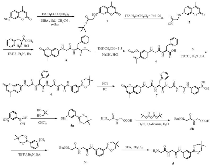
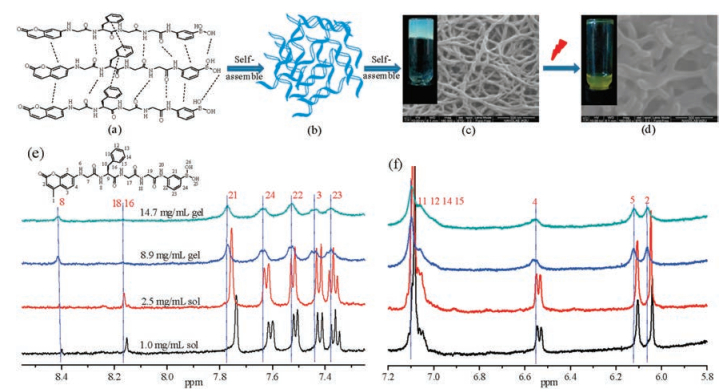
The synthetic route of gelator was presented in Scheme 1, the characterization of the gelator was showed in Figs. S1-S27 (Supporting information). The designed gelator had good gelation by immobilizing the solvents in the three-dimensional network. The critical gelation concentration (CGC) of the gelator was 2.7 mg/mL in PEG200:H2O = 1:2. SEM image of the gel indicated that the gel consisted of spiral-shaped fibers (30-60 nm wide), and these nanofibers further assembled into three-dimensional network (Fig. 1c). 1H NMR spectra showed the gradual high-field shift of protons upon increasing the concentration of gelator (Figs. 1e and f). The protons at 7.73 ppm, 7.61 ppm, 7.50 ppm, 7.41 ppm, 7.35 ppm and 6.52 ppm attributed to benzene units shifted downfield to 7.77 ppm, 7.64 ppm, 7.53 ppm, 7.44 ppm, 7.38 ppm and 6.55 ppm, respectively, with increasing concentration from 1.0 mg/mL (sol) to 14.7 mg/mL (gel), for the presence of π-π stacking. The π conjugated moieties of the molecular structure containing coumarin and phenylalanine, which strengthened intermolecular π-π stacking to self-assemble into network to immobilize a large volume of solvent in the gel [27]. The π-π stacking interaction between gelators was also characterized by UV spectra (Fig. S28 in Supporting information), the red-shift occurred as the concentration increasing [28]. Moreover, the cleavage peaks of hydrogen at positions 3, 4, 22, 23, 24 on the aromatic ring disappeared, as the sol-gel transition. The protons at 8.15 ppm and 8.40 ppm attributed to amino in peptide bonds also shifted downfield with increasing gelator concentration for the intermolecular hydrogen bond. The formation of hydrogen bonds between these peptide bonds might be responsible for the formation of spiral-shaped fibers in the gel (Fig. 1c). These results implied that the self-assembly of gelator was driven by hydrogen bond as well as π-π interaction during the gelation [29].

|
Download:
|
| Scheme 1. The synthesis of gelator (compound 7). | |

|
Download:
|
| Fig. 1. (a) gelator, (b) self-assembly of gelator, (c) photo and SEM images of gel, (d) photo and SEM images of gel, upon laser irradiation, (e, f) 1H NMR spectra of gel with different concentration. | |
The photocleavable property of the gel was investigated as shown in Fig. 2. The gelator was responsive to 365 nm irradiation, but stable under 420 nm, 630 nm and natural light, as shown in Fig. 2A(a). The gel gradually transformed into sol under UV irradiation (365 nm, 77.5 mW/cm2), and the sol gradually turned yellow (Fig. 2A(c)), the ultraviolet and fluorescence intensity gradually weakened (Figs. 2B and C). The photosensitive material based on coumarin has been reported to realize the light response usually through photoinduced dimerization [30]. Dimerisation of the gelator molecule often led to the molecules being less soluble due to the molecule doubling in size and so becoming more hydrophobic. This decrease in solubility can lead to destruction of the gel or a decrease in the rheological properties due to the gel network being disruption of the gel network [31]. Different from traditional photodimerisation of coumarin gel, the light response of this gel was mainly achieved by the photocleavage. After exposing to light, this gel was photodegraded into sol. The sol was tested by high performance liquid chromatography (HPLC). Two peaks with the retention time Rt = 3.4 min and Rt = 8.3 min were found (Fig. 2D). The corresponding compound with Rt = 3.4 min was detected by 1H NMR as coumarin, as show in Fig. S29 in Supporting information. The results demonstrated the gelator was photocleaved gradually into the coumarin in the exposure to light. It was also found that gelator in solution was more sensitive to light than that in gel (Fig. 2E), more coumarin was cleaved from the gelator in solution. The photocleavage speed of gel was much slow, this might owe to the enhanced molecular interaction in gel state, which weakened the degradation speed of gelator exposing to light.

|
Download:
|
| Fig. 2. (A) Images of gelator in solution and gel with or without exposure to laser irradiation: (a) gelator in solution with exposure to different laser irradiation (1, in dark; 2, with natural light; 3, with 630 nm irradiation; 4, with 420 nm irradiation; 5, with 365 nm irradiation for 10 min; 6, with 365 nm irradiation for 20 min), (b) gel under natural light for different time, (c) gel with laser irradiation for different time. (B) UV spectra and (C) fluorescence spectra of gel during light-exposure with different time, insert image is the fluorescent intensity ration I/I0, I = fluorescent intensity of gelator with different irradiation time, I0 = fluorescent intensity of gelator. (D) HPLC spectra of gel after the laser irradiation (365 nm, 77.5 mW/cm2) with different time. (E) the concentration of coumarin photocleaved from the gelator in gel or in solution during exposing to the light. | |
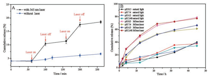
In order to evaluate the photo-sensitive controlled release, cytarabine hydrochloride was selected as the model drug, a chemotherapy medication used to treat acute myeloid leukemia, acute lymphocytic leukemia, and chronic myelogenous leukemia [32]. Since cytarabine hydrochloride is less sensitive to light, which can avoid the effect of light on drug during evaluating the photosensitive controlled release. The cytarabine hydrochloride was often given by intravenous injection, but it quickly disappeared and eliminated from the blood, and T1/2 was only 3-15 min. There was a liposomal formulation for cytarabine reported to outcomes the quick elimination [33]. In this study, the molecular hydrogel was used to photo-triggered delivery for cytarabine. In vitro release of cytarabine from drug-loaded hydrogel was carried out, as shown in Fig. 3. With the comparison of cytarabine releasing with and without laser irradiation (Fig. 3A), the concentration of cytarabine increased from 39.41 μg/mL to 104.27 μg/mL with an increment of 64.86 μg/mL under 40 min laser irradiation at the time point of 60 min, and nearly no increase of cytarabine with no laser irradiation at the same time. It was clear that laser could accelerate the photocleavage of gelator, and the cleavage of the C-N bond could further destroy the architecture of gel and accelerated the release of cytarabine. The release of drug-loaded gel in phosphate buffer with different pH values was also investigated, as shown in Fig. 3B. The hydrogel exhibited promising controlled release behavior with sustained release of cytarabine in buffer solutions. Few cytarabine was released from the gel under natural light than that with laser irradiation. Upon laser irradiation, few cytarabine was released from the gel in physiological environment (pH 7.4), and the release rate increased in PBS with acidic or alkaline environment (pH 8.5). This drug-loaded hydrogels with stability and sustaining drug release could be used intratumoral injection or other target injection, which is a research focus in the tumor treatment now.

|
Download:
|
| Fig. 3. (A) the released profile of cytarabine hydrochloride-loaded gel in PBS (7.4) with or without laser irradiation (365 nm, 77.5 mW/cm2). Means + SD (n = 3). (B) the release profile of cytarabine hydrochloride-loaded gel in different media. All the drug-loaded gels were prepared by adding 5.0 mg gelator and 1.0 mg cytarabine hydrochloride in 3 mL PBS (pH 7.4). | |
In this study, a new coumarin derivative based molecular hydrogel has been fabiricated, which exhibited satisfied photosensitivity. This gel went through a photocleavage of C-N bond in 7-amino coumarin upon the UV light irradiation based on a new photosensitive broken site of 7-amino coumarin derivative. The designed gelator had good gelation by immobilizing the solvents in the three-dimensional network. The critical gelation concentration was 2.7 mg/mL in PEG200:H2O = 1:2. The cytarabine hydrochloride was selected as the model drug to evaluate the photo-triggered drug delivery. The hydrogel exhibited promising controlled release behavior with laser to accelerate the photocleavage of C-N bond of gelator, which could destroy the architecture of gel and accelerated the release of cytarabine. This drug-loaded hydrogels could be used intratumoral injection or other target injection with a non-invasive light stimulus triggered release, which is a research focus in the tumor treatment now.
AcknowledgmentThe authors thank for the financial support from the National Natural Science Foundation of China (Nos. 21672164, 21372177)
Appendix A. Supplementary dataSupplementary material related to this article can befound, in the online version, at doi: https://doi.org/10.1016/j.cclet.2018.06.009.
| [1] |
W.C. Liao, Y.S. Sohn, R. Nechushtai, et al., J. Am. Chem. Soc. 138 (2016) 8936-8945. DOI:10.1021/jacs.6b04773 |
| [2] |
S. Theis, A. Iturmendi, C. Gorsche, et al., Angew. Chem. Int. Ed. 56 (2017) 1-5. DOI:10.1002/anie.201610955 |
| [3] |
S.O. Poelma, S.S. Oh, S. Helmy, et al., Chem. Commun. 52 (2016) 10525-10528. DOI:10.1039/C6CC04127B |
| [4] |
P. Xiao, J. Zhang, J. Zhao, M.H. Stenzel, Prog, Polym. Sci. 74 (2017) 1-33. DOI:10.1016/j.progpolymsci.2017.06.002 |
| [5] |
H. Li, X. Yang, Z. Zhou, J. Control Release 261 (2017) 126-137. DOI:10.1016/j.jconrel.2017.06.029 |
| [6] |
C. Lin, S. Maisonneuve, R. Metivier, J. Xie, Chem. -Eur. J. 23 (2017) 14996-15001. DOI:10.1002/chem.201703461 |
| [7] |
Y. Zhou, S. Yang, J. Li, et al., Dalton Trans. 45 (2016) 18308-18312. DOI:10.1039/C6DT03489F |
| [8] |
Y. Kang, X. Tang, H. Yu, et al., Chem. Sci. 8 (2017) 8357-8361. DOI:10.1039/C7SC04125J |
| [9] |
S. Son, E. Shin, B.S. Kim, Biomacromolecules 15 (2014) 628-634. DOI:10.1021/bm401670t |
| [10] |
H.M. Lin, W.K. Wang, P.A. Hsiung, S.G. Shyu, Acta Biomater. 6 (2010) 3256-3263. DOI:10.1016/j.actbio.2010.02.014 |
| [11] |
T.K. Claus, S. Telitel, A. Welle, et al., Chem. Commun. 53 (2017) 1599-1602. DOI:10.1039/C6CC09897E |
| [12] |
J.E. Lee, E. Ahn, J.M. Bak, et al., Polymer 55 (2014) 1436-1442. DOI:10.1016/j.polymer.2014.01.026 |
| [13] |
T. Nishimura, M. Takara, S. Mukai, et al., Chem. Commun. 52 (2016) 1222-1225. DOI:10.1039/C5CC08416D |
| [14] |
P. Wei, H. Wang, K. Jie, F. Huang, Chem. Commun. 53 (2017) 1688-1691. DOI:10.1039/C6CC10089A |
| [15] |
H. He, M. Feng, Q. Chen, X. Zhang, H. Zhan, Angew. Chem. Int. Ed. 55 (2016) 936-940. DOI:10.1002/anie.201508355 |
| [16] |
E.R. Draper, T.O. McDonald, D.J. Adams, Chem.Commun. 51 (2015) 12827-12830. DOI:10.1039/C5CC03817K |
| [17] |
S.H. Kim, Y. Sun, J.A. Kaplan, M.W. Grinstaff, J.R. Parquette, New J. Chem. 39 (2015) 3225-3228. DOI:10.1039/C5NJ00038F |
| [18] |
C. de Gracia Lux, J. Lux, G. Collet, et al., Biomacromolecules 16 (2015) 3286-3296. DOI:10.1021/acs.biomac.5b00950 |
| [19] |
N. Fomina, C.L. McFearin, M. Sermsakdi, J.M. Morachis, A. Almutairi, Macromolecules 44 (2011) 8590-8597. DOI:10.1021/ma201850q |
| [20] |
J. Cao, S. Huang, Y. Chen, et al., Biomaterials 34 (2013) 6272-6283. DOI:10.1016/j.biomaterials.2013.05.008 |
| [21] |
L.L. Fedoryshin, A.J. Tavares, E. Petryayeva, S. Doughan, U.J. Krull, ACS Appl, Mater. Interfaces 6 (2014) 13600-13606. DOI:10.1021/am503039f |
| [22] |
G. Yu, W. Yu, Z. Mao, C. Gao, F. Huang, Small 11 (2015) 919-925. DOI:10.1002/smll.v11.8 |
| [23] |
S. Kumar, J.F. Allard, D. Morris, et al., J. Mater. Chem. 22 (2012) 7252-7257. DOI:10.1039/c2jm16380b |
| [24] |
Q. Lin, C. Bao, Y. Yang, et al., Adv. Mater. 25 (2013) 1981-1986. DOI:10.1002/adma.201204455 |
| [25] |
T.M. Guardado-Alvarez, L.S. Devi, et al., Chem. Soc. 135 (2013) 14000-14003. DOI:10.1021/ja407331n |
| [26] |
Y. Liang, W. Gao, X. Peng, et al., Biomaterials 100 (2016) 76-90. DOI:10.1016/j.biomaterials.2016.05.023 |
| [27] |
T.B. Wei, J.F. Chen, X.B. Cheng, et al., Polym. Chem. 8 (2017) 2005-2009. DOI:10.1039/C7PY00335H |
| [28] |
H. Fang, H. Gao, T. Wang, et al., Dyes and Pigments 147 (2017) 190-198. DOI:10.1016/j.dyepig.2017.07.045 |
| [29] |
A. Dawn, H. Kumari, Chem. -Eur. J. 23 (2017) 1-16. DOI:10.1002/chem.201605561 |
| [30] |
Y.F. Han, G.X. Jin, C.G. Daniliuc, F.E. Hahn, Angew. Chem. Int. Ed. 54 (2015) 4958-4962. DOI:10.1002/anie.201411006 |
| [31] |
Z. Zheng, J. Hu, H. Wang, et al., ACS Appl. Mater. Interfaces 9 (2017) 24511-24517. DOI:10.1021/acsami.7b07204 |
| [32] |
A.D. Tullio, K. Rouault-Pierre, A. Abarrategi, et al., Nat. Commun. (2017) 1679. |
| [33] |
S. Kim, S. Khatibi, S.B. Howell, et al., Cancer Res. 53 (1993) 1596. |