 2019, Vol. 30


b Laboratory of Controllable Preparation and Application of Nanomaterials CAS Key Laboratory of Cryogenics Technical Institute of Physics and Chemistry Chinese Academy of Sciences (CAS), Beijing 100190, China;
c Second Clinical Medical College of Jinan University, Shenzhen People's Hospital, Shenzhen 518020, China
Cancers could bring serious pain around the world. Although surgical resection was the frequently-used method for the cancer therapy [1], unfortunately, it is not enough to treat the large and heteromorpic tumor [2, 3]. Recently, microwave (MW) thermal therapy, a treatment method to destroy solid tumour by some heating sources [4], was an efficient complement to surgical resection, factitiously improving the temperatures of tumor tissue based on the poor resistance of the tumor cells on heat [5]. MW thermal therapy with selective heat in tumor tissues is urgently necessary, which should produce sufficient temperature and reduce side effect at the same time [6].
Some developed nanomaterials for delivering heat to tumor tissue were applied in antitumor based on the enhanced permeability and retention effect (EPR) [7, 8]. The size, shape and porosity of nanomaterial would influence their MW heating efficacy [9]. In our previous work, these nanomaterials, as MW sensitizers, were used to selectively heat the tumors as following procedure [10, 11]. For example, ionic liquids were encapsulated in hollow ZrO2 nanospheres [12] and PLGA microspheres [13], where their shell could confine the motion range of ions induced by MW irradiation. Thus the collision among the ions were enhanced, resulting in an enhanced heating temperature. However, ionic liquids, which are liquid at room temperature and their components were different kinds of ions such as positive ions [14], including quaternary phosphonium salt, quaternary ammonium salt, pyrrole and imidazole salts, and negative ions, including four fluorine boric acid root, halogen ions and six fluoride phosphate ions [15], were poisonous to the human body [16]. So it is necessary to develop a more biosecurity MW sensitizer with efficient MW selective heating for tumor therapy.
Recently, as the rapidly developing nanomaterials, metalorganic frameworks (MOF) show a remarkable prospect in the biological and chemical applications due to the combination of their unique features [17-19]. Zirconium metal-organic frameworks (ZrMOFs), with Zr and p-phthalic acid (H2BDC), were prepared for the applications in catalysis [20], sensing, durg release [21] and photothermal therapy [22] due to their abundant micropores and large specific surface area [23]. The coordination mode of ZrMOFs was Zr6O4(OH)4(CO2)12 [24], and their coordination structure also maintained stability in the sub-acidic environment of the tumor. ZrMOFs with the property of flexible structure [25], were regarded as soft porous crystals, similar to organic molecular chain with the crystalline order. Selective heating of MW sensitizers was based on the confined effect [26], namely, compared with in free space, the MW heating of ions in the micropores of ZrMOFs was more obvious.
In this work, ZrMOF NCs were synthesize by one-pot solvothermal method [27] and their thermal efficiency under MW was investigated, and effect of their reaction time on the formation of ZrMOF NCs crystal structure was also analyzed. In addition, polyvinyl pyrrolidone (PVP) was introduced into the synthesis system to improve the dispersion of ZrMOF NCs. Then polyethylene glycol (PEG) was linked on the surface of ZrMOF NCs (ZrMOF-PEG NCs) in order to increase their circulation time in the body. Finally, the physicochemical properties of ZrMOF-PEG NCs were investigated, and the MW heating, anticancer efficacy and acute toxicity test of ZrMOF-PEG NCs were also analyzed. The details of material synthesis, cell experiments in vitro and animal test can be found in Supporting information.
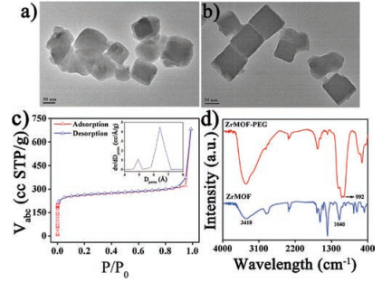
In order to obtain the regular and well-dispersed ZrMOF NCs, the addition of PVP and reaction time were analyzed to tune the micro-structure of ZrMOF NCs. In transmission electron microscopy (TEM) images of reaction time of 8 h with PVP, ZrMOF NCs presented the irregular ellipsoid and larger average particle size (Fig. 1a and Fig. S1 in Supporting information), due to the short reaction time for coordination between Zr and H2BDC, and reaction time of 10 h resulted in ZrMOF NCs agglomeration without PVP (Figs. S2 and S3 in Supporting information). However, the longer reaction time of 10 h and the introduce of PVP could improve the dispersion and microscopic shape of ZrMOF NCs (Fig. 1b and Fig. S4 in Supporting information), showing regular cubic particles with the size of~50 nm, and this cube structure would favor the cell motility [28]. Therefore, ZrMOF NCs synthesized with 10 h reaction time and PVP were used for subsequent experiments. High resolution transmission electron microscopy (HRTEM)-mapping images (Fig. S5 in Supporting information) showed the presence of carbon (C, red), oxygen (O, yellow) and zirconium (Zr, magenta) in ZrMOF NCs, and the quantitative results in scanning electron microscope-energy dispersive spectrum (SEM-EDS) showed that there were 38.26 at% C, 46.87 at% O and 4.11 at% Zr in ZrMOF NCs (Fig. S6 in Supporting information). N2 isothermal adsorption and desorption curve (Fig. 1c) showed that the specific surface area and average micropore diameter of ZrMOF NCs were 1109 m2/g and ~6.34 Å, respectively (Fig. 1c inset). FTIR spectra showed that the peak at 3418 cm-1 was the stretching vibration of O—H [29] and the peak at 1040 cm-1 was the stretching vibration of C—O [30] in ZrMOF NCs (Fig. 1d), in which an obvious increase was observed after the PEGylation. In addition, a new peak at 992 cm-1 was the rocking vibration of N—H [31], indicating that PEG was successfully coupled with ZrMOF NCs via the amide bonds.

|
Download:
|
| Fig. 1. TEM of ZrMOF NCs obtained in different reaction time: (a) 8 h and (b) 10 h with PVP. (c) N2 isothermal adsorption-desorption and the micropore size distribution curves (inset) of ZrMOF NCs. (d) FTIR spectra of ZrMOF NCs and ZrMOF-PEG NCs dispersed in water, respectively. | |
In MW heating experiments of ZrMOF NCs, effect of the ions on the enhancement of the MW heating of ZrMOF NCs was first investigated. 10 mg of ZrMOF NCs were dispersed into 1 mL of water and 0.9% NaCl solution, respectively, then two suspensions were irradiated under MW with 450 MHz and 1.8 W/cm2 power for 5 min. At the same time, an infrared thermal imager was used to record the real-time temperature value of ZrMOF NCs suspension. As shown in Fig. 2a, under MW irradiation, no obvious temperature increase in water and water + ZrMOF NCs groups appeared, and their temperature changes were only 0.5 ℃ and 0.8 ℃, respectively (Fig. 2b). When 0.9% NaCl solution was under MW irradiation, the suspension temperature was increased 28 ℃. While the temperature of 0.9% NaCl suspension under MW irradiation reached to 56 ℃, the temperature changed 36 ℃ after ZrMOF NCs were added into the 0.9% NaCl solution. These results indicated that the ions were necessary for the MW heating, and ZrMOF NCs could enhance MW heating of the 0.9% NaCl solution due to their ability of converting MW electromagnetic energy into thermal energy.
Because the photothermal conversion efficiency was an important index of nanomaterials, the efficiencies of MW conversion into thermal energy of four groups were quantitatively assessed, including water, water + ZrMOF NCs, 0.9% NaCl solution and ZrMOF NCs + 0.9% NaCl solution groups. Their results showed that the MW conversion efficiency of ZrMOF NCs + 0.9% NaCl solutionwas highest than the other three groups (Fig. 2c). A computational model was fully utilized with some significant changes. As shown in Fig. 2d, the typical MW heating and cooling curves of ZrMOF NCs were divided into two stages. Stage Ⅰ represented the increase process that the temperature of the 0.9% NaCl solution and reached to a steady state, and stage Ⅱ indicated the temperature decrease of the 0.9% NaCl solution after MW source was removed. The thermal energy of the 0.9% NaCl solution was calculated according to stage Ⅰ. Then a fitting line was acquired according to thementioned stage Ⅱ to calculate the lost thermal energy, due to the convection heat between air and the 0.9% NaCl solution (Fig. 2e).

|
Download:
|
| Fig. 2. (a) MW heating curves of water, 0.9% NaCl solution, ZrMOF NCs dispersed in water and 0.9% NaCl solution. (b) The temperature changes of ZrMOF NCs dispersed in water and 0.9% NaCl solution. (c) MW conversion efficiencies of ZrMOF NCs dispersed in water and 0.9% NaCl solution. (d) Heating and cooling curves of ZrMOF NCs dispersed in DMEM under MW irradiation. (e) The linear fitting of cooling curve for ZrMOF NCs. | |
Effect of concentration of ZrMOF NCs, power of MW irradiation and concentration of NaCl solution on MW heating were evaluated. First, 20 mg of ZrMOF NCs were dispersed in 1 mL of 0.9% NaCl solution, and the suspension was irradiated with MW irradiation of different power (1.8, 1.5, 1.2 and 0.9 W/cm2) and 450 MHz frequency for 5 min. As shown in Fig. S7 (Supporting information), the temperature was increased when the power of MW irradiation increased from 0.9 W/cm2 to 1.8 W/cm2. The temperature changes of 0.9% NaCl solution raised from 3.8 ℃ to 9.3 ℃ after 5 min of MW irradiation (Fig. S8 in Supporting information). Then 20, 10 and 5 mg of ZrMOF NCs were dispersed in 1 mL of 0.9% NaCl solution, and irradiated for 5 min with 1.8 W/cm2 of power. As shown in Fig. S9 (Supporting information), the temperature increased with the concentration of ZrMOF NCs suspension. The temperature of 0.9% NaCl solution raised from 3.4 ℃ to 9.3 ℃ after 5 min of MW irradiation (Fig. S10 in Supporting information). Finally, effect of different concentration of NaCl solution (0.09%, 0.3%, 0.9% and 1.5%) on MW heating was investigated. As shown in Fig. S11 (Supporting information), when the ion concentration of NaCl solution increased, the MW heating curves of NaCl solution also gradually rose, the temperatures of NaCl solution with the ion concentration of 0.09%, 0.3%, 0.9% and 1.5% were 12.2, 17.9, 36.4 and 38.3 ℃, respectively (Fig. S12 in Supporting information), suggesting that more surrounding ions could diffuse into ZrMOF NCs from the higher concentration of NaCl solution, and producing more violent ions collision. So ZrMOF NCs obviously improved the MW heating efficiency.
To use ZrMOF-PEG NCs in clinic, the in vivo biosafety of ZrMOFPEG NCs needed to be evaluated via an acute toxicity test, and the normal mice were used as the animal model. The results of blood routine test of the mice intravenously injected with different concentrations of ZrMOF-PEG NCs showed that the examined parameters of the mice were normal (Fig. S13 in Supporting information) and had no significant difference with that of the control group. In addition, the body weight curve of the mice showed no significant changes during the test period of 20 days (Fig. S14 in Supporting information), indicating that ZrMOF-PEG NCs did not cause detectable systemic toxicity on the mice. Moreover, no noticeable organ damage was detected by histological analysis (Fig. S15 in Supporting information), further confirming the favorable biocompatibility of ZrMOF-PEG NCs. Altogether, ZrMOF-PEG NCs were biosafe microwave sensitizers.
In order to analyze tumor therapy in vivo, the cytotoxicity of ZrMOF-PEG NCs was first evaluated by the standard methyl thiazolyl tetrazolium (MTT) assay. The viability of HUVE and HepG2 cells incubated with ZrMOF-PEG NCs for 24 h were both over 70% (Figs. 3a and b) even at the concentrationof 100 μg/mL, respectively [32]. Then H22 tumor-bearing (HTB) mice were taken as the animal model for in vivo tumor therapy. It is clearly seen in Fig. 3c that the temperature (51 ℃) of tumor region in MW group was obviously lower than 63 ℃ of ZrMOF-PEG NCs + MWgroup, indicating that the higher temperatures were achieved in ZrMOF-PEG NCs + MW group, and ZrMOF-PEG NCs could be used as a MW sensitizer to selectively raise the temperature of tumor. After 20 days treatment, the tumor volumes of PBS group (control group) were ~5.8 ± 1.0 times of the initial tumors (Fig. 3d), and ZrMOF-PEG NCs groupwere ~6.1 ± 1.2 times of the initial tumors. MW group exhibited a few tumor inhibition effect, and the tumor volumes were ~3.8 ±0.8 times of the initial tumors. However, an obvious treatment effect was observed in ZrMOF-PEG NCs + MW group, namely, the mean volume of tumor was only~0.9 ± 0.4 times of the initial one after 20 days treatment. Moreover, the safety of different treatments was also investiged, and the body weight curves of mice suggested that the treatment of HTB mice with ZrMOF-PEGNCs + MW didnot cause continuous weight loss (Fig. 3e). Moreover, the survival rate of mice in control group, MW group and ZrMOF NCs + MW group was 0%, 66.6% and 33.3% (n = 3, Fig. 3f), respectively. The survival rate in ZrMOF-PEG NCs + MW group reached to 100% (n = 3) after 20 days treatment, suggesting that the treatment with ZrMOF-PEG NCs + MW on HTB mice was safety.

|
Download:
|
| Fig. 3. After incubation with (a) normal HUVE and (b) HepG2 cells for 24 h, the cytotoxicity of ZrMOF-PEG NCs at different concentration (1, 25, 50 and 100 mg/mL). (c) The temperature change curves of the tumor region in MW, and ZrMOF-PEG NCs + MW groups. (d) The assessment of anti-tumor in vivo of HTB mice in different groups with the concentration of 40 mg/kg at 8 h of intravenous post-injection. (e) The body weight change curves of the mice in different groups. (f) Survival rate of mice in different groups after 20 days of treatment. The different groups were control (PBS), MW, ZrMOF-PEG NCs and ZrMOF-PEG NCs + MW groups, respectively. MW irradiation was at 1.8 W/cm2 for 5 min, n = 3. | |
H&E-stained tissue images of the main organs suggested that there was no obvious difference among the four groups (Fig. S16 in Supoprting information). Therefore, ZrMOF-PEG NCs could effectively inhibit the tumor growth of the HTB mice, without systemic toxicity under MW irradiation, becaue ZrMOF-PEG NCs could preferentially accumulate at the tumor site via EPR effect, which improved the antitumor efficiency of MW irradiation.
In summary, the biosafety MW sensitizers, ZrMOF NCs, was synthesized for improving the selective of tumor region and enhancing MW thermal anticancer therapy. The prepared ZrMOF NCs without the ionic liquids showed good MW sensitization due to their abundant micropores and large specific surface area. The viability of HUVE and HepG2 cells incubated with ZrMOF-PEG NCs for 24 h were both over 70% even at the concentration of 100 μg/mL, respectively. Compared with MW group, ZrMOF-PEG NCs + MW group presented a higher temperature in the tumor region, because ZrMOF-PEG NCs could preferentially accumulate in the tumor region via the EPR effect, suggesting a better anti-tumor effect in MW thermal therapy. These results indicated that ZrMOF NCs could selectively raise the temperature of tumor region, and increasing the bioavailability of MW sensitizers for enhancing MW thermal therapy against tumors.
AcknowledgmentsThe authors acknowledge the financial support from the National Science Foundation of China (Nos. 61671435 and 81630053), Beijing Natural Science Foundation (No. 4161003).
Appendix A. Supplementary dataSupplementary material related to this article can befound, in the online version, at doi: https://doi.org/10.1016/j.cclet.2018.06.016.
| [1] |
P. Therasse, S.G. Arbuck, E.A. Eisenhauer, et al., J. Natl. Cancer Inst. 92 (2000) 205-216. DOI:10.1093/jnci/92.3.205 |
| [2] |
D. Hanahan, R.A. Weinberg, Cell 100 (2011) 57-70. |
| [3] |
L.M. Coussens, Z. Werb, Nature 420 (2002) 860-867. DOI:10.1038/nature01322 |
| [4] |
H. Shi, M. Niu, L. Tan, et al., Chem. Sci. 6 (2016) 5016-5027. |
| [5] |
D. Long, T. Liu, L. Tan, et al., ACS Nano 10 (2016) 9516-9528. DOI:10.1021/acsnano.6b04749 |
| [6] |
H. Zhou, C. Fu, X. Chen, et al., Biomater. Sci.-UK 6 (2018) 1535-1545. DOI:10.1039/C8BM00142A |
| [7] |
T. Gordon, M. Cargnello, T. Paik, J. Am. Chem. Soc. 134 (2012) 6751-6761. DOI:10.1021/ja300823a |
| [8] |
M. Voinov, J. Pagan, E. Morrison, J. Am. Chem. Soc. 134 (2011) 35-41. |
| [9] |
Y. Shi, X. Deng, S. Bao, et al., Chem. Asian J. 12 (2017) 2183-2193. DOI:10.1002/asia.v12.17 |
| [10] |
L. Tan, S. Wang, K. Xu, et al., Small 15 (2016) 2046-2050. |
| [11] |
S.H. Wang, A. Riedinger, H.B. Li, et al., ACS Nano 9 (2015) 1788-1800. DOI:10.1021/nn506687t |
| [12] |
L. Tan, T. Liu, C. Fu, et al., J. Mater. Chem. B 4 (2016) 859-866. DOI:10.1039/C5TB02205C |
| [13] |
S. Tang, H. Zhou, Q. Wu, et al., J. Mater. Chem. B 5 (2017) 9025-9032. DOI:10.1039/C7TB01472D |
| [14] |
T.R. Cook, Y. Zheng, P.J. Stang, Chem. Rev. 113 (2013) 734-777. DOI:10.1021/cr3002824 |
| [15] |
A. Schneemann, V. Bon, I. Schwedler, et al., Chem. Soc. Rev. 43 (2014) 6062-6096. DOI:10.1039/C4CS00101J |
| [16] |
M. Hu, Y. Ju, K. Liang, et al., Adv. Funct. Mater. 26 (2016) 5827-5834. DOI:10.1002/adfm.v26.32 |
| [17] |
J. Li, R. Kuppler, H. Zhou, Chem. Soc. Rev. 38 (2009) 1477-1504. DOI:10.1039/b802426j |
| [18] |
J. Lee, O. Farha, J. Roberts, et al., Chem. Soc. Rev. 38 (2009) 1450-1459. DOI:10.1039/b807080f |
| [19] |
M. Allendorf, C. Bauer, R. Bhakta, et al., Chem. Soc. Rev. 38 (2009) 1330-1352. DOI:10.1039/b802352m |
| [20] |
T. Rodenas, I. Luz, G. Prieto, et al., Nat. Mater. 14 (2015) 48-55. DOI:10.1038/nmat4113 |
| [21] |
M. Wu, Y. Yang, Adv. Mater. 29 (2017) 23-32. |
| [22] |
T.D. Bennett, A.K. Cheetham, A.H. Fuchs, F. Coudert, Nat. Chem. 9 (2017) 11-16. |
| [23] |
P. Horcajada, C. Serre, G. Maurin, et al., J. Am. Chem. Soc. 130 (2008) 6774-6780. DOI:10.1021/ja710973k |
| [24] |
J.W. Nichols, Y.H. Bae, Nano Today 7 (2012) 606-618. DOI:10.1016/j.nantod.2012.10.010 |
| [25] |
M. Mossalam, A.S. Dixon, C.S. Lim, Ther. Deliv. 1 (2010) 169-193. DOI:10.4155/tde.10.8 |
| [26] |
L. Rajendran, H.J. Knolker, K. Simons, Nat. Rev. Drug Discov. 9 (2010) 29-42. DOI:10.1038/nrd2897 |
| [27] |
J.H. Cavka, S. Jakobsen, U. Olsbye, et al., J. Am. Chem. Soc. 130 (2008) 13850-13851. DOI:10.1021/ja8057953 |
| [28] |
S. Shi, S. Lin, X. Shao, et al., Cell Prolif. 50 (2017) e12368. DOI:10.1111/cpr.2017.50.issue-5 |
| [29] |
Y.S. Huang, K. Li, G.H. Yang, et al., Small 2 (2018) 31-42. |
| [30] |
J. Lu, M. Liong, I. Jeffrey, F. Tamanoi, Small 8 (2007) 1341-1346. |
| [31] |
Y. Zhang, J. Liu, W. Gao, W. Chen, Nanoscale 10 (2018) 907-913. DOI:10.1039/C7NR06674K |
| [32] |
D. Zhao, C. Xue, Q. Li, et al., J. Cell. Physiol. 233 (2018) 3407-3417. DOI:10.1002/jcp.v233.4 |